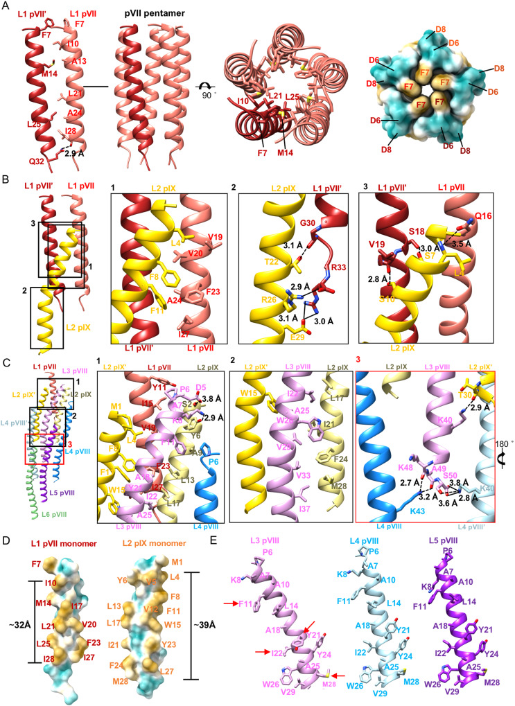
Fig. 2. Structures and detailed interactions in the top cap.
A Ribbon diagrams and surface-rendered representations showing the symmetric plug-like pVII pentamer in side and top views. In the ribbon diagrams, one pVII molecule is colored wine red, while other four pVII moleucles are colored salmon pink. Residues involved in the key contacts are shown in sticks with the C, N, O atoms colored salmon/wine red, blue and red, respectively. The surface is colored cyan for hydrophilic residues and mustard yellow for hydrophobic residues. B Ribbon diagrams showing the interactions between on pIX and two pVIIs. The two pVII molecules are colored salmon pink and wine red, respectively. Residues involved in the key contacts are shown in sticks with the N, O atoms colored blue and red, respectively. The C atoms in the sticks are colored the same as the corresponding ribbon diagrams. The hydrogen bonds and salt bridges are represented with dash lines and solid lines, respectively. C Ribbon diagrams showing the detailed interactions between the pVIIIs in layers 3–6 and the pVII-pIX complex. The zoom-in views at the right showing detailed contacts at different interfaces. The key contacting residues are shown in sticks with the N, O atoms colored salmon, blue and red, respectively. The C atoms in the sticks are colored the same as the corresponding ribbon diagrams. D Surface-rendered representations showing the hydrophobic segments of pVII and pIX. Hydrophilic residues are colored cyan and hydrophobic residues are colored mustard yellow. E Ribbon diagrams showing the key residues involved in the hydrophobic interactions among pVIIIs in layers 3–5. PVIII in layer 3 is colored plum, pVIII in layer 4 is colored light blue and pVIII in layer 5 is colored purple. The key contacting residues are shown in sticks with the N, O atoms colored blue and red, respectively. The C atoms in the sticks are colored the same as the corresponding ribbon diagrams.

