Fig. 2.
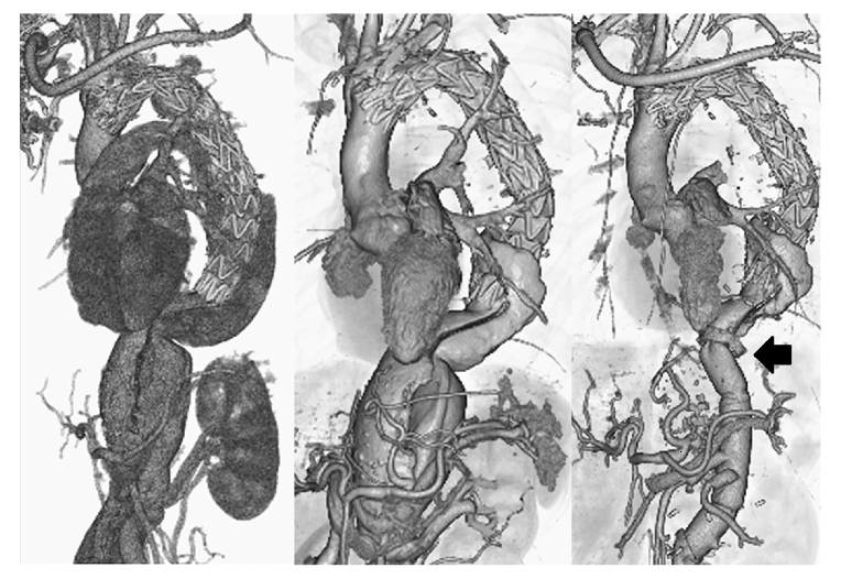
Contrast-enhanced CT images (three-dimensional reconstructed images). Left: Contrast-enhanced CT immediately after 1-debranching TEVAR. Blood flow from the entry immediately below the left subclavian artery has disappeared.
Middle: Contrast-enhanced CT before thoracoabdominal aortic replacement 3 months after the initial surgery. Although blood flow into the false lumen near the stent graft placement site has visibly reduced, the residual false lumen of the thoracoabdominal aorta has enlarged. The aortic diameter was 46.9 mm on the distal side of the left subclavian artery bifurcation, 51.2 mm× 64.0 mm at the pulmonary artery bifurcation, 48.6 mm× 56.2 mm at the left pulmonary vein bifurcation, 45.4 mm× 51.4 mm at the celiac artery bifurcation, 35.7 mm× 38.7 mm above the renal artery, and 14.3 mm× 16.0 mm below the renal artery.
Right: Contrast-enhanced CT after thoracoabdominal aortic replacement. Contrast agent inflow into the false lumen from the proximal anastomosis (Black Marker) of the thoracoabdominal replacement is evident.

