Figure 3.
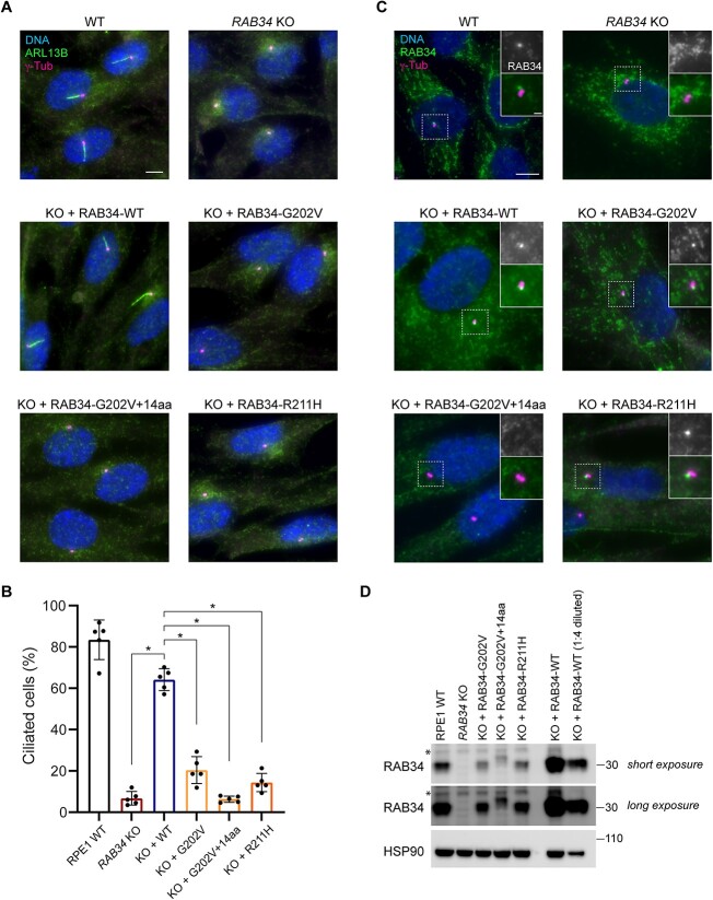
Functional characterization of OFDS-associated RAB34 mutations. (A) Cilia (marked by ARL13B) and centrioles (marked by γ-tubulin) were stained in WT RPE1 cells and RAB34 KO RPE1 cells stably expressing the indicated RAB34 cDNAs. Scale bar: 5 μm. (B) Quantification of ciliogenesis in RPE1 cells shown in (A); bars indicate means, dots show values from > 70 cells analyzed in each of N = 5 independent experiments and error bars show standard deviation across independent experiments. * indicates P < 0.00001. (C) RAB34 was stained in RAB34 KO RPE cells transduced with the indicated RAB34 cDNAs (insets show enlargement of centriolar region). Scale bar: 5 μm (insets: 1 μm). (D) Western blot of the indicated RPE1 cell lines showing expression levels of WT RAB34 and RAB34 variants in addition to HSP90 loading control. Asterisk indicates nonspecific band detected by anti-RAB34 antibody.

