
Figure 1:
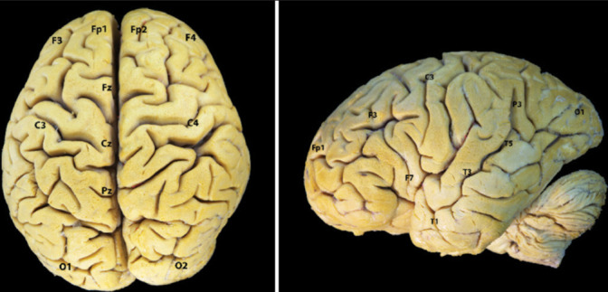
Correlation between electroencephalogram electrodes and cortical anatomy. Superior and lateral view of the brain surface (left side). Fp1: L Superior frontal gyrus, F3: L Middle frontal gyrus, F7: L Inferior frontal gyrus (TP), C3: L Precentral/Postcentral gyrus, T3: L Middle/Superior temporal gyrus*, T5: L Middle temporal gyrus, P3: L Angular gyrus, O1: L Middle occipital gyrus, Fz: Supplementary motor area, Cz: Paracentral lobe, Fp2: R Middle/ Superior frontal G, F4: R Middle Frontal gyrus, F8: R Inferior Frontal gyrus (TP), C4: R Precentral/Postcentral gyrus, T4: R Middle/superior temporal gyrus*, T6: R Middle temporal G **, P4: R Angular gyrus, O2: R Middle occipital gyrus, Pz: Superior parietal gyrus, L: Left, R: Right, *: Rostro-caudal location-posterior to rolandic fissure; **: Caudal to termination of sylvian fissure/posterior part. TP: Triangular part