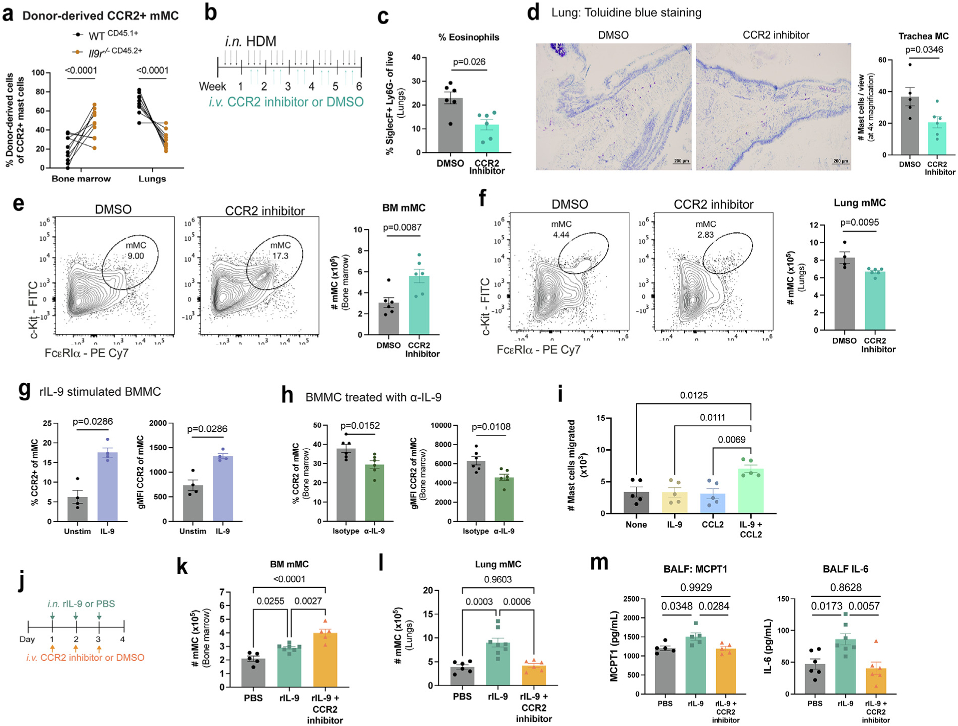
Fig. 4.

IL-9 promotes CCR2-dependent MC migration from the bone marrow to the allergic lung. (A) WT (CD45.1+) and Il9r−/− (CD45.2+) bone marrow cells were transferred to lethally irradiated BoyJ x C57BL/6J F1 mice, allowed to reconstitute immune cells, and treated with HDM for 6 weeks. Flow cytometry analysis of CCR2+ lung MC. (n = 10) Data are representative of two independent experiments with similar results. (B–F) HDM-treated WT mice were treated with CCR2 inhibitor or DMSO control as indicated in (B); C, eosinophil (CD11b+ SiglecF+ Ly6G−) frequencies were determined by flow cytometry. (D) MC in the trachea were assessed using toluidine blue staining. MC numbers were assessed in the bone marrow (E) and lungs (F) by flow cytometry (n = 6). Data are representative of two independent experiments with similar results. (G) Flow cytometry analyses of CCR2 expression in BMMC treated with IL-9 (40 ng/ml) for 2 hours (n = 4). Data are representative of two independent experiments. (H) Flow cytometry analyses of CCR2 expression in bone marrow MC from HDM-treated WT mice treated with anti-IL-9 or an isotype control (n = 6). Data are representative of three independent experiments with similar results. (I) BMMC migration assay toward cytokines and/or chemokines (n = 5). Data are representative of three independent experiments with similar results. (J–M) WT mice, treated intranasally with IL-9 for 3 days, were also intravenously treated with CCR2 inhibitor for 3 days indicated in (J). Flow cytometry analysis of bone marrow (K) and lung (L) MC. (M) MCPT1 and IL-6 protein expression was determined via enzyme-linked immunosorbent assay (n = 5–8). Data are representative of two independent experiments with similar results. Error bars indicate ± standard error of mean. Statistical significance was determined by analysis of variance, followed by Tukey’s multiple comparison test (A, I, K, and L) or Mann-Whitney U test (C, E–H, and M). CD = clusters of differentiation; DMSO = dimethyl sulfoxide; gMFI = geometric mean fluorescence intensity; HDM = house dust mite; IL = interleukin; MC = mast cell; MCp = MC progenitors; MCPT 1 = MC protease 1; mMC = mature MC; ns = not significant; PBS = Phosphate buffered saline; PE = R-phycoerythrin; r = recombinant; SSC = side scatter; WT, wild type.
