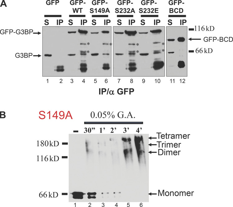
Figure 5.
G3BP oligomerization in vitro. (A) Whole-cell extracts prepared from Cos cells transfected with GFP, GFP-G3BP wild type, phosphorylation mutants S149A, S232A, S232E, and BCD-mutant lacking A domain were immunoprecipitated with anti-GFP antibodies. Proteins were revealed by immunoblot analysis using anti-G3BP antibody. S, supernatant; IP, immunoprecipitation. Asterisks correspond to proteolytic fragments of G3BP-GFP that can be detected by anti-GFP antibodies, whereas the band at the level of G3BP is only seen with the antiG3BP antibody. (B) Glutaraldehyde crosslinking analysis of purified G3BP phosphorylation mutants S149A. Proteins (0.2 g each) were incubated for the indicated time with glutaraldehyde (G.A.) and were then analyzed by Western blotting with an anti-G3BP.

