
Fig. 6.
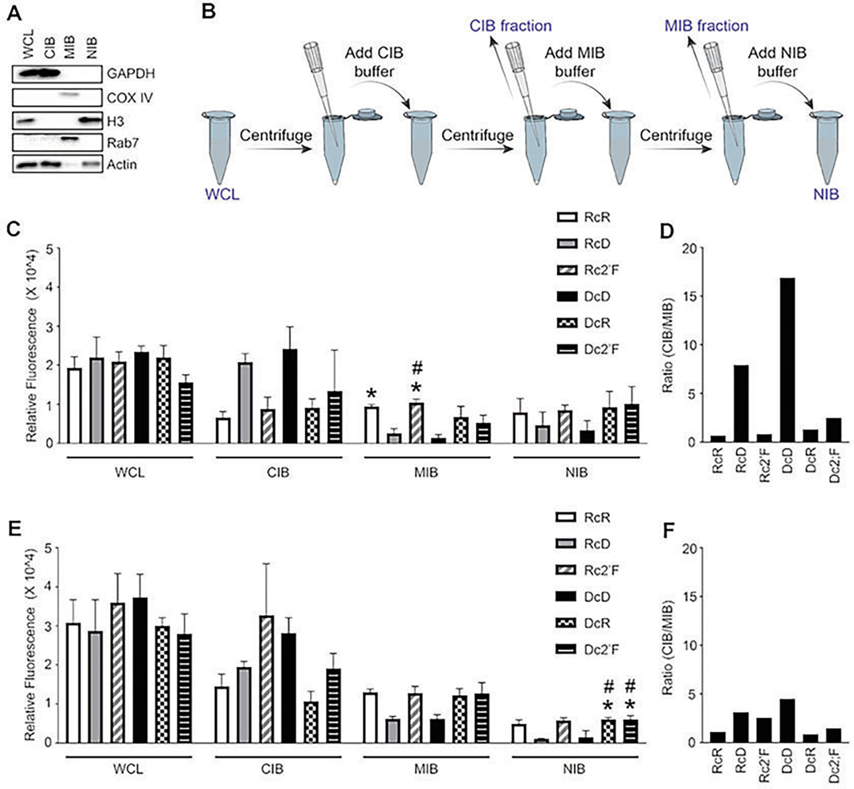
Cellular fractionation into cytosolic (cytosolic isolation buffer, CIB), membrane/organelle (membrane/organelle isolation buffer, MIB), and nuclear/cytoskeleton (nuclear/cytoskeleton isolation buffer, NIB) fractions. (a) Immunoblot analysis for evaluation protein expression of GAPDH, COX IV, histone 3 (H3), Rab7, and actin in fractions. (b) Illustration of cell fractionation protocol. Fluorescence of cell fractions at (c) 2 h and (e) 4 h. The (CIB/MIB) ratio showed at (d) 2 h and (f) 4 h. (Reproduced from Ref. [5])