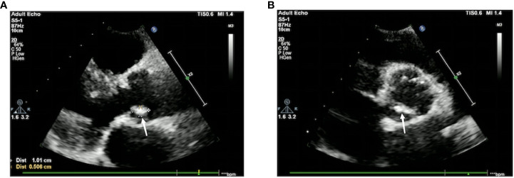
Figure 3.
Cardiac color Doppler ultrasound. There is a slightly echogenic mass measuring approximately 10 mm × 5mm on the aortic valve, with low mobility. (A) The long-axis view of the left ventricle next to the sternum shows an attached vegetation on the aortic valve without coronary valve involvement. (B) The short-axis view of the left ventricle next to the sternum shows the formation of a vegetation on the aortic valve without coronary valve involvement. A small amount of fluid-filled dark area is visible in the pericardial cavity, with an anterior–posterior diameter of 3 mm.

