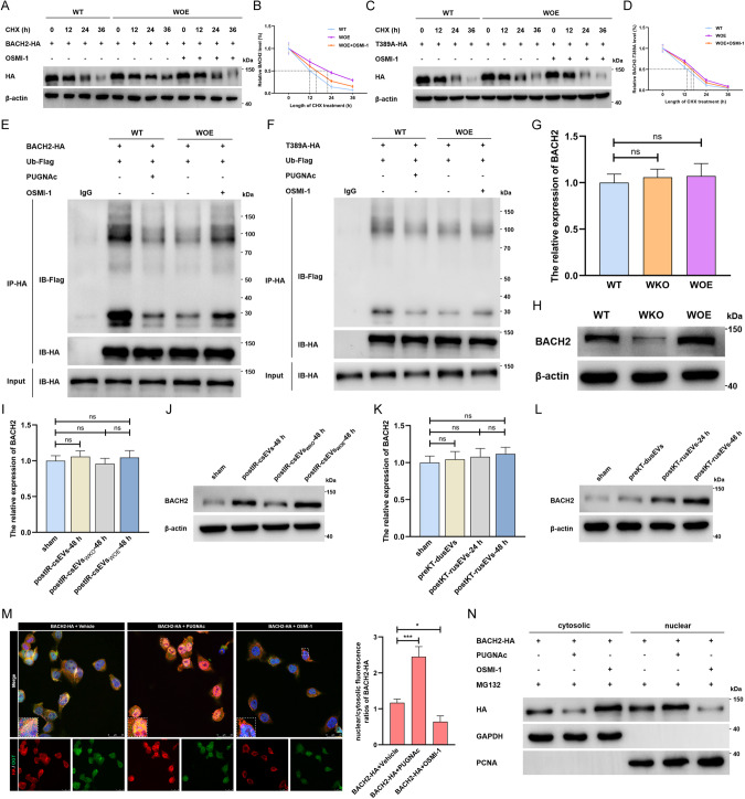
Fig. 5. LncRNA-WAC-AS1-faciliated BACH2 O-GlcNAcylation enhances protein stability and nuclear translocation of BACH2.
A Protein stability detection and half-life analysis of parental BACH2-HA (A, B) and BACH2-Thr389A-HA mutant (C, D) in WT HK-2 cells and WOE HK-2 cells treated with vehicle or 50 μM OSMI-1. The ubiquitination assays of the parental BACH2 (E) and BACH2-Thr389A mutant (F) in WT and WOE HK-2 cells co-transfected with Flag-tagged ubiquitin (Ub-Flag). The transcriptional (G) and translational (H) levels of BACH2 in HK-2 cells with different endogenous lncRNA-WAC-AS1 expression (n = 6 group−1); one-way ANOVA followed by Tukey’s test. I–L The expression of BACH2 in normal HK-2 cells after treatments with IRI-sEVs containing distinct levels of the exogenous lncRNA-WAC-AS1; one-way ANOVA followed by Tukey’s test. M IF (left) and quantitative assays (right) showing the expression, subcellular localization and colocalization of BACH2-HA (red) and OGT (green) in HK-2 cells transfected with BACH2-HA following administration of vehicle, 50 μM PUGNAc or 50 μM OSMI-1; scale bar: 25 μm. N IB results illustrating the cytosolic and nuclear levels of HA-tagged BACH2 in HK-2 cells treated with 40 μM MG132 and 50 μM PUGNAc either or 50 μM OSMI-1. ***p < 0.001 and *p < 0.05 represent significant differences between two groups; ns represents no significant difference.

