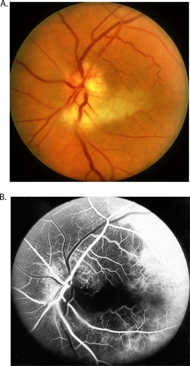
Fig. 11.

Fundus photograph (A) and fluorescein fundus angiogram (B), of left eye of a GCA patient with arteritic AION, and a cilioretinal artery occlusion. A Fundus photograph shows a classical appearance of arteritic AION, i.e., chalky white optic disc oedema with some hyperaemia. B Fluorescein fundus angiogram shows normal filling of the area supplied by the lateral PCA, but no filling of the area supplied by the medial PCA (including the entire optic disc, with no perception of light).
