
Fig. 12.
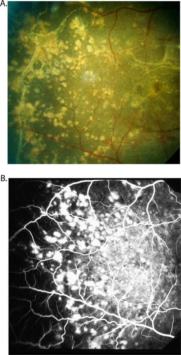
Fundus photograph (A) and fluorescein angiogram (during the late phase) (B) of right eye of a rhesus monkey with malignant arterial hypertension (day 78; blood pressure 190 mmHg). A It shows multiple acute focal retinal pigment epithelial lesions with serous retinal detachment (B). It shows fluorescein staining of the acute focal retinal pigment epithelial lesions.