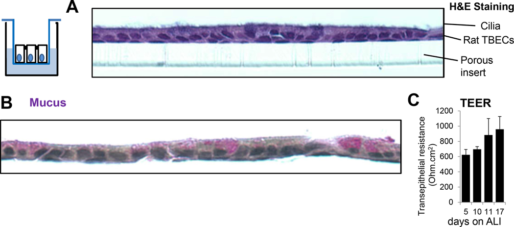
Figure 1. Air-liquid interface cultures of polarized, differentiated rat tracheal epithelial cells provide an excellent in vitro model of the bronchial epithelium.
(A) H&E staining shows formation of a ciliated monolayer of polarized cells after 17 days of culture on ALI. One representative results, n=3. (B) Mayer’s mucicarmine staining detects mucins. These large glycoproteins were identified by positive staining (purple) originating from mucin-producing Goblet cells. One representative result, n=3. (C) Transepithelial resistance (Ohm.cm2) was measured on days 5, 10, 11, 17 post ALI using a voltohmmeter. Mean+/−S.E.M., n=7. TEER, transepithelial electrical resistance; TBEC, tracheobronchial epithelial cell; ALI, air-liquid interface.

