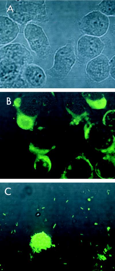
FIG. 2.
DFA staining of J774.1 cell monolayers. The cell monolayers were infected with L. pneumophila SG1 80-045 at 1.6 × 10⁶ CFU/well and were cultured for another 72 h. At the indicated time intervals, the culture medium was discarded and the cell monolayers were stained with fluorescein-conjugated anti-L. pneumophila SG1 antibody. The stained samples were examined under a confocal laser scanning microscope (original magnification, ×600). (A) Control at the time of infection of J774.1 cell monolayers with L. pneumophila; (B) 36 h after infection; note the presence of replicating bacteria within the cytoplasms of the J774.1 cells; (C) 72 h after infection. Note that the majority of the J774.1 cells are destroyed and that only a few remaining cells are present. Note also that these cells are filled with bacteria.

