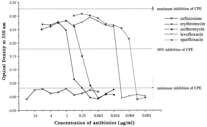
FIG. 6.
Dose-response curves demonstrating the effect of different concentrations of various antimicrobial agents on the cytopathic effect (CPE) of L. pneumophila 80-045 SG1. J774.1 cells (2 × 10⁴/well) were cultured in 96-well microplates for 12 h. The cell monolayers were infected with L. pneumophila 80-045 SG1 (at 8 × 10⁵ CFU/well; 20 times the CPED50) for 12 h. The contents of the microplates were decanted to remove extracellular medium and bacteria after culture at 37°C for 12 h, and then new culture medium (200 μl) containing twofold dilution series of different antibiotics was added to each well. The viability of the J774.1 cells was quantified 72 h later by the MTT assay and was expressed as the optical density at 550 nm. Maximum inhibition of the cytopathic effect was obtained when J774.1 cells were treated with levofloxacin (4 μg/ml); minimum inhibition of the cytopathic effect was recorded when no antibiotic was used. The MIEC resulting in >50% inhibition of the bacterial cytopathic effect was defined as the MIEC of each drug, representing an indicator of the intracellular activity of the antibiotic.

