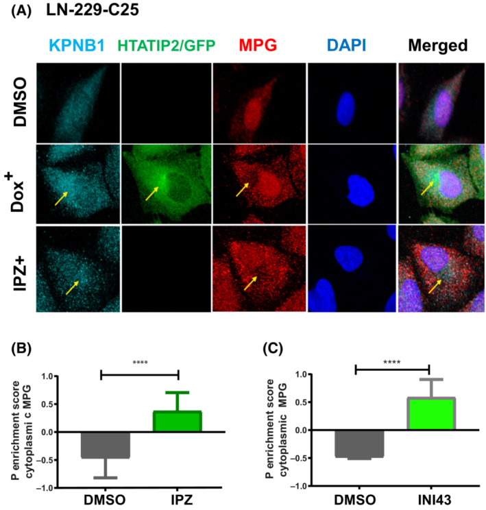
Fig. 3.

HTATIP2 depletion favors nuclear localization of MPG. (A) LN‐229‐C25‐HTATIP2Dox cells were either induced with Dox to express HTATIP2 or treated with 8 nm importazole (IPZ) for 48 h. Cells were visualized by confocal microscopy image using four channels: KPNB1‐Alexa 647 (far red), GFP (488, green), MPG‐Alexa 555 (red), and DAPI (blue). HTATIP2‐GFP localizes mainly in the cytoplasm, with a focus in proximity to the nuclear membrane. HTATIP2 co‐localized with importin β1 (KBNB1) at the location where MPG was excluded. Areas of interest are indicated by arrows. (B, C) Quantification of the P cytoplasmic enrichment score for MPG upon treatment with the pharmacologic inhibitors of KPNB1: (B) IPZ (8 nm); (C) INI‐43 (8 nm). INI‐43 treatment: n = 486 cells, t‐test: ****P < 0.0001. IPZ treatment: n = 263 cells, t‐test: ****P < 0.0001. Representative read‐out of one of three biological replicates, mean ± SD.
