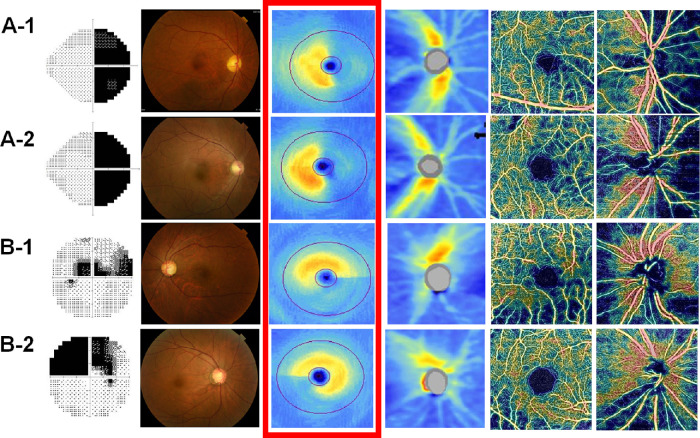
Figure 2.
Representative cases of CON and NTG. (A-1) A 60-year-old female patient 3.4 years after undergoing surgery for craniopharyngioma. The average GCIPL thickness and peripapillary RNFL thickness and the average peripapillary VD and macular VD were 64 µm, 69 µm, 51.5%, and 44.8%, respectively. (A-2) A 48-year-old male patient 3.2 years after surgery for a pituitary adenoma. The average GCIPL thickness and peripapillary RNFL thickness and the average peripapillary VD and macular VD were 64 µm, 64 µm, 49.7%, and 44.1%, respectively. (B-1) A 56-year-old female patient with NTG. The average GCIPL thickness and peripapillary RNFL thickness and the average peripapillary VD and macular VD were 64 µm, 68 µm, 56.6%, and 44.4%, respectively. (B-2) A 46-year-old female patient with NTG. The average GCIPL thickness and peripapillary RNFL thickness and the average peripapillary VD and macular VD were 64 µm, 65 µm, 56.4%, and 50.7%, respectively. The images are presented as automated VF test grayscale maps, color fundus photographs, macular GCIPL thickness maps, peripapillary RNFL thickness maps, macular en face superficial retinal capillary plexus VD maps, and peripapillary en face radial peripapillary capillary VD maps from left to right.

