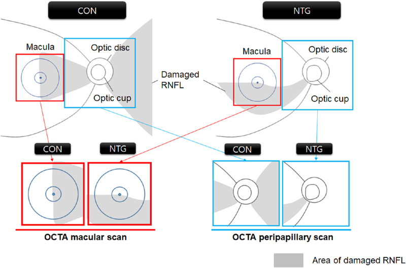
Figure 3.
Different distribution of RNFL damage in CON and NTG detected by macular and peripapillary OCTA scans. The OCTA macula scans detected a 3 × 3 mm2 area centered on the macula (red boxes), and the peripapillary scans detected a 4.5 × 4.5 mm2 area centered on the optic disc (blue boxes). The proportional area of the damaged RNFL (gray shadows) is distributed differently in the macular (red boxes) and peripapillary areas (blue boxes), similarly in the macular area, and markedly differently in the peripapillary area. Note that the diagram, albeit not encompassing all cases, reflects the general conditions.

