Figure 6.

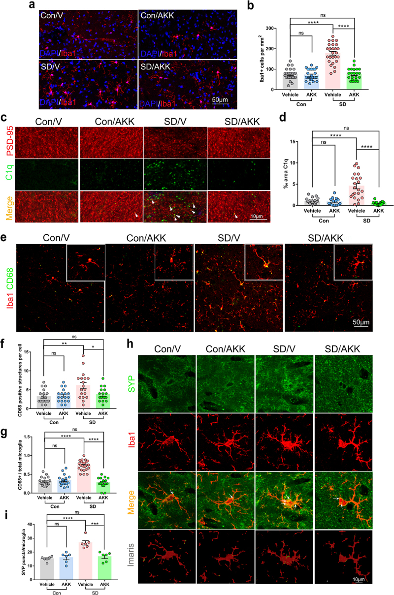
A. muciniphila supplement inhibited extensive microglial activation and synaptic engulfment in the hippocampus of SD mice.
(a) Representative immunofluorescence images of microglia (Iba1+) in the hippocampus of Con/V, Con/AKK, SD/V and SD/AKK mice. Scale bar = 50 μm. n = 22-27 areas per group. (b) The density of Iba1+ cells in the hippocampus of each group.(c) Representative images of C1q (Green) immunoreactivity in the hippocampus of each group and (d) quantification. Scale bar = 10 μm. (e, f and g) Iba1 and CD68 co-stain in the hippocampus of each group. Scale bar = 50 μm. n = 17-18 areas per group. (h and i) Confocal and Imaris reconstruction images showing SYP puncta within Iba1+ microglia in the hippocampus in each group (white arrows). n = 6 cells per group. (One-way ANOVA with Tukey’s multiple comparison tests, *P < .05, **P < .01, ***P < .001, ****P < .0001, ns, no significant difference).
