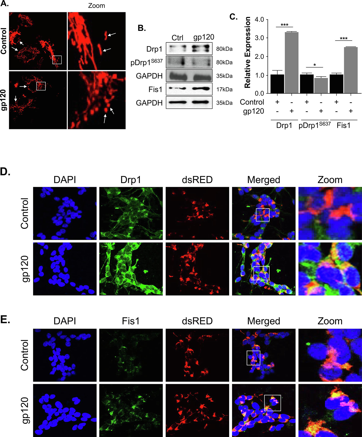
Figure 3. HIV-1 gp120 alters the mitochondrial shape and Drp1 and Fis1 expression levels.

(A) SH-SY5Y cells were transfected with a pDsRed2-Mito expression plasmid for 24 hours and then differentiated for 3 days. The cells were then treated with 100ng/ml of recombinant gp120 protein for an additional 24 hours. Images of the mitochondria were captured using Leica EL600 DMI3000 confocal microscopy 63x lenses. The rod-shaped elongated mitochondria are the healthy mitochondria and the rounded mitochondria seen in gp120-treated cells indicate the swelling of mitochondria and possible increased fission. (B) The western blot analysis represents the protein expressions of total Drp1, phosphor-Drp1S637, and Fis1. GAPDH was used as a loading control. (C) Quantitative analysis of the western blot was presented along with the bands. Data represent the mean ± S.D. Results were judged statistically significant if p<0.05 by analysis of variance. (*p<0.05; **p<0.01; ***p<0.001). (D, E) SH-SY5Y cells were plated on the glass chamber slides followed by transfection with pDsRed2-Mito to label the mitochondria, differentiated for 3 days, then treated with gp120 for an additional 24 hours. An immunocytochemistry assay was performed using anti-Drp1 (D) or anti-Fis1 (E) antibodies. Mitochondria are labeled in red, Drp1 or Fis1 are green, and DAPI (blue) was used to label the nuclei. Colocalization of the proteins with mitochondria is indicated by the presence of an orange or yellow signal (zoom). (Control = Ctrl)
