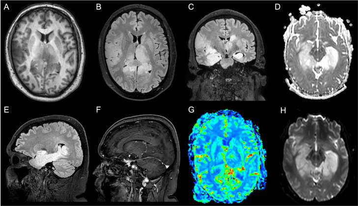
Figure 1.
Patient 1 MRI scan of the brain shows diffuse cortico-subcortical T2 and Fluid Attenuated Inversion Recovery (FLAIR) images hyperintense lesions involving the bilateral hippocampal, the fusiform gyri, the right frontoparietal cortex, the left thalamus, and the right pulvinar. (A) Axial T1-weighted image; (B) Axial FLAIR image; (C) Coronal FLAIR image; (D) Axial apparent diffusion coefficient (ADC) image; (E) Sagittal FLAIR image; (F) Sagittal made of contrast positive T1-weighted image; (G) Axial perfusion weighted (PWI) image; (H) Axial diffusion weighted image (DWI).

