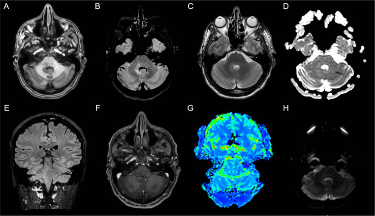
Figure 3.
Patient 2 MRI scans of the brain shows T2-weighted and Fluid Attenuated Inversion Recovery (FLAIR) images hyperintense blurred lesion on the right pontine-bulbar portion and the ipsilateral superior and middle cerebellar peduncles (A) Axial T1-weighted image; (B) Axial FLAIR image; (C) Axial T2-weighted image; (D) Axial apparent diffusion coefficient (ADC) image; (E) Sagittal FLAIR image; (F) Axial made of contrast positive T1-weighted image; (G) Axial perfusion weighted (PWI) image; (H)) Axial diffusion weighted image (DWI).

