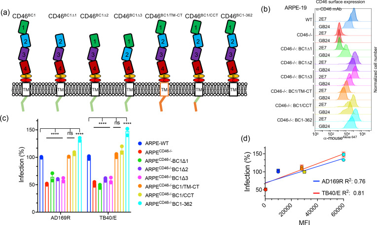
Fig. 4.
The CD46 ectodomain plays a role in HCMV infection. (a) Schematic depicting CD46-BC1 mutant constructs: BC1 lacking SCR1 (Δ1), SCR2 (Δ2), SCR3 (Δ3), CD46-BC1 transmembrane and cytoplasmic tail (TM-CT) replaced with TM-CT of CD4 (BC1/TM-CT), CD46-BC1 whose carboxy-terminal end was replaced with the CD4 carboxy-terminal end (BC1/CCT), and CD46-BC1 carboxy-terminal truncation mutant (BC1-362). (b) Cell surface expression of CD46 molecules on WT ARPE-19, ARPECD46-/-, ARPECD46-/-BC1Δ1, ARPECD46-/-BC1Δ2, ARPECD46-/-BC1Δ3, ARPECD46-/-BC1/TM-CT, ARPECD46-/-BC1/CCT, and ARPECD46-/-BC1-362 was assessed by fluorescence-based flow cytometry with mAb 2E7 (anti-SCR1) and conformational mAb GB24 (anti-SCR4). The normalized cell number was plotted based on Alexa647 fluorescence intensity. (c) HCMV AD169R and TB40/E infection (MOI: 0.25) of ARPE-19 cells, ARPECD46-/- cells, and ARPECD46-/ cells expressing the CD46 BC1 mutants were assessed at 24hpi by Celigo cytometer. Percent infection was measured by an infectivity assay and WT ARPE-19 cells were used to normalize infection to 100 %. s.d. is depicted by errors bars. (d) A linear regression model was used to calculate the Goodness of Fit (R2 ) using infection (%) and mean fluorescence intensity (MFI) of CD46 surface expression of AD169R and TB40/E infected cells. ****P<0.0001 (two-way ANOVA, Sidak’s multiple comparison test) values are compared to ARPE-WT infection.

