
Figure 2.
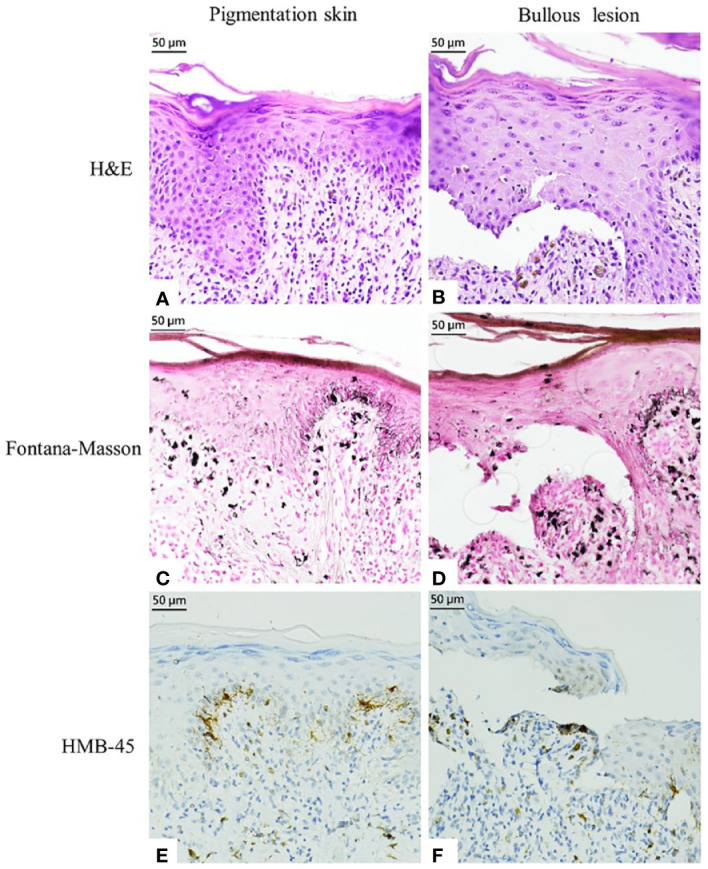
Histopathological examination and immunostaining finding of pigmented markers in the pigmented skin and bullous lesion. (A, B) Hematoxylin and eosin staining. (C, D) Fontana–Masson Staining. (E, F) HMB-45 staining.
Official websites use .gov
A
.gov website belongs to an official
government organization in the United States.
Secure .gov websites use HTTPS
A lock (
) or https:// means you've safely
connected to the .gov website. Share sensitive
information only on official, secure websites.
Histopathological examination and immunostaining finding of pigmented markers in the pigmented skin and bullous lesion. (A, B) Hematoxylin and eosin staining. (C, D) Fontana–Masson Staining. (E, F) HMB-45 staining.