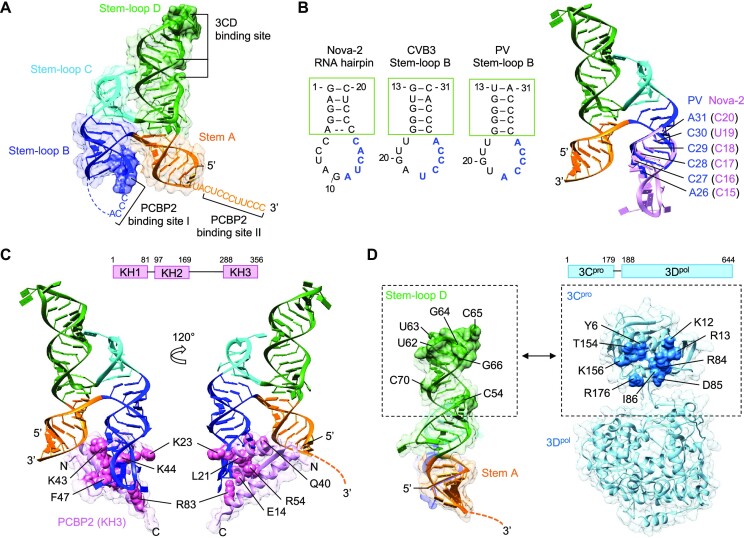
Figure 5.
Model of the ternary complex containing cloverleaf RNA, PCBP2, and 3CDpro. (A) Locations of PCBP2 and 3CDpro binding sites in cloverleaf RNA. Cloverleaf RNA is shown as ribbon diagram in a transparent surface and colored as in Figure 1. The nucleotides indicated in PCBP2 binding in stem–loop B (blue surface) and 3CDpro binding in stem–loop D (green surface) are depicted with darker colors in PV cloverleaf. The 3′ poly-rC site adjacent to stem A (PCBP2 binding site II) is also indicated. (B, C) Model of the cloverleaf-PCBP2 complex. The cloverleaf-PCBP2 (KH3 domain) model was generated using the Nova-2 KH3 domain structure complexed with a 20 nt RNA hairpin (PDB code 1EC6). The nova-2 RNA sequence that interacts with the KH3 domain (11AUCAC15, blue) corresponds to 22UCCCA26 and 22ACCCA26 in CVB3 and PV cloverleaf, respectively (B). The cloverleaf stem–loop B and nova-2 RNA hairpins were overlaid by the stem (green box) to generate a complete cloverleaf structure. Superposed cloverleaf and Nova-2 nucleotides are shown in blue and pink, respectively. For clarity, the PCBP2 KH3 domain is omitted. The cloverleaf-PCBP2 (KH3 domain) complex (C). The KH3 domain residues that directly interact with RNA is shown in pink spheres and labeled. The schematic of the PCBP2 domains is shown on top. (D) Cloverleaf and 3CDpro interaction. Stem-loop D nucleotides and the 3Cpro residues implicated in the cloverleaf and 3Cpro interaction are labeled and shown as green surface on the cloverleaf and blue spheres on the 3CDpro structure (PDB code 2IJD), respectively. 3Dpol also contributes to cloverleaf interaction, but its interaction site on cloverleaf is not known. The schematic of the 3CDpro domains is shown on top.

