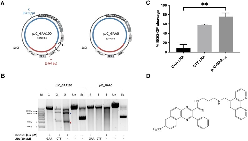
Figure 5.
BQQ-OP mediated DNA cleavage of the (GAA)100•(TTC)100 repeat in the presence of LNA oligomers. (A) Schematic illustration of pJC-GAA100 and pJC-GAA0. The H-DNA forming site in pJC-GAA100 is indicated as [GAA100]; there is no H-DNA forming site in pJC-GAA0 plasmid. The two DNA fragments generated triplex cleavage by BQQ-OP (benzoquinoquinoxaline 1,10-ortho-phenanthroline) followed by unique site restriction digestion are indicated as X (8431 bp) and Y (3997 bp) in pJC-GAA•TTC and the same reaction would result in a linearized fragment only in pJC-GAA0. (B) Representative agarose gel for pJC-GAA100 and pJC-GAA0 plasmids incubated with 10 μM GAA (LNA-DNA) mixmer (lanes 1 and 4 respectively), or CTT (LNA-DNA) mixmer (lanes 2 and 5 respectively), or in the absence of LNAs (lanes 3 and 6 respectively). BQQ-OP-mediated triplex specific cleavage of pJC-GAA100 and pJC-GAA0 was performed in the presence of Cu2+ and 3-mercaptopropionic acid (MPA) followed by unique site restriction digestion with SacI. As controls, supercoiled (Sc) and linearized (Lin) variants of both plasmids and molecular weight DNA ladder (M) are shown. (C) Graph showing the percentage of BQQ-OP-mediated triplex specific cleavage of pJC-GAA100 in the presence of GAA and CTT (LNA-DNA) mixmers or in the absence of LNA-DNA mixmers (pJC-GAA100). The values represent the ratio between the intensity of DNA double strand cleavage (X + Y) to the total band intensity of the particular lane and are shown as mean with S.D. (n = 2). No cleavage was obtained in pJC-GAA0 and not included in the graph. ** indicate P ≤ 0.01 compared to the plasmid in the absence of LNA oligomers. (D) Chemical structure of benzoquinoquinoxaline 1,10-ortho-phenanthroline (BQQ-OP).

