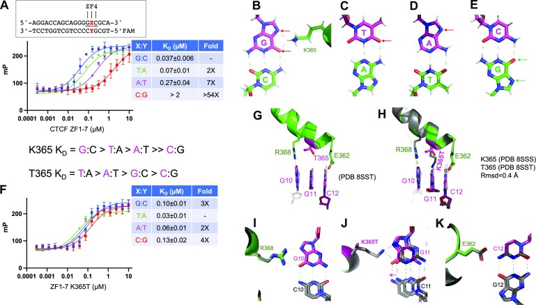
Figure 5.
Structure of K365T mutant in complex with DNA. (A) DNA binding of ZF1–ZF7 (wild type) was quantified by fluorescence polarization. (B) Lys365 provides a proton donor to either N7 nitrogen or O6 oxygen of the guanine ring (indicated by two red arrows). The hydrogen atoms (in gray) are depicted for illustration. (C–E) Base pairs of T:A, A:T and C:G with recognition base in magenta, and opposite pairing base in green. The proton acceptors located in the DNA major groove side are indicated by the arrows and hydrogen atoms are included for illustration. (F) DNA binding affinities of K365T were measured against four possible base pairs at cognate triplet. (G) Structure of K365T of ZF4 in complex with 5′-GCG-3′ triplet. (H) Superimposition of ZF4 wild type and K365T mutant. (I–K) Superimposition of ZF4 wild type and K365T mutant with the cognate triplet.

