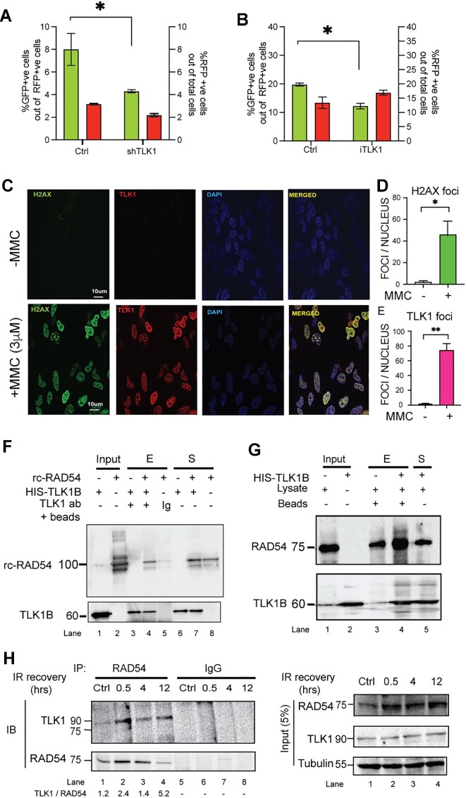
Figure 1.
TLK1 regulates HRR and interacts with RAD54. (A) TLK1 depletion in HeLa-DRGFP using shTLK1 (shRNA#1 shown) and (B) TLK1 inhibition in U2OS-DRGFP cells (by iTLK1) lead to reduced in HRR efficiency. %GFP +ve cells (green bars corresponding to left y-axis) and %RFP +ve cells (red bars corresponding to right y-axis) as SceI transfected cell population in all cell lines (the results are mean ± SEM from three independent experiments. *P< 0.1; P-values were calculated from One-tailed Student's t-tests (unpaired) with Welch's correction). (C–E) TLK1 foci show strong correlation with γH2A.X (labelled-H2A.X) foci upon treatment with MMC (3 μM). One hundred nuclei from three independent experiments were assessed. Results are mean ± SEM. *P< 0.05, **P< 0.01; Unpaired Student's t-tests with Welch's correction. Scale bar is 10 μm. (F) TLK1B interacts with RAD54 (rc-RAD54); immunoprecipitation of recombinant TLK1B incubated with rcRAD54 protein using anti-TLK1- or IgG coated beads. (G) HIS pulldown assay of HIS-TLK1B using Ni-NTA beads incubated with HeLa cell lysate. Interaction between HIS-TLK1B and RAD54 probed by immunoblotting. Upper panel, IB showing RAD54 enriched in TLK1B pulldown sample (lane 4, +HIS-TLK1B). Endogenous RAD54 (75kD) level in lysate (lane 3, - HIS-TLK1B). Lower panel, showing TLK1B band (60 kDa). Input lanes of lysate and TLK1B shown in lane 1 and 2. (H) TLK1 interaction with endogenous RAD54 increases post-irradiation, as shown by co-immunoprecipitation (IP) reaction of RAD54 from HeLa cells treated with IR and allowed to recover for indicated times. TLK1 level normalized to RAD54 pulldown added in bottom panel. Right panel shows the input amounts for each reaction. Note, RAD54 expression is induced by DNA damage.

