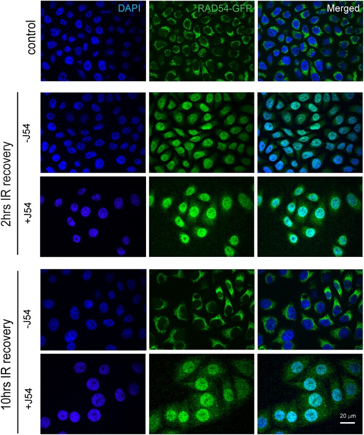
Figure 4.
RAD54-GFP localization changes with TLK1 inhibition after IR (10 Gy). At 2 h IR recovery, RAD54-GFP is predominantly in nucleus (middle panel) and at 10 h after IR (lower panel) when HRR attenuates, RAD54-GFP re-localizes to cytoplasm. With TLK1 inhibition followed by IR and recovery for 2 h (2 h, +J54), RAD54GFP localizes in nucleus. At increased time of IR recovery (10 h, +J54) with TLK1 inhibition, RAD54GFP re-localization to cytoplasm is hindered. Control panel shows RAD54-GFP localization in non-irradiated cells. Images acquired from fixed cell microscopy with DAPI stained nucleus. Scale bar is 20 μm.

