Figure 3.
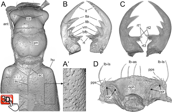
Volume rendering of the holotype specimen of Palaeoiresina cassolai Wiesner et al., 2017 (A, B, D) and Horn’s tiger beetle fossil (C). (A) anterior part of beetle body, dorsal view, with (A’), enlarged section of part of the elytra, showing puncture. (B, C) mandibles, dorsal view. (D) ventral surface of labrum. Abbreviations: ant, antenna; bfb, basal face brush of mandible; ce, compound eye; eph, epipharynx, dorsal surface; hu, humerus; it, incisor tooth; la-as, -ls, apical resp. lateral setae of labrum; pn, pronotum; ppr, parapedial ridge; pps, parapedial setae; rt, retinacular tooth; rt1, 2, 3, retinacular tooth, cusp 1, 2, 3; sc, scutellum (highlighted by dotted line); su, elytral suture; tta, ttb, anterior resp. basal terebral tooth; vm, ventral microtrichia of mandible. The interactive PDF of the microCT reconstruction of the forebody of the holotype specimen of P. cassolai as an embedded 3D image is available at Supplementary figure 3.

