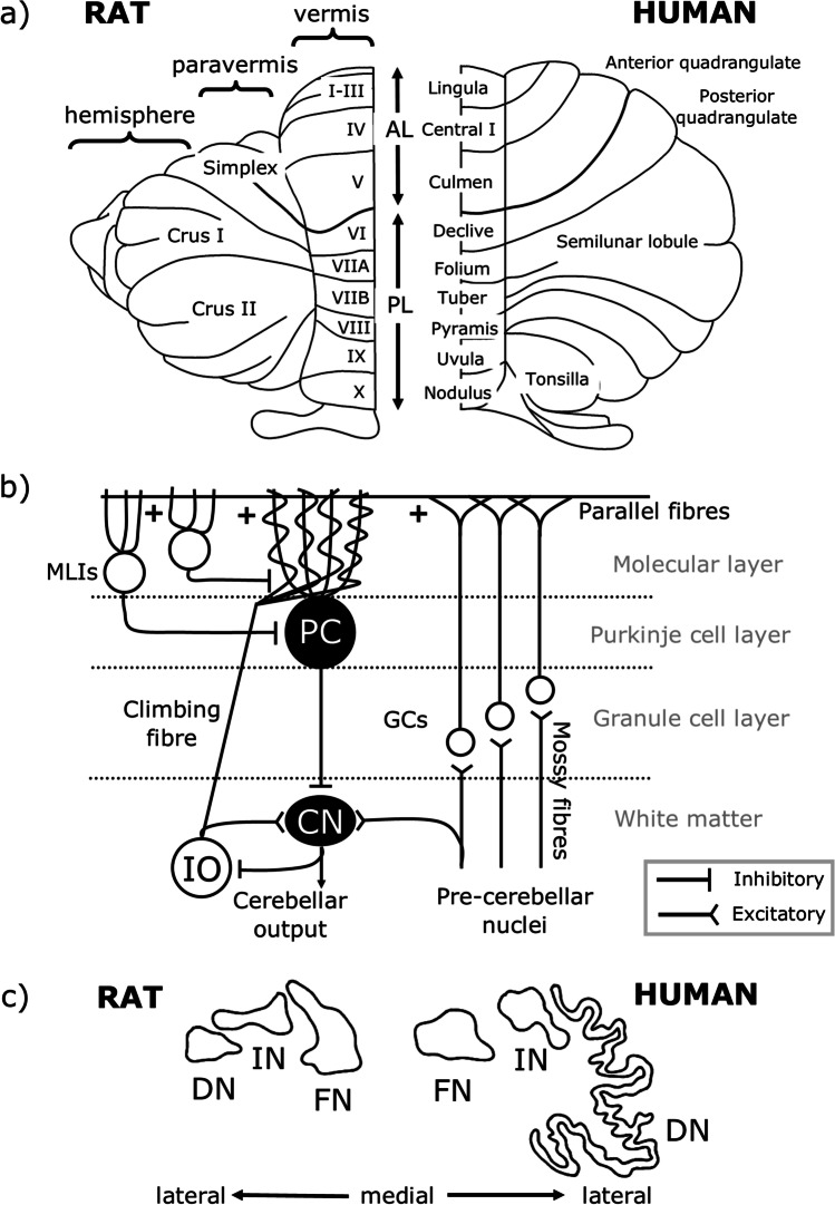
Fig. 1.
Cerebellar anatomical organisation. a Dorsal view of the rat (left) and human (right) cerebellum. There are three main longitudinal compartments of the cerebellar cortex, from medial to lateral the vermis, paravermis and hemisphere. AL, anterior lobe; PL, posterior lobe. b Simplified cerebellar circuitry. Inputs to the cerebellum are from mossy fibres of various pre-cerebellar nuclei and climbing fibres of the inferior olive (IO), both of which are glutamatergic. Mossy fibres synapse onto granule cells (GCs) which form bifurcating axons, known as parallel fibres, targeting Purkinje cell (PC) dendrites, and climbing fibres synapse onto PC dendrites directly. Both mossy fibres and climbing fibres also form collaterals targeting neurons of the cerebellar nuclei (CN). PCs are the sole output neuron of the cerebellar cortex, and these GABAergic neurons target neurons of the CN which form cerebellar output. Several types of interneurons also act within the cerebellar cortex, including molecular layer interneurons (MLIs), not all of which are shown. c Outlines of the rat (left) and human (right) cerebellar nuclei. The vermis, paravermis and hemispheres of the cerebellar cortex project to the fastigial nuclei (FN, also known as medial nuclei), interpositus nuclei (IN) and dentate nuclei (DN, also known as lateral nuclei), respectively. Scaled so that FN is a similar size in both species.
Adapted from Altman and Bayer [26]

