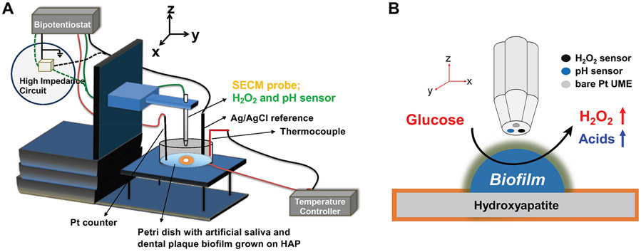
Figure 5.
Schematic of SECM experimental setup with an SECM probe tip, including the H2O2 and pH sensors. (A) SECM stage with Petri dish with dental plaque biofilm grown on hydroxyapatite. The dish is filled with the artificial saliva solution, and the tip is immersed in it to control the distance between the tip and the biofilm. (B) The hydroxyapatite was covered with a layer of 40 μm thickness Kapton tape to expose only 1.5 mm diameter of its surface on which dental plaque biofilm grows. The SECM tip was fixed at 20 μm above the mature biofilm and used to map the chemical profiles of H2O2 and pH in 3D.

