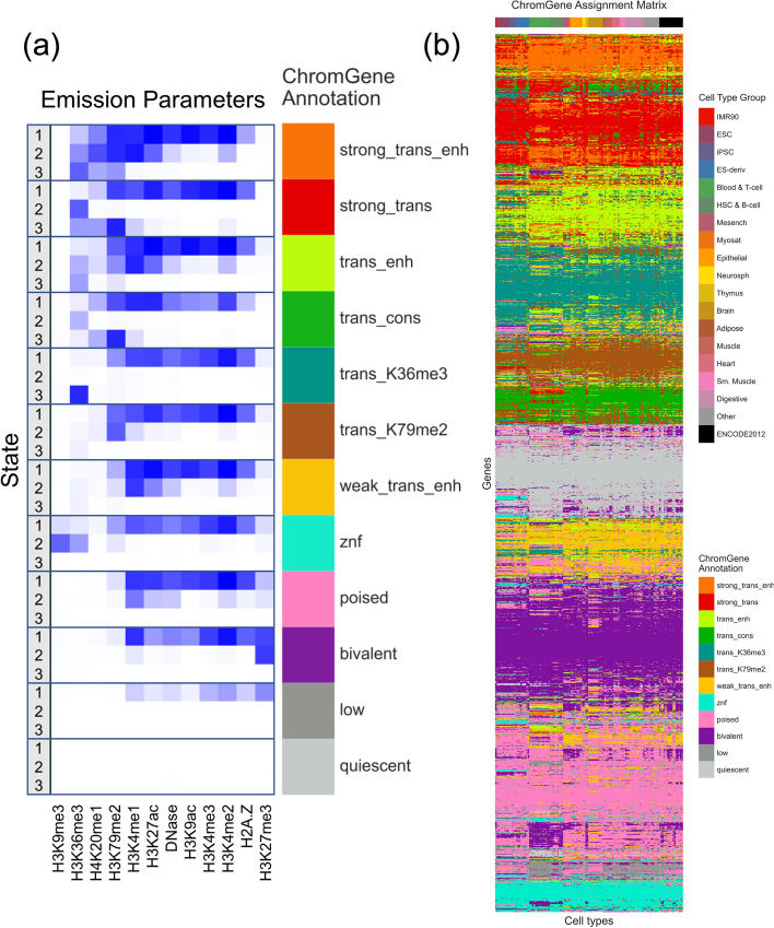
Fig. 2.
The emission parameters and assignment of genes to ChromGene annotations. a Heatmap of emission parameters with blue corresponding to a higher probability and white a lower. The ChromGene annotations are labeled on the right and the states within each annotation are labeled on the left. Annotations are ordered from top to bottom by decreasing expression, and states within each annotation are ordered by decreasing enrichment at the gene TSS. Marks are ordered from left to right as previously done [27]. Transition probabilities are pictured (Additional File 1: Fig. S2); per-state emission probabilities and enrichments, along with transition probabilities, are also reported (Additional File 2: Table S1). b Graphical representation of the ChromGene assignment matrix. Columns correspond to cell types, which are ordered as previously done [1], and their tissue group is indicated by the top colorbar (upper right legend). Rows correspond to 2000 subsampled genes (approximately 10% of all genes). Rows were ordered by hierarchical clustering (“Methods”). Each cell is colored by ChromGene annotation for the corresponding cell type and gene (lower right legend)

