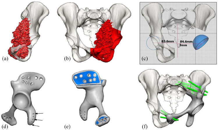
Figure 2.
3D computer renders for the design of a 3DPPI for a patient with a left sided pelvic chondrosarcoma: (a) frontal view of the left hemi-pelvis, with the chondrosarcoma highlighted [red], (b) frontal view of pelvis with the planned P2-3 resection highlighted [red], including a part of the contralateral os pubis, (c) frontal view of the pelvis with the planned centre of hip rotation for the 3DPPI, based on the contralateral side, (d) lateral view of the completed 3DPPI design including suture holes for soft tissue reattachment (arrows), (e) medial view of the 3DPI design, indicating the porous surface areas highlighted [blue], and (f) frontal view of the pelvis with the 3DPPI in place and locking head screw locations and trajectories highlighted in green.
Images used with permission of OSSIS Limited, Christchurch, New Zealand, all rights reserved.

