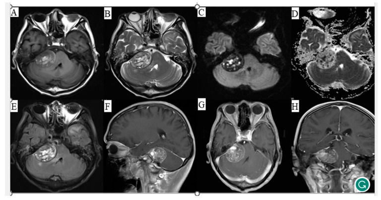
Figure 1.
Magnetic resonance imaging (MRI) of the patient’s head with corresponding image sequences. (A) T1-weighted imaging, (B) T2-weighted imaging, (C) Diffusion-weighted imaging, (D) Apparent diffusion coefficient, (E) Fluid attenuated inversion recovery, (F) Sagittal contrast-enhanced scan, (G) Axial contrast-enhanced scan, (H) Coronal contrast-enhanced scan.

