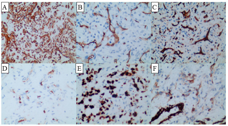
Figure 2.
Immunohistochemical staining of the lesion demonstrated positive reactivity for vimentin (A) throughout and revealed scattered weak positivity for SMA (B) in the vascular regions. Additionally, the vascular regions exhibited positive staining for CD31 (C), ERG (D), Ki-67 (E), and CD34 (F). The original magnification is 20X.

