Persoonia : Molecular Phylogeny and Evolution of Fungi logoLink to Persoonia : Molecular Phylogeny and Evolution of Fungi
. 2021 Dec 24;47:178–374. doi: 10.3767/persoonia.2021.47.06

Fungal Planet description sheets: 1284–1382

PW Crous 1,2, ER Osieck 3, Ž Jurjević 4, J Boers 5, AL van Iperen 1, M Starink-Willemse 1, B Dima 6, S Balashov 4, TS Bulgakov 7, PR Johnston 8, OV Morozova 9, U Pinruan 10, S Sommai 10, P Alvarado 11, CA Decock 12, T Lebel 13, S McMullan-Fisher 14, G Moreno 15, RG Shivas 16, L Zhao 1, J Abdollahzadeh 17, M Abrinbana 18, DV Ageev 19, G Akhmetova 6, AV Alexandrova 20, A Altés 15, AGG Amaral 21, C Angelini 22,23, V Antonín 23, F Arenas 24, P Asselman 25, F Badali 18, A Baghela 26,27, A Bañares 28, RW Barreto 29, IG Baseia 30, J-M Bellanger 31, A Berraf-Tebbal 32, AYu Biketova 33,34, NV Bukharova 35, TI Burgess 36, J Cabero 37, MPS Câmara 21, JF Cano-Lira 38, P Ceryngier 39, R Chávez 40, DA Cowan 41, AF de Lima 21, RL Oliveira 42, S Denman 43, QN Dang 44, F Dovana 45, IG Duarte 21, A Eichmeier 32, A Erhard 4, F Esteve-Raventós 15, A Fellin 46, G Ferisin 47, RJ Ferreira 48, A Ferrer 49, P Finy 50, E Gaya 51, ADW Geering 52, C Gil-Durán 40, K Glässnerová 53, AM Glushakova 20,54, D Gramaje 55, FE Guard 56, AL Guarnizo 24, D Haelewaters 25,57, RE Halling 58, R Hill 51, Y Hirooka 59, V Hubka 53,60, VA Iliushin 9, DD Ivanova 61, NE Ivanushkina 62, P Jangsantear 63, A Justo 64, AV Kachalkin 20,62, S Kato 59, P Khamsuntorn 65, IY Kirtsideli 9, DG Knapp 6, GA Kochkina 62, O Koukol 66, GM Kovács 6, J Kruse 67, TKA Kumar 68, I Kušan 69, T Læssøe 70, E Larsson 71, R Lebeuf 72, G Levicán 40, M Loizides 73, P Marinho 74, JJ Luangsa-ard 10, EG Lukina 75, V Magaña-Dueñas 38, G Maggs-Kölling 76, EF Malysheva 9, VF Malysheva 9, B Martín 77, MP Martín 78, N Matočec 69, AR McTaggart 79, M Mehrabi-Koushki 80,81, A Mešić 69, AN Miller 82, P Mironova 25, P-A Moreau 83, A Morte 24, K Müller 84, LG Nagy 33, S Nanu 68, A Navarro-Ródenas 24, WJ Nel 2, TH Nguyen 44, TF Nóbrega 29, ME Noordeloos 85, I Olariaga 86, BE Overton 87, SM Ozerskaya 62, P Palani 88, F Pancorbo 89, V Papp 90, J Pawłowska 91, TQ Pham 44, C Phosri 92, ES Popov 9, A Portugal 93,94, A Pošta 69, K Reschke 95, M Reul 96, GM Ricci 87, A Rodríguez 24, J Romanowski 39, N Ruchikachorn 97, I Saar 98, A Safi 80, B Sakolrak 63, F Salzmann 99, M Sandoval-Denis 1, E Sangwichein 100, L Sanhueza 49, T Sato 101, A Sastoque 38, B Senn-Irlet 102, A Shibata 59, K Siepe 103, S Somrithipol 10, M Spetik 32, P Sridhar 88, AM Stchigel 38, K Stuskova 32, N Suwannasai 104, YP Tan 105, R Thangavel 106, I Tiago 93, S Tiwari 26,27, Z Tkalčec 69, MA Tomashevskaya 62, C Tonegawa 59, HX Tran 44, NT Tran 52, J Trovão 93, VE Trubitsyn 62, J Van Wyk 107, WAS Vieira 21, J Vila 108, CM Visagie 2, A Vizzini 109, SV Volobuev 9, DT Vu 110, N Wangsawat 111, T Yaguchi 60, E Ercole 112, BW Ferreira 29, AP de Souza 113, BS Vieira 113, JZ Groenewald 1
PMCID: PMC10486635  PMID: 37693795

Abstract

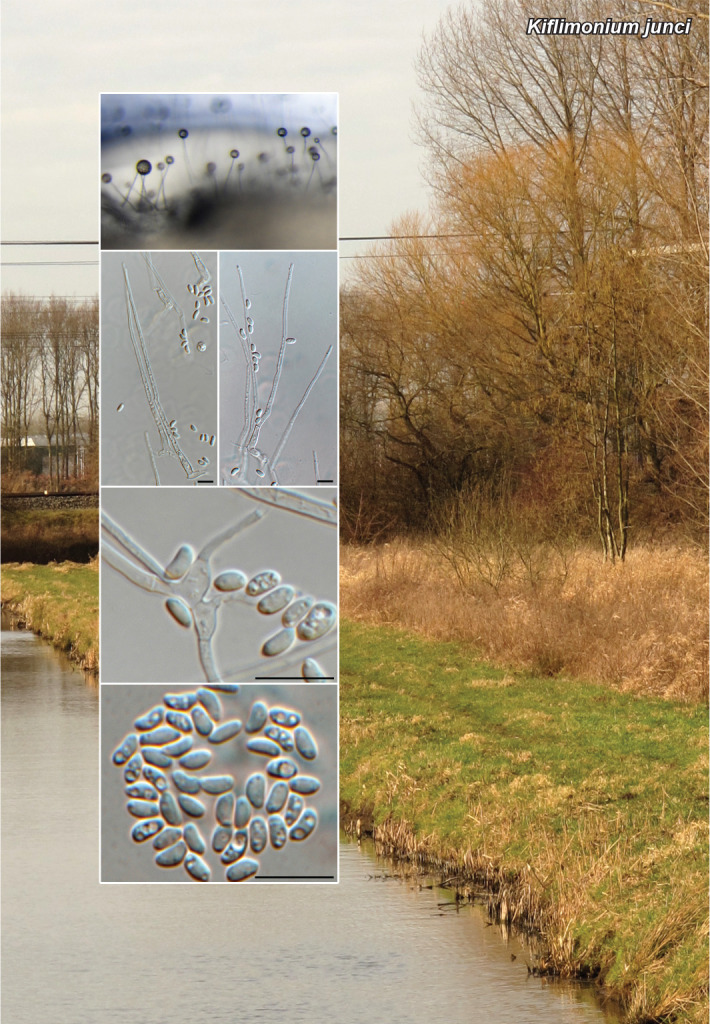
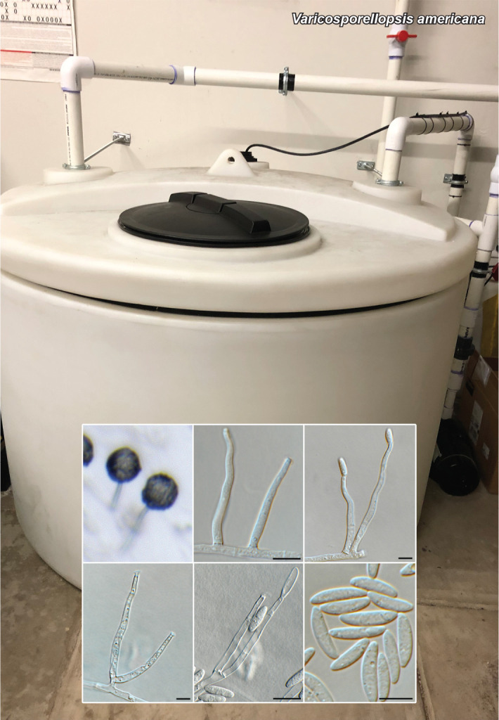
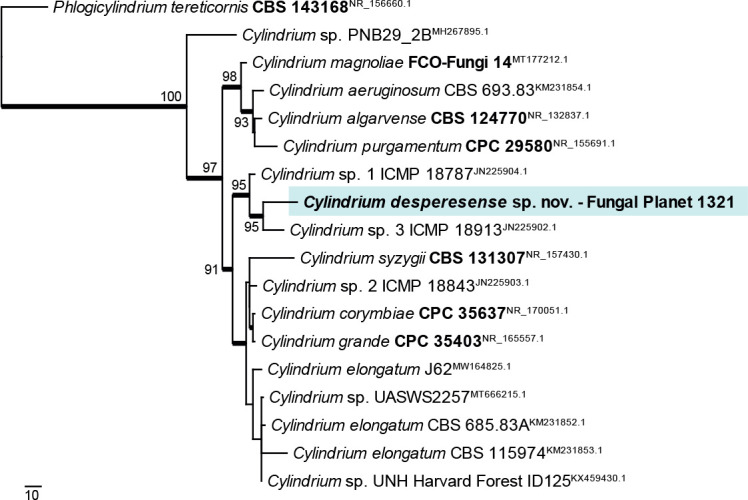
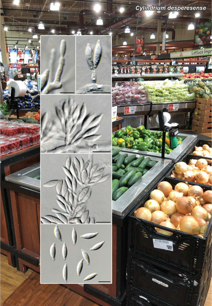
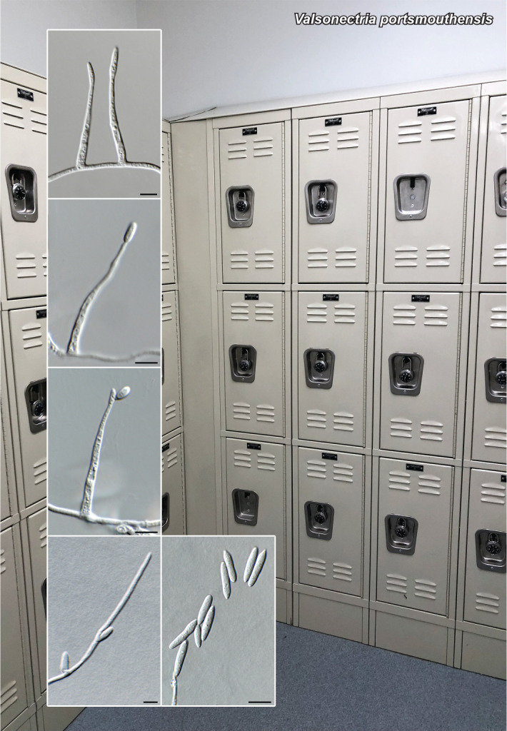
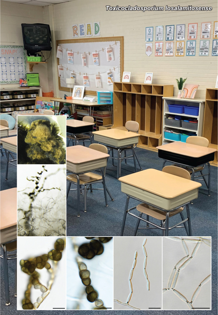
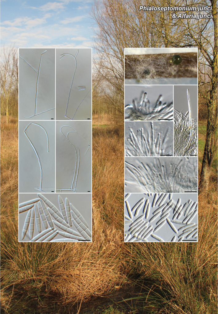
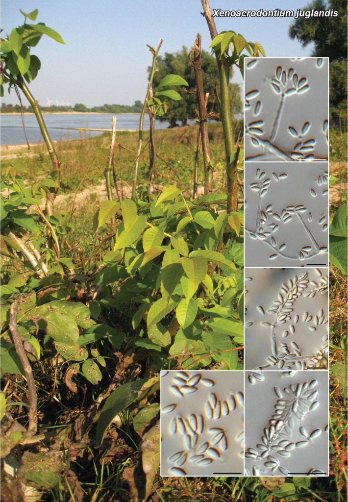
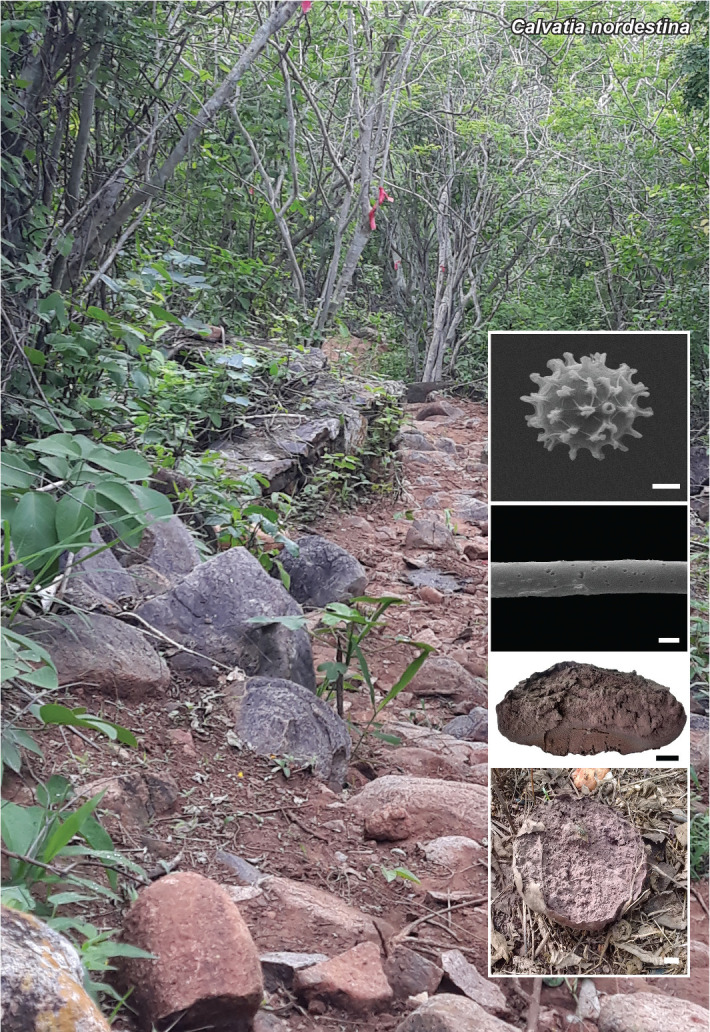
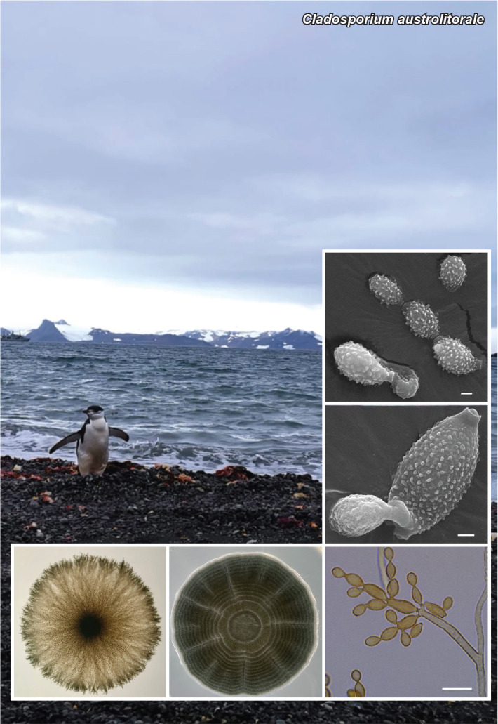
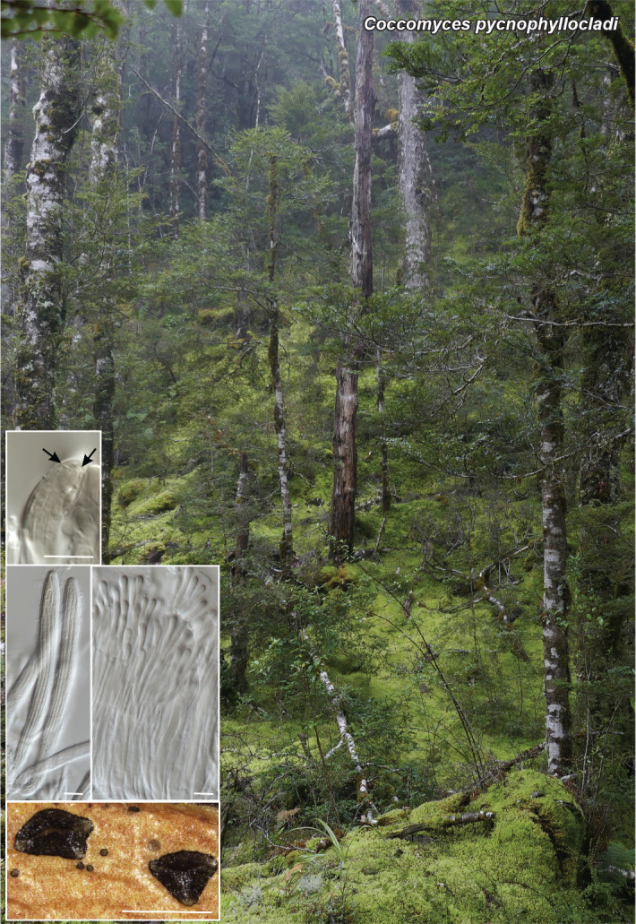
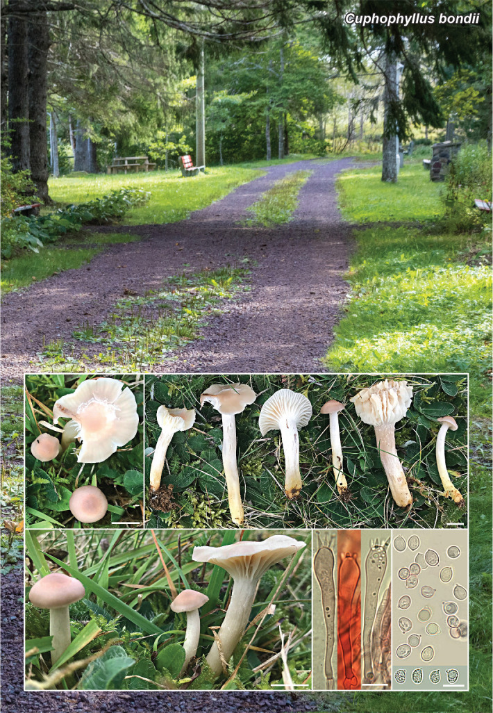
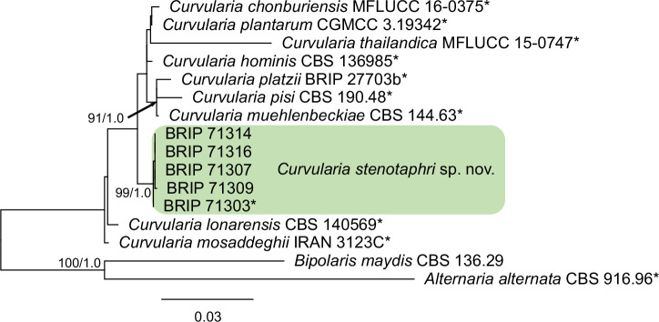
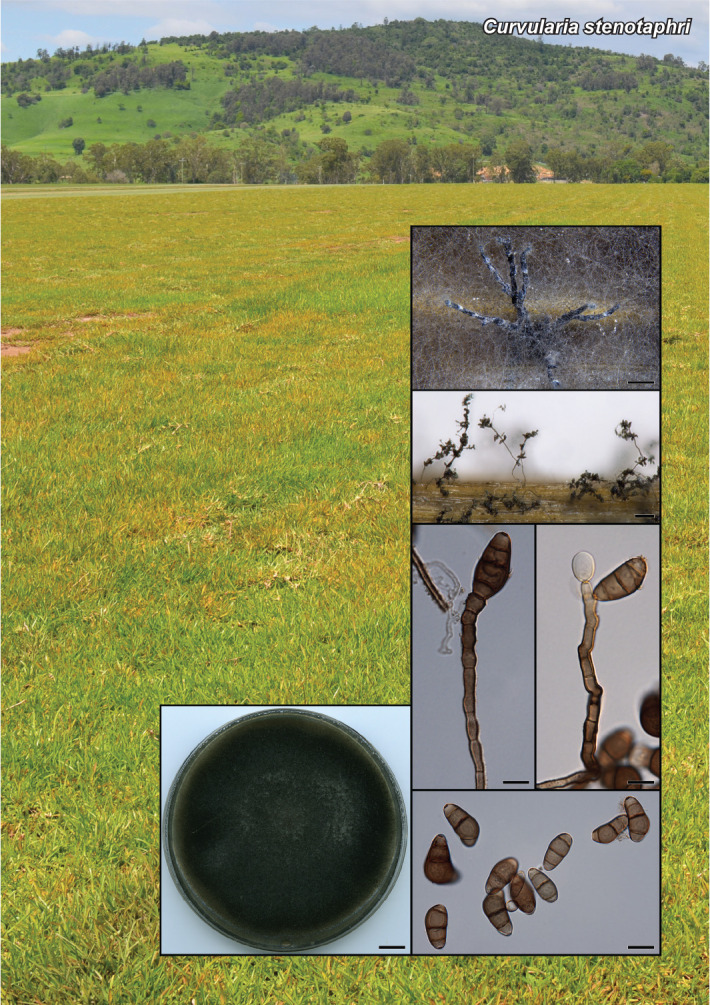
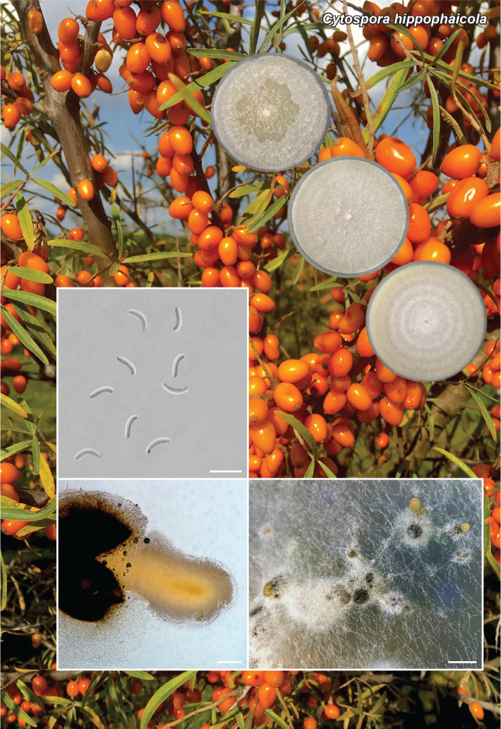
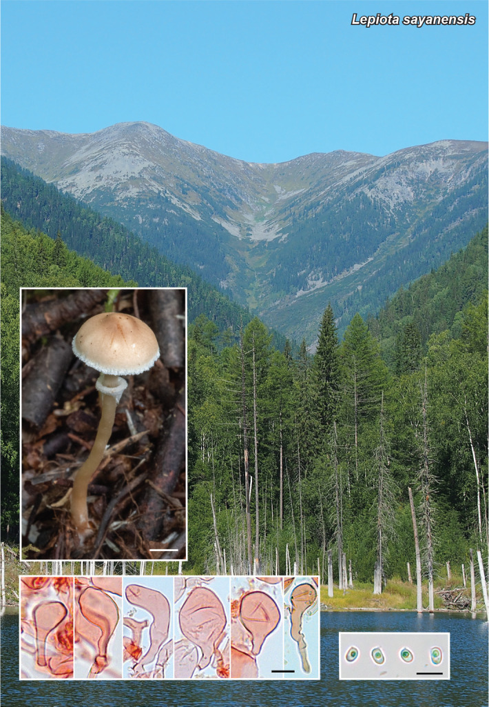
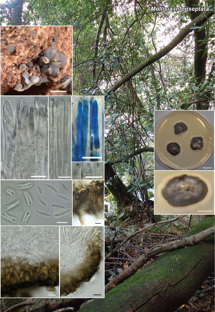
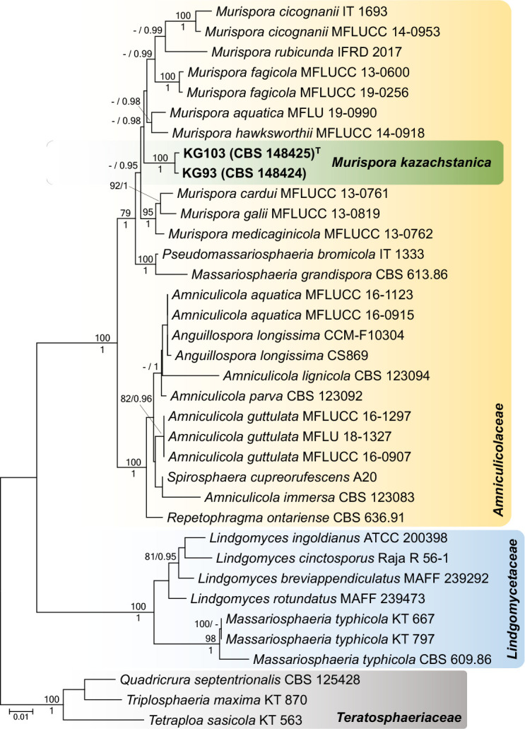
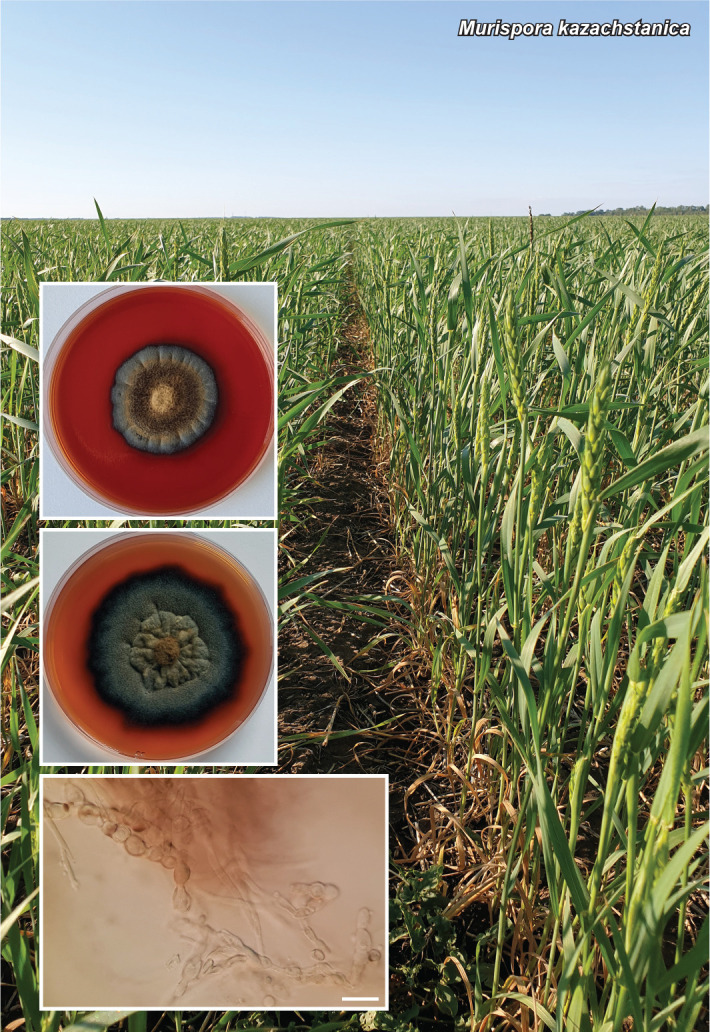
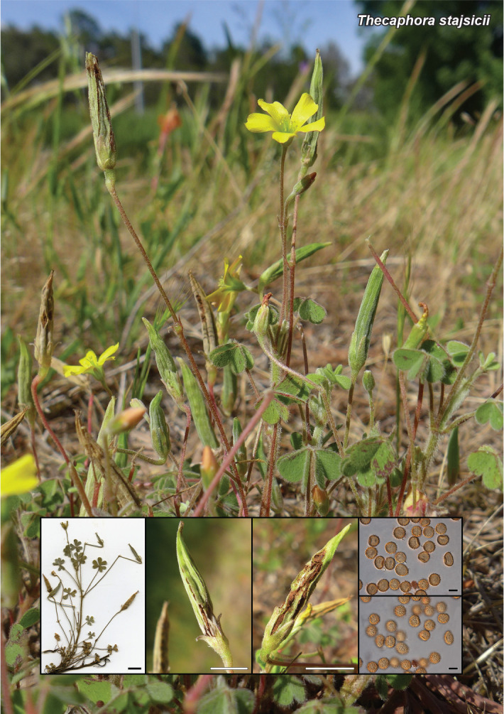
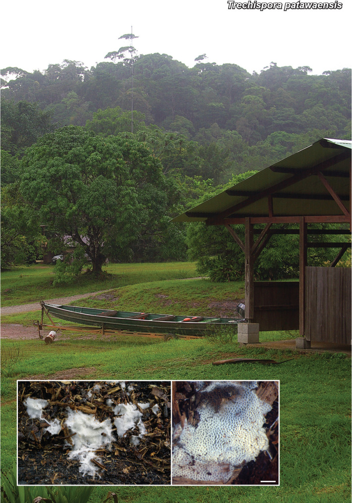
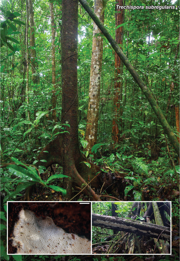
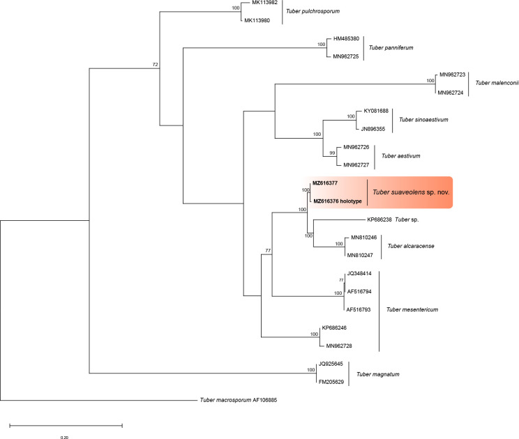
Novel species of fungi described in this study include those from various countries as follows: Antartica, Cladosporium austrolitorale from coastal sea sand. Australia, Austroboletus yourkae on soil, Crepidotus innuopurpureus on dead wood, Curvularia stenotaphri from roots and leaves of Stenotaphrum secundatum and Thecaphora stajsicii from capsules of Oxalis radicosa. Belgium, Paraxerochrysium coryli (incl. Paraxerochrysium gen. nov.) from Corylus avellana. Brazil, Calvatia nordestina on soil, Didymella tabebuiicola from leaf spots on Tabebuia aurea, Fusarium subflagellisporum from hypertrophied floral and vegetative branches of Mangifera indica and Microdochium maculosum from living leaves of Digitaria insularis. Canada, Cuphophyllus bondii from a grassland. Croatia, Mollisia inferiseptata from a rotten Laurus nobilis trunk. Cyprus, Amanita exilis on calcareous soil. Czech Republic, Cytospora hippophaicola from wood of symptomatic Vaccinium corymbosum. Denmark, Lasiosphaeria deviata on pieces of wood and herbaceous debris. Dominican Republic, Calocybella goethei among grass on a lawn. France (Corsica), Inocybe corsica on wet ground. France (French Guiana), Trechispora patawaensis on decayed branch of unknown angiosperm tree and Trechispora subregularis on decayed log of unknown angiosperm tree. Germany, Paramicrothecium sambuci (incl. Paramicrothecium gen. nov.) on dead stems of Sambucus nigra. India, Aureobasidium microtermitis from the gut of a Microtermes sp. termite, Laccaria diospyricola on soil and Phylloporia tamilnadensis on branches of Catunaregam spinosa. Iran, Pythium serotinoosporum from soil under Prunus dulcis. Italy, Pluteus brunneovenosus on twigs of broadleaved trees on the ground. Japan, Heterophoma rehmanniae on leaves of Rehmannia glutinosa f. hueichingensis. Kazakhstan, Murispora kazachstanica from healthy roots of Triticum aestivum. Namibia, Caespitomonium euphorbiae (incl. Caespitomonium gen. nov.) from stems of an Euphorbia sp. Netherlands, Alfaria junci, Myrmecridium junci, Myrmecridium juncicola, Myrmecridium juncigenum, Ophioceras junci, Paradinemasporium junci (incl. Paradinemasporium gen. nov.), Phialoseptomonium junci, Sporidesmiella juncicola, Xenopyricularia junci and Zaanenomyces quadripartis (incl. Zaanenomyces gen. nov.), from dead culms of Juncus effusus, Cylindromonium everniae and Rhodoveronaea everniae from Evernia prunastri, Cyphellophora sambuci and Myrmecridium sambuci from Sambucus nigra, Kiflimonium junci, Sarocladium junci, Zaanenomyces moderatricis-academiae and Zaanenomyces versatilis from dead culms of Juncus inflexus, Microcera physciae from Physcia tenella, Myrmecridium dactylidis from dead culms of Dactylis glomerata, Neochalara spiraeae and Sporidesmium spiraeae from leaves of Spiraea japonica, Neofabraea salicina from Salix sp., Paradissoconium narthecii (incl. Paradissoconium gen. nov.) from dead leaves of Narthecium ossifragum, Polyscytalum vaccinii from Vaccinium myrtillus, Pseudosoloacrosporiella cryptomeriae (incl. Pseudosoloacrosporiella gen. nov.) from leaves of Cryptomeria japonica, Ramularia pararhabdospora from Plantago lanceolata, Sporidesmiella pini from needles of Pinus sylvestris and Xenoacrodontium juglandis (incl. Xenoacrodontium gen. nov. and Xenoacrodontiaceae fam. nov.) from Juglans regia. New Zealand, Cryptometrion metrosideri from twigs of Metrosideros sp., Coccomyces pycnophyllocladi from dead leaves of Phyllocladus alpinus, Hypoderma aliforme from fallen leaves Fuscopora solandri and Hypoderma subiculatum from dead leaves Phormium tenax. Norway, Neodevriesia kalakoutskii from permafrost and Variabilispora viridis from driftwood of Picea abies. Portugal, Entomortierella hereditatis from a biofilm covering a deteriorated limestone wall. Russia, Colpoma junipericola from needles of Juniperus sabina, Entoloma cinnamomeum on soil in grasslands, Entoloma verae on soil in grasslands, Hyphodermella pallidostraminea on a dry dead branch of Actinidia sp., Lepiota sayanensis on litter in a mixed forest, Papiliotrema horticola from Malus communis, Paramacroventuria ribis (incl. Paramacroventuria gen. nov.) from leaves of Ribes aureum and Paramyrothecium lathyri from leaves of Lathyrus tuberosus. South Africa, Harzia combreti from leaf litter of Combretum collinum ssp. sulvense, Penicillium xyleborini from Xyleborinus saxesenii, Phaeoisaria dalbergiae from bark of Dalbergia armata, Protocreopsis euphorbiae from leaf litter of Euphorbia ingens and Roigiella syzygii from twigs of Syzygium chordatum. Spain, Genea zamorana on sandy soil, Gymnopus nigrescens on Scleropodium touretii, Hesperomyces parexochomi on Parexochomus quadriplagiatus, Paraphoma variabilis from dung, Phaeococcomyces kinklidomatophilus from a blackened metal railing of an industrial warehouse and Tuber suaveolens in soil under Quercus faginea. Svalbard and Jan Mayen, Inocybe nivea associated with Salix polaris. Thailand, Biscogniauxia whalleyi on corticated wood. UK, Parasitella quercicola from Quercus robur. USA, Aspergillus arizonicus from indoor air in a hospital, Caeliomyces tampanus (incl. Caeliomyces gen. nov.) from office dust, Cippumomyces mortalis (incl. Cippumomyces gen. nov.) from a tombstone, Cylindrium desperesense from air in a store, Tetracoccosporium pseudoaerium from air sample in house, Toxicocladosporium glendoranum from air in a brick room, Toxicocladosporium losalamitosense from air in a classroom, Valsonectria portsmouthensis from air in men’s locker room and Varicosporellopsis americana from sludge in a water reservoir. Vietnam, Entoloma kovalenkoi on rotten wood, Fusarium chuoi inside seed of Musa itinerans, Micropsalliota albofelina on soil in tropical evergreen mixed forests and Phytophthora docyniae from soil and roots of Docynia indica. Morphological and culture characteristics are supported by DNA barcodes.

Citation: Crous PW, Osieck ER, Jurjević Ž, et al. 2021. Fungal Planet description sheets: 1284–1382. Persoonia 47: 178–374. https://doi.org/10.3767/persoonia.2021.47.06.

Keywords: ITS nrDNA barcodes, LSU, new taxa, systematics

Acknowledgments

P.R. Johnston thanks J. Sullivan (Lincoln University) for the habitat image of Kowai Bush, Duckchul Park (Manaaki Whenua – Landcare Research) for the DNA sequencing, and the New Zealand Department of Conservation for permission to collect the specimens; this research was supported through the Manaaki Whenua – Landcare Research Biota Portfolio with funding from the Science and Innovation Group of the New Zealand Ministry of Business, Innovation and Employment. V. Hubka was supported by the Czech Ministry of Health (grant number NU21-05-00681), and is grateful for the support from the Japan Society for the Promotion of Science – grant-in-aid for JSPS research fellow (grant no. 20F20772). K. Glässnerová was supported by the Charles University Grant Agency (grant No. GAUK 140520). J. Trovão and colleagues were financed by FEDER- Fundo Europeu de Desenvolvimento Regional funds through the COMPETE 2020 – Operational Programme for Competitiveness and Internationalisation (POCI), and by Portuguese funds through FCT – Fundação para a Ciência e a Tecnologia in the framework of the project POCI-01-0145-FEDER-PTDC/EPH-PAT/3345/2014. This work was carried out at the R&D Unit Centre for Functional Ecology – Science for People and the Planet (CFE), with reference UIDB/04004/2020, financed by FCT/MCTES through national funds (PIDDAC). J. Trovão was also supported by POCH – Programa Operacional Capital Humano (co-funding by the European Social Fund and national funding by MCTES), through a ‘FCT – Fundação para a Ciência e Tecnologia’ PhD research grant (SFRH/BD/132523/2017). D. Haelewaters acknowledges support from the Research Foundation – Flanders (Junior Postdoctoral Fellowship 1206620N). M. Loizides and colleagues are grateful to Y. Cherniavsky for contributing collections AB A12-058-1 and AB A12-058-2, and Á. Kovács and B. Kiss for their help with molecular studies of these specimens. C. Zmuda is thanked for assisting with the collection of ladybird specimens infected with Hesperomyces parexochomi. A.V. Kachalkin and colleagues were supported by the Russian Science Foundation (grant No. 19-74-10002). The study of A.M. Glushakova was carried out as part of the Scientific Project of the State Order of the Government of Russian Federation to Lomonosov Moscow State University No. 121040800174-6. S. Nanu acknowledges the Kerala State Council for Science, Technology and Environment (KSCSTE) for granting a research fellowship and is grateful to the Chief Conservator of Forests and Wildlife for giving permission to collect fungal samples. A. Bañares and colleagues thank L. Monje and A. Pueblas of the Department of Drawing and Scientific Photography at the University of Alcalá for their help in the digital preparation of the photographs, and J. Rejos, curator of the AH herbarium for his assistance with the specimens examined in the present study. The research of V. Antonín received institutional support for long-term conceptual development of research institutions provided by the Ministry of Culture (Moravian Museum, ref. MK000094862). The studies of E.F. Malysheva, V.F. Malysheva, O.V. Morozova, and S.V. Volobuev were carried out within the framework of a research project of the Komarov Botanical Institute RAS, St Petersburg, Russia (AAAA-A18-118022090078-2) using equipment of its Core Facility Centre ‘Cell and Molecular Technologies in Plant Science’.The study of A.V. Alexandrova was carried out as part of the Scientific Project of the State Order of the Government of Russian Federation to Lomonosov Moscow State University No. 121032300081-7. The Kits van Waveren Foundation (Rijksherbariumfonds Dr E. Kits van Waveren, Leiden, Netherlands) contributed substantially to the costs of sequencing and travelling expenses for M.E. Noordeloos. The work of B. Dima was partly supported by the ÚNKP-20-4 New National Excellence Program of the Ministry for Innovation and Technology from the source of the National Research, Development and Innovation Fund. The work of L. Nagy was supported by the ‘Momentum’ program of the Hungarian Academy of Sciences (contract No. LP2019-13/2019 to L.G.N.). G.A. Kochkina and colleagues acknowledge N. Demidov for the background photograph, and N. Suzina for the SEM photomicrograph. The research of C.M. Visagie and W.J. Nel was supported by the National Research Foundation grant no 118924 and SFH170610239162. C. Gil-Durán acknowledges Agencia Nacional de Investigación y Desarrollo, Ministerio de Ciencia, Tecnología, Conocimiento e Innovación, Gobierno de Chile, for grant ANID – Fondecyt de Postdoctorado 2021 – N° 3210135. R. Chávez and G. Levicán thank DICYT-USACH and acknowledges the grants INACH RG_03-14 and INACH RT_31-16 from the Chilean Antarctic Institute, respectively. S. Tiwari and A. Baghela would like to acknowledge R. Avchar and K. Balasubramanian from the Agharkar Research Institute, Pune, Maharashtra for helping with the termite collection. S. Tiwari is also thankful to the University Grants Commission, Delhi (India) for a junior research fellowship (827/(CSIR-UGC NET DEC.2017)). R. Lebeuf and I. Saar thank D. and H. Spencer for collecting and photographing the holotype of C. bondii, and R. Smith for photographing the habitat. A. Voitk is thanked for helping with the colour plate and review of the manuscript, and the Foray Newfoundland and Labrador for providing the paratype material. I. Saar was supported by the Estonian Research Council (grant PRG1170) and the European Regional Development Fund (Centre of Excellence EcolChange). M.P.S. Câmara acknowledges the ‘Conselho Nacional de Desenvolvimento Científico e Tecnológico – CNPq’ for the research productivity fellowship, and financial support (Universal number 408724/2018-8). W.A.S. Vieira acknowledges the ‘Coordenação de Aperfeiçoamento Pessoal de Ensino Superior – CAPES’ and the ‘Programa Nacional de Pós-Doutorado/CAPES – PNPD/CAPES’ for the postdoctoral fellowship. A.G.G. Amaral acknowledges CNPq, and A.F. Lima and I.G. Duarte acknowledge CAPES for the doctorate fellowships. F. Esteve-Raventós and colleagues were financially supported by FEDER/Ministerio de Ciencia, Innovación y Universidades – Agencia Estatal de Investigación (Spain)/ Project CGL2017-86540-P. The authors would like to thank L. Hugot and N. Suberbielle (Conservatoire Botanique National de Corse, Office de l’Environnement de la Corse, Corti) for their help. The research of E. Larsson is supported by The Swedish Taxonomy Initiative, SLU Artdatabanken, Uppsala. Financial support was provided to R.J. Ferreira by the National Council for Scientific and Technological Development (CNPq), and to I.G. Baseia, P.S.M. Lúcio and M.P. Martín by the National Council for Scientific and Technological Development (CNPq) under CNPq-Universal 2016 (409960/2016-0) and CNPq-visiting researcher (407474/2013-7). J. Cabero and colleagues wish to acknowledge A. Rodríguez for his help to describe Genea zamorana, as well as H. Hernández for sharing information about the vegetation of the type locality. S. McMullan-Fisher and colleagues acknowledge K. Syme (assistance with illustrations), J. Kellermann (translations), M. Barrett (collection, images and sequences), T. Lohmeyer (collection and images) and N. Karunajeewa (for prompt accessioning). This research was supported through funding from Australian Biological Resources Study grant (TTC217-06) to the Royal Botanic Gardens Victoria. The research of M. Spetik and co-authors was supported by project No. CZ.02.1.01/0.0/0.0/16_017/0002334. N. Wangsawat and colleagues were partially supported by NRCT and the Royal Golden Jubilee Ph.D. programme, grant number PHD/0218/2559. They are thankful to M. Kamsook for the photograph of the Phu Khiao Wildlife Sanctuary and P. Thamvithayakorn for phylogenetic illustrations. The study by N.T. Tran and colleagues was funded by Hort Innovation (Grant TU19000). They also thank the turf growers who supported their surveys and specimen collection. N. Matočec, I. Kušan, A. Pošta, Z. Tkalčec and A. Mešić thank the Croatian Science Foundation for their financial support under the project grant HRZZ-IP-2018-01-1736 (ForFungiDNA). A. Pošta thanks the Croatian Science Foundation for their support under the grant HRZZ-2018-09-7081. A. Morte is grateful to Fundación Séneca – Agencia de Ciencia y Tecnología de la Región de Murcia (20866/PI/18) for financial support. The research of G. Akhmetova, G.M. Kovács, B. Dima and D.G. Knapp was supported by the National Research, Development and Innovation Office, Hungary (NKFIH KH-130401 and K-139026), the ELTE Thematic Excellence Program 2020 supported by the National Research, Development and Innovation Office (TKP2020-IKA-05) and the Stipendium Hungaricum Programme. The support of the János Bolyai Research Scholarship of the Hungarian Academy of Sciences and the Bolyai+ New National Excellence Program of the Ministry for Innovation and Technology to D.G. Knapp is highly appreciated. F.E. Guard and colleagues are grateful to the traditional owners, the Jirrbal and Warungu people, as well as L. and P. Hales, Reserve Managers, of the Yourka Bush Heritage Reserve. Their generosity, guidance, and the opportunity to explore the Bush Heritage Reserve on the Einasleigh Uplands in far north Queensland is greatly appreciated. The National Science Foundation (USA) provided funds (DBI#1828479) to the New York Botanical Garden for a scanning electron microscope used for imaging the spores. V. Papp was supported by the ÚNKP-21-5 New National Excellence Program of the Ministry for Innovation and Technology from the National Research, Development and Innovation Fund of Hungary. A.N. Miller thanks the WM Keck Center at the University of Illinois Urbana – Champaign for sequencing Lasiosphaeria deviata. J. Pawłowska acknowledges support form National Science Centre, Poland (grant Opus 13 no 2017/25/B/NZ8/00473). The research of T.S. Bulgakov was carried out as part of the State Research Task of the Subtropical Scientific Centre of the Russian Academy of Sciences (Theme No. 0492-2021-0007). K. Bensch (Westerdijk Fungal Biodiversity Institute, Utrecht) is thanked for correcting the spelling of various Latin epithets.

REFERENCES

  1. Abad ZG, Burgess TI, Bienapfl JC, et al. 2019. IDphy: Molecular and morphological identification of Phytophthora based on the types. USDA APHIS PPQ S&T Beltsville Lab, USDA APHIS PPQ S&T ITP, Centre for Phytophthora Science and Management, and World Phytophthora Collection. [Google Scholar]
  2. Abdollahzadeh J, Groenewald JZ, Coetzee MPA, et al. 2020. Evolution of lifestyles in Capnodiales. Studies in Mycology 95: 381–414. [DOI] [PMC free article] [PubMed] [Google Scholar]
  3. Alvarado P, Cabero J, Moreno G, et al. 2014. Species diversity of Genea (Ascomycota, Pezizales) in Europe. Ascomycete.org 6: 41–51. [Google Scholar]
  4. Alvarado P, Cabero J, Moreno G, et al. 2016. Phylogenetic overview of the genus Genea (Pezizales, Ascomycota) with an emphasis on European taxa. Mycologia 108: 441–456. [DOI] [PubMed] [Google Scholar]
  5. Alvarado P, Pérez J-B, Van Vooren N, et al. 2020. Genea coronata (Pyronemataceae, Pezizales), a cryptic new species in a highly polymorphic genus. Sydowia 72: 231–242. [Google Scholar]
  6. Anonymous. 1969. Flora of British Fungi – Colour Identification Chart. Her Majesty’s Stationery Office, Edinburgh. [Google Scholar]
  7. Antonín V, Noordeloos M. 2010. A monograph of marasmioid and collybioid fungi in Europe. IHW Verlag. [Google Scholar]
  8. Arzanlou A, Groenewald JZ, Gams W, et al. 2007. Phylogenetic and morphotaxonomic revision of Ramichloridium and allied genera. Studies in Mycology 58: 57–93. [DOI] [PMC free article] [PubMed] [Google Scholar]
  9. Aveskamp MM, Verkley GJM, De Gruyter J, et al. 2009. DNA phylogeny reveals polyphyly of Phoma section Peyronellaea and multiple taxonomic novelties. Mycologia 101: 363–382. [DOI] [PubMed] [Google Scholar]
  10. Bandini D, Oertel B, Ploch S, et al. 2019. Revision of some central European species of Inocybe (Fr.: Fr.) Fr. subgenus Inocybe, with description of five new species. Mycological Progress 18: 247–294. [Google Scholar]
  11. Bandini D, Sesli E, Oertel B, et al. 2020. Inocybe antoniniana, a new species of Inocybe section Marginatae with nodulose spores. Sydowia 72: 95–106. [Google Scholar]
  12. Bao DF, Wanasinghe DN, Luo ZL, et al. 2019. Murispora aquatica sp. nov. and Murispora fagicola, a new record from freshwater habitat in China. Phytotaxa 416: 1–13. [Google Scholar]
  13. Baral H-O. 1987. Lugol’s solution / IKI versus Melzer’s reagent: hemiamyloidity, a universal feature of the ascus wall. Mycotaxon 29: 399–450. [Google Scholar]
  14. Bates ST, Roberson RW, Desjardin DE. 2009. Arizona gasteroid fungi I: Lycoperdaceae (Agaricales, Basidiomycota). Fungal Diversity 37: 153–207. [Google Scholar]
  15. Bensch K, Groenewald JZ, Braun U, et al. 2015. Common but different: The expanding realm of Cladosporium. Studies in Mycology 82: 23–74. [DOI] [PMC free article] [PubMed] [Google Scholar]
  16. Bensch K, Groenewald JZ, Meijer M, et al. 2018. Cladosporium species in indoor environments. Studies in Mycology 89: 177–301. [DOI] [PMC free article] [PubMed] [Google Scholar]
  17. Berkeley MJ. 1845. Decades of Fungi: Australian Fungi. The London Journal of Botany 4: 42–73. [Google Scholar]
  18. Bertault R. 1965. Amanites du Maroc. Bulletin trimestriel de la Société mycologique de France 81: 345–371. [Google Scholar]
  19. Bezerra JDP, Sandoval-Denis M, Paiva LM, et al. 2017. New endophytic Toxicocladosporium species from cacti in Brazil, and description of Neocladosporium gen. nov. IMA Fungus 8: 77–97. [DOI] [PMC free article] [PubMed] [Google Scholar]
  20. Bien S, Kraus C, Damm U. 2020. Novel collophorina-like genera and species from Prunus trees and vineyards in Germany. Persoonia 45: 46–67. [DOI] [PMC free article] [PubMed] [Google Scholar]
  21. Boerema GH, De Gruyer J, Noordeloos ME, et al. 2004. Phoma identification manual. Differentiation of specific and intra-specific and intra-specific taxa in culture. CABI Publishing, Wallingford, UK. [Google Scholar]
  22. Bose T, Wingfield MJ, Roux J, et al. 2021. Phytophthora species associated with roots of native and non-native trees in natural and managed forests Microbial Ecology 81: 122–133. [DOI] [PubMed] [Google Scholar]
  23. Braun U. 1998. A monograph of Cercosporella, Ramularia and allied genera (phytopathogenic hyphomycetes), Vol. 2, IHW-Verlag, Eching, Munich, Germany. [Google Scholar]
  24. Buisman CJ. 1927. Root rots caused by Phycomycetes. Mededelingen uit het Phytopathologisch laboratorium Willie Commelin Scholten 11: 1–65. [Google Scholar]
  25. Bukharova NV, Zmitrovich IV. 2014. Aphyllophoroid fungi of the Bastak Reserve. Mikologiya i fitopatologiya 48: 343–354. [Google Scholar]
  26. Castañeda-Ruiz RF. 1984. Nuevos taxones de Deuteromycotina: Arnoldiella robusta gen. et sp. nov.; Roigiella lignicola gen. et sp. nov.; Sporidesmium pseudolmediae sp. nov. y Thozetella havanensis sp. nov. Revista del Jardín Botánico Nacional Universidad de la Habana 5: 57–87. [Google Scholar]
  27. Castañeda-Ruiz RF, Gams W, Saikawa M. 1997. Three new conidial fungi (hyphomycetes) from Cuba. Nova Hedwigia 64: 473–483. [Google Scholar]
  28. Chen C, Verkley GJM, Sun G, et al. 2016. Redefining common endophytes and plant pathogens in Neofabraea, Pezicula, and related genera. Fungal Biology 120: 1291–1322. [DOI] [PubMed] [Google Scholar]
  29. Chen Q, Hou LW, Duan WJ, et al. 2017. Didymellaceae revisited. Studies in Mycology 87: 105–159. [DOI] [PMC free article] [PubMed] [Google Scholar]
  30. Chen Q, Jiang JR, Zhang GZ, et al. 2015. Resolving the Phoma enigma. Studies in Mycology 82: 137–217. [DOI] [PMC free article] [PubMed] [Google Scholar]
  31. Chernomor O, von Haeseler A, Minh BQ. 2016. Terrace aware data structure for phylogenomic inference from supermatrices. Systematic Biology 65: 997–1008. [DOI] [PMC free article] [PubMed] [Google Scholar]
  32. Contu M. 1985a ‘1984’. Novitates (2). Documents Mycologiques 14: 26. [Google Scholar]
  33. Contu M. 1985b. Amanita mairei Foley sensu Foley. A. mairei sensu Bertault: Nome nuovo per la specie di Bertault: Amanita bertaultii sp. nov. Funghie Ambiente 1: 11–13. [Google Scholar]
  34. Contu M. 2000. Chiave per la determinazione delle specie europee del genere Amanita sez. Vaginatae. Bollettino del gruppo micologico G. Bresadola 43: 233–240. [Google Scholar]
  35. Contu M, Pacioni G. 1998. Amanita cistetorum and Psathyrella liciosae, two new Mediterranean species. Mycotaxon 69: 437–446. [Google Scholar]
  36. Corriol G. 2003. Pluteus diverticulatus sp. nov., une espece nouvelle de la sous-section Mixtini. Bulletin Trimestriel de la Societe Mycologique de France 119: 231–244. [Google Scholar]
  37. Corriol G, Moreau PA, Bellanger JM. 2017. Calocybe hymenoderma sp. nov., Calocybella juncicola comb. nov. et les contours taxinomiques de la section Rugosomyces. Errotari 14: 35–46. [Google Scholar]
  38. Crandall BS. 1947. A new Phytophthora causing root and collar rot of cinchona in Peru. Mycologia 39: 218–223. [PubMed] [Google Scholar]
  39. Crous PW, Braun U, McDonald BA, et al. 2020a. Redefining genera of cereal pathogens: Oculimacula, Rhynchosporium and Spermospora. Fungal Systematics and Evolution 7: 67–98. [DOI] [PMC free article] [PubMed] [Google Scholar]
  40. Crous PW, Braun U, Wingfield MJ, et al. 2009. Phylogeny and taxonomy of obscure genera of microfungi. Persoonia 22: 139–161. [DOI] [PMC free article] [PubMed] [Google Scholar]
  41. Crous PW, Carnegie AJ, Wingfield MJ, et al. 2019a. Fungal Planet description sheets: 868–950. Persoonia 42: 291–473. [DOI] [PMC free article] [PubMed] [Google Scholar]
  42. Crous PW, Cowan DA, Maggs-Kölling G, et al. 2021a. Fungal Planet description sheets: 1182–1283. Persoonia 46: 313–528. [DOI] [PMC free article] [PubMed] [Google Scholar]
  43. Crous PW, Groenewald JZ, Mansilla JP, et al. 2004. Phylogenetic reassessment of Mycosphaerella spp. and their anamorphs occurring on Eucalyptus. Studies in Mycology 50: 195–214. [DOI] [PMC free article] [PubMed] [Google Scholar]
  44. Crous PW, Lombard L, Sandoval-Denis M, et al. 2021b. Fusarium: more than a node or a foot-shaped basal cell. Studies in Mycology 98: 100116. [DOI] [PMC free article] [PubMed] [Google Scholar]
  45. Crous PW, Luangsa-ard JJ, Wingfield MJ, et al. 2018a. Fungal Planet description sheets: 785–867. Persoonia 41: 238–417. [DOI] [PMC free article] [PubMed] [Google Scholar]
  46. Crous PW, Schumacher RK, Akulov A, et al. 2019b. New and Interesting Fungi. 2. Fungal Systematics and Evolution 3: 57–134. [DOI] [PMC free article] [PubMed] [Google Scholar]
  47. Crous PW, Schumacher RK, Wingfield MJ, et al. 2018b. New and interesting fungi. 1. Fungal Systematics and Evolution 1: 169–215. [DOI] [PMC free article] [PubMed] [Google Scholar]
  48. Crous PW, Shivas RG, Quaedvlieg W, et al. 2014a. Fungal Planet description sheets: 214–280. Persoonia 32: 184–306. [DOI] [PMC free article] [PubMed] [Google Scholar]
  49. Crous PW, Summerell BA, Shivas RG, et al. 2011. Fungal Planet description sheets: 92–106. Persoonia 27: 130–162. [DOI] [PMC free article] [PubMed] [Google Scholar]
  50. Crous PW, Verkley GJM, Christensen M, et al. 2012. How important are conidial appendages? Persoonia 28: 126–137. [DOI] [PMC free article] [PubMed] [Google Scholar]
  51. Crous PW, Wingfield MJ, Burgess TI, et al. 2017. Fungal Planet description sheets: 625–715. Persoonia 39: 270–467. [DOI] [PMC free article] [PubMed] [Google Scholar]
  52. Crous PW, Wingfield MJ, Cheewangkoon R, et al. 2019c. Foliar pathogens of eucalypts. Studies in Mycology 94: 125–298. [DOI] [PMC free article] [PubMed] [Google Scholar]
  53. Crous PW, Wingfield MJ, Chooi Y-H, et al. 2020b. Fungal Planet description sheets: 1042–1111. Persoonia 44: 301–459. [DOI] [PMC free article] [PubMed] [Google Scholar]
  54. Crous PW, Wingfield MJ, le Roux JJ, et al. 2015. Fungal Planet description sheets: 371–399. Persoonia 35: 264–327. [DOI] [PMC free article] [PubMed] [Google Scholar]
  55. Crous PW, Wingfield MJ, Lombard L, et al. 2019d. Fungal Planet description sheets: 951–1041. Persoonia 43: 223–425. [DOI] [PMC free article] [PubMed] [Google Scholar]
  56. Crous PW, Wingfield MJ, Richardson DM, et al. 2016. Fungal Planet description sheets: 400–468. Persoonia 36: 316–458. [DOI] [PMC free article] [PubMed] [Google Scholar]
  57. Crous PW, Wingfield MJ, Schumacher RK, et al. 2014b. Fungal Planet description sheets 281–319. Persoonia 33: 212–289. [DOI] [PMC free article] [PubMed] [Google Scholar]
  58. Crous PW, Wingfield MJ, Schumacher RK, et al. 2020c. New and interesting fungi 3. Fungal Systematics and Evolution 6: 157–231. [DOI] [PMC free article] [PubMed] [Google Scholar]
  59. Darriba D, Taboada GL, Doallo R, et al. 2012. jModelTest 2: more models, new heuristics and parallel computing. Nature Methods 9: 772. [DOI] [PMC free article] [PubMed] [Google Scholar]
  60. De Gruyter J, Noordeloos ME. 1992. Contributions towards a monograph of Phoma (Coelomycetes) – I. 1. Section Phoma: Taxa with very small conidia in vitro. Persoonia 15: 71–92. [Google Scholar]
  61. De Gruyter J, Noordeloos ME, Boerema GH. 1993. Contributions towards a monograph of Phoma (Coelomycetes) – I. 2. Section Phoma: Additional taxa with very small conidia and taxa with conidia up to 7 μm long. Persoonia 15: 369–400. [Google Scholar]
  62. De Hoog GS. 1977. Rhinocladiella and allied genera. Studies in Mycology 15: 1–140. [Google Scholar]
  63. De Hoog GS, Mayser P, Haase G, et al. 2000. A new species, Phialophora europaea, causing superficial infections in humans. Mycoses 43: 409–416. [DOI] [PubMed] [Google Scholar]
  64. Denk T, Grimm GW. 2010. The oaks of western Eurasia: Traditional classifications and evidence from two nuclear markers. Taxon 59: 351–366. [Google Scholar]
  65. Diederich P, Braun U. 2009. First lichenicolous record of Acremonium hypholomatis (anamorphic Ascomycota). Bulletin de la Société des naturalistes luxembourgeois 110: 97–99. [Google Scholar]
  66. Domsch KH, Gams W, Anderson TH. 1993. Compendium of soil fungi. Reprinted by IHW-Verlag with supplement. [Google Scholar]
  67. Domsch KH, Gams W, Anderson T-H. 2007. Compendium of soil fungi. 2nd ed. IHW-Verlag, Eching, Germany. [Google Scholar]
  68. Dong W, Hyde KD, Jeewon R, et al. 2021. Towards a natural classification of annulatascaceae-like taxa II: introducing five new genera and eighteen new species from freshwater. Mycosphere 12: 1–88. [Google Scholar]
  69. Drummond AJ, Suchard MA, Xie D, et al. 2012. Bayesian phylogenetics with BEAUti and the BEAST 1.7. Molecoluar Biology and Evolution 29: 1969–1973. [DOI] [PMC free article] [PubMed] [Google Scholar]
  70. Duan JX, Wu WP, Liu XZ. 2007. Dinemasporium (coelomycetes). Fungal Diversity 26: 205–218. [Google Scholar]
  71. Edler D, Klein J, Antonelli A, et al. 2020. raxmlGUI 2.0: a graphical interface and toolkit for phylogenetic analyses using RAxML. Methods in Ecology and Evolution 12: 373–377. [Google Scholar]
  72. Egidi E, De Hoog GS, Isola D, et al. 2014. Phylogeny and taxonomy of meristematic rock-inhabiting black fungi in the Dothideomycetes based on multi-locus phylogenies. Fungal Diversity 65: 127–165. [Google Scholar]
  73. Ekanayaka AH, Hyde KD, Gentekaki E, et al. 2019. Preliminary classification of Leotiomycetes. Mycosphere 10: 310–489. [Google Scholar]
  74. Esteve-Raventós F, Moreno G, Bizio E, et al. 2015. Inocybe flavobrunnescens, a new species in section Marginatae. Mycological Progress 14: 14. [Google Scholar]
  75. Farr DF, Rossman AY. Fungal Databases, U.S. National Fungus Collections, ARS, USDA. Retrieved October 14, 2021, from https://nt.ars-grin.gov/fungaldatabases/. [Google Scholar]
  76. Fassatiová O. 1986. Moulds and filamentous fungi in technical microbiology. Elsevier, Amsterdam. [Google Scholar]
  77. Fechner N, Bonito G, Bougher NL, et al. 2017. New species of Austroboletus (Boletaceae) in Australia. Mycological Progress 16: 769–775. [Google Scholar]
  78. Feng P, Lu Q, Najafzadeh MJ, et al. 2014. Cyphellophora and its relatives in Phialophora biodiversity and possible role in human infection. Fungal Diversity 65: 17–45. [Google Scholar]
  79. Fries E. 1821. Systema mycologicum 1. Ex officina Berlingiana, Lund, Sweden. [Google Scholar]
  80. Gams W. 1971. Cephalosporium-artige Schimmelpilze (Hyphomycetes), Fischer, Stuttgart, Germany. [Google Scholar]
  81. Gams W. 1975. Cephalosporium-like Hyphomycetes: Some tropical species. Transactions of the British Mycological Society 64: 389–404. [Google Scholar]
  82. Gams W. 1976. Some new or noteworthy species of Mortierella. Persoonia 9: 111–140. [Google Scholar]
  83. Giraldo A, Gené J, Sutton DA, et al. 2015. Phylogeny of Sarocladium (Hypocreales). Persoonia 34: 10–24. [DOI] [PMC free article] [PubMed] [Google Scholar]
  84. Gonçalves MFM, Santos L, Silva BMV, et al. 2019. Biodiversity of Penicillium species from marine environments in Portugal and description of Penicillium lusitanum sp. nov., a novel species isolated from sea water. International Journal of Systematic and Evolutionary Microbiology 69: 3014–3021. [DOI] [PubMed] [Google Scholar]
  85. Gouy M, Guindon S, Gascuel O. 2010. SeaView version 4: A multiplatform graphical user interface for sequence alignment and phylogenetic tree building. Molecular Biology and Evolution 27: 221–224. [DOI] [PubMed] [Google Scholar]
  86. Gräfenhan T, Schroers H-J, Nirenberg HI, et al. 2011. An overview of the taxonomy, phylogeny, and typification of nectriaceous fungi in Cosmospora, Acremonium, Fusarium, Stilbella, and Volutella. Studies in Mycology 68: 79–113. [DOI] [PMC free article] [PubMed] [Google Scholar]
  87. Gryzenhout M, Tarigan M, Clegg PA, et al. 2010. Cryptometrion aestuescens gen. sp. nov. (Cryphonectriaceae) pathogenic to Eucalyptus in Indonesia. Australasian Plant Pathology 39: 161–169. [Google Scholar]
  88. Guindon S, Lethiec F, Duroux P. et al. 2010. PHYML Online – a web server for fast maximum likelihood-based phylogenetic inference. Nucleic Acids Research 33: W557–W559. [DOI] [PMC free article] [PubMed] [Google Scholar]
  89. Guzmán-Dávalos L, Pradeep CK, Vrinda KB, et al. 2017. A new stipitate species of Crepidotus from India and Thailand, with notes on other tropical species. Mycologia 109: 804–814. [DOI] [PubMed] [Google Scholar]
  90. Haelewaters D, De Kesel A. 2020. Checklist of thallus-forming Laboulbeniomycetes from Belgium and the Netherlands, including Hesperomyces halyziae and Laboulbenia quarantenae spp. nov. MycoKeys 71: 23–86. [DOI] [PMC free article] [PubMed] [Google Scholar]
  91. Haelewaters D, De Kesel A, Pfister DH. 2018. Integrative taxonomy reveals hidden species within a common fungal parasite of ladybirds. Scientific Reports 8: 15966. [DOI] [PMC free article] [PubMed] [Google Scholar]
  92. Hall TA. 1999. BioEdit: A user-friendly biological sequence alignment editor and analysis program for Windows 95/98/NT. Nucleic Acids Symposium Series 41: 95–98. [Google Scholar]
  93. Halling RE. 1996. Notes on Collybia V. Gymnopus section Levipedes in tropical South America, with comments on Collybia. Brittonia 48: 487–494. [Google Scholar]
  94. Hanss J-M, Moreau P-A. 2020. Une Reìvision des Amanites «vagineìes» (Amanita sect. Vaginatae) en Europe, 1re partie: quelques Amanites argenteìes. Bulletin de la Socieìteì mycologique de France 133: 67–141. [Google Scholar]
  95. Hashimoto A, Sato G, Matsuda T, et al. 2015. Taxonomic revision of Pseudolachnea and Pseudolachnella and establishment of Neopseudolachnella and Pseudodinemasporium gen. nov. Mycologia 107: 383–408. [DOI] [PubMed] [Google Scholar]
  96. Hawksworth DL. 1972. A large-spored species of Acremonium sect. Nectrioidea. Transactions of the British Mycological Society 58: 510–512. [Google Scholar]
  97. Hawksworth DL. 1979. The lichenicolous hyphomycetes. Bulletin of the British Museum for Natural History 6: 183–300. [Google Scholar]
  98. He M-Q, Hyde KD, Ratchadawan C, et al. 2020. Two new species of Micropsalliota (Agaricaceae/Agaricales) from Thailand. Phytotaxa 453: 140–141. [Google Scholar]
  99. Heinemann P, Leelavathy KM. 1991. The genus Micropsalliota (Agaricaceae) in Kerala State, India. Mycological Research 95: 343–344. [Google Scholar]
  100. Heinemann P, Little Flower S. 1983. Micropsalliota de Kerala (Inde). Bulletin du Jardin Botanique National de Belgique 53: 75–84. [Google Scholar]
  101. Hernández-Restrepo M, Groenewald JZ, Crous PW, et al. 2016a. Taxonomic and phylogenetic re-evaluation of Microdochium, Monographella and Idriella. Persoonia 36: 57–82. [DOI] [PMC free article] [PubMed] [Google Scholar]
  102. Hernández-Restrepo M, Groenewald JZ, Elliott ML, et al. 2016b. Take-all or nothing. Studies in Mycology 83: 19–48. [DOI] [PMC free article] [PubMed] [Google Scholar]
  103. Hipp AL, Manos PS, Hahn M, et al. 2020. Genomic landscape of the global oak phylogeny. New Phytologist 226: 1198–1212. [DOI] [PubMed] [Google Scholar]
  104. Hjortstam K, Ryvarden L. 2007. Checklist of corticioid fungi (Basidiomycotina) from the tropics, subtropics and the southern hemisphere. Synopsis Fungorum 22: 27–146. [Google Scholar]
  105. Hoang DT, Chernomor O, von Haeseler A, et al. 2018. UFBoot2: Improving the ultrafast bootstrap approximation. Molecular Biology and Evolution 35: 518–522. [DOI] [PMC free article] [PubMed] [Google Scholar]
  106. Holubová-Jechová V. 1974. A revision of the genus Olpitrichum Atk. Folia Geobotanica et Phytotaxonomica 9: 425–432. [Google Scholar]
  107. Hongo T. 1971. Notulae mycologicae. Mem Shiga University 21: 62–68. [Google Scholar]
  108. Horak E. 1976. On cuboid spored species of Entoloma. Sydowia 28: 171–236. [Google Scholar]
  109. Horak E. 1977. Additions to ‘On cuboid spored species of Entoloma’. Sydowia 29: 289–299. [Google Scholar]
  110. Horak E. 1978 ‘1977’. Crepidotus episphaeria and related species from the Southern Hemisphere. Berichte der Schweizerischen Botanischen Gesellschaft 87: 227–235. [Google Scholar]
  111. Horak E. 2018. Agaricales (Basidiomycota) of New Zealand 2. Brown spored genera p.p. Crepidotus, Flammulaster, Inocybe, Phaeocollybia, Phaeomarasmius, Pleuroflammula, Pyrrhoglossum, Simocybe, Tubaria and Tympanella. Fungi of New Zealand Nga Hekaheka o Aotearoa Volume 6. Westerdijk Biodiversity Series 16: 1–205. Westerdijk Fungal Biodiversity Institute, Utrecht, the Netherlands. [Google Scholar]
  112. Hou C-L, Lin Y-R, Piepenbring M. 2005. Species of Rhytismataceae on needles of Juniperus spp. from China. Canadian Journal of Botany 83: 37–46. [Google Scholar]
  113. Hou LW, Groenewald JZ, Pfenning LH, et al. 2020a. The phoma-like dilemma. Studies in Mycology 96: 309–396. [DOI] [PMC free article] [PubMed] [Google Scholar]
  114. Hou LW, Hernandez-Restrepo M, Groenewald JZ, et al. 2020b. Citizen science project reveals high diversity in Didymellaceae (Pleosporales, Dothideomycetes). MycoKeys 65: 49–99. [DOI] [PMC free article] [PubMed] [Google Scholar]
  115. Houbraken J, Kocsube S, Visagie CM, et al. 2020. Classification of Aspergillus, Penicillium, Talaromyces and related genera (Eurotiales): an overview of families, genera, subgenera, sections, series and species. Studies in Mycology 95: 5–169. [DOI] [PMC free article] [PubMed] [Google Scholar]
  116. Huang S, Xia J, Zhang X, et al. 2020. Two new species of Microdochium from Indocalamus longiauritus in south-western China. MycoKeys 72: 93–108. [DOI] [PMC free article] [PubMed] [Google Scholar]
  117. Hubka V, Dudová Z, Kubátová A, et al. 2017. Taxonomic novelties in Aspergillus section Fumigati: A. tasmanicus sp. nov., induction of sexual state in A. turcosus and overview of related species. Plant Systematics and Evolution 303: 787–806. [Google Scholar]
  118. Hubka V, Peterson SW, Frisvad JC, et al. 2013. Aspergillus waksmanii sp. nov. and Aspergillus marvanovae sp. nov., two closely related species in section Fumigati. International Journal of Systematic and Evolutionary Microbiology 63: 783–789. [DOI] [PubMed] [Google Scholar]
  119. Huelsenbeck JP, Ronquist F. 2001. MrBayes: Bayesian inference of phylogenetic trees. Bioinformatics 17: 754–755. [DOI] [PubMed] [Google Scholar]
  120. Huson DH, Kloepper TH. 2005. Computing recombination networks from binary sequences. Bioinformatics, 21: ii159–ii165. [DOI] [PubMed] [Google Scholar]
  121. Hyde KD, Dong Y, Phookamsak R, et al. 2020. Fungal diversity notes 1151–1276: taxonomic and phylogenetic contributions on genera and species of fungal taxa. Fungal Diversity 100: 5–277. [Google Scholar]
  122. Hyde KD, Norphanphoun C, Abreu VP, et al. 2017. Fungal diversity notes 603–708: taxonomic and phylogenetic notes on genera and species. Fungal Diversity 87: 1–235. [Google Scholar]
  123. Hyde KD, Tennakoon DS, Jeewon R. et al. 2019. Fungal diversity notes 1036–1150: taxonomic and phylogenetic contributions on genera and species of fungal taxa. Fungal Diversity 96: 1–242. [Google Scholar]
  124. Hymphries Z, Seifert KA, Hirooka Y, et al. 2017. A new family and genus in Dothideales for Aureobasidium-like species isolated from house dust. IMA Fungus 8: 299–315. [DOI] [PMC free article] [PubMed] [Google Scholar]
  125. Irwin JAG. 1991. Phytophthora macrochlamydospora, a new species from Australia. Mycologia 83: 517–519. [Google Scholar]
  126. Jiang H-B, Hyde KD, Yang E-F, et al. 2021. Morphological and phylogenetic appraisal of Ophioceras (Ophioceraceae, Magnaporthales). PLoS ONE 16: e0253853. [DOI] [PMC free article] [PubMed] [Google Scholar]
  127. Jiang N, Fan X, Tian C, et al. 2020. Reevaluating Cryphonectriaceae and allied families in Diaporthales. Mycologia 112: 267–292. [DOI] [PubMed] [Google Scholar]
  128. Johnston PR. 1986. Rhytismataceae in New Zealand 1. Some foliicolous species of Coccomyces de Notaris and Propolis (Fries) Corda. New Zealand Journal of Botany 24: 89–124. [Google Scholar]
  129. Johnston PR. 1994. Ascospore sheaths of some Coccomyces, Hypoderma, and Lophodermium species (Rhytismataceae). Mycotaxon 52: 221–239. [Google Scholar]
  130. Johnston PR, Gamundi IJ. 2000. Torrendiella (Ascomycota, Helotiales) on Nothofagus. New Zealand Journal of Botany 38: 493–513. [Google Scholar]
  131. Ju Y-M, Rogers JD. 2001. New and interesting Biscogniauxia taxa, with a key to the world species. Mycological Research 9: 1123–1133. [Google Scholar]
  132. Kalyaanamoorthy S, Minh BQ, Wong TFK, et al. 2017. ModelFinder: fast model selection for accurate phylogenetic estimates. Nature Methods 14: 587–589. [DOI] [PMC free article] [PubMed] [Google Scholar]
  133. Karstedt F, Baroni TJ, Largent DL, et al. 2019. Phylogenetic and morphological analyses of species of the Entolomataceae (Agaricales, Basidiomycota) with cuboid basidiospores. Phytotaxa 391: 001–027. [Google Scholar]
  134. Katoh K, Rozewicki J, Yamada KD. 2019. MAFFT online service: multiple sequence alignment, interactive sequence choice and visualization. Briefings in Bioinformatics 20: 1160–1166. [DOI] [PMC free article] [PubMed] [Google Scholar]
  135. Katoh K, Standley DM. 2013. MAFFT multiple sequence alignment software version 7: improvements in performance and usability. Molecular Biology and Evolution 30: 772–780. [DOI] [PMC free article] [PubMed] [Google Scholar]
  136. Khalabuda TV. 1973. Fungi of the genus Mortierella Coemans. Institut mikrobiologii (Akademiia nauk SSSR). [Google Scholar]
  137. Kirk PM. 1982. New or interesting microfungi VI. Sporidesmiella gen. nov. (hyphomycetes). Transactions of the British Mycological Society 79: 479–489. [Google Scholar]
  138. Klaubauf S, Tharreau D, Fournier E, et al. 2014. Resolving the polyphyletic nature of Pyricularia (Pyriculariaceae). Studies in Mycology 79: 85–120. [DOI] [PMC free article] [PubMed] [Google Scholar]
  139. Knapp DG, Kovács GM, Zajta E, et al. 2015. Dark septate endophytic pleosporalean genera from semiarid areas. Persoonia 35: 87–100. [DOI] [PMC free article] [PubMed] [Google Scholar]
  140. Kobayashi T, Courtecuisse R. 2000. Two new species of Inocybe, section Marginatae (Agaricales, Cortinariaceae) from Japan. Mycoscience 41: 161–166. [Google Scholar]
  141. Kornerup A, Wanscher JH. 1967. Methuen Handbook of Colour. Methuen & Co Ltd, London, England. [Google Scholar]
  142. Kornerup A, Wanscher JH. 1978. Methuen Handbook of Colour, 3rd edn. Eyre Methuen, Ltd., London. [Google Scholar]
  143. Kornerup A, Wanscher JH. 1983. Methuen Handbook of Colour, 3rd edn. Eyre Methuen, Ltd., London. [Google Scholar]
  144. Kozlov AM, Darriba D, Flouri T, et al. 2019. RAxML-NG: a fast, scalable, and user-friendly tool for maximum likelihood phylogenetic inference. Bioinfomatics 35: 4453–4455. [DOI] [PMC free article] [PubMed] [Google Scholar]
  145. Kruys A, Huhndorf SM, Miller AN. 2015. Coprophilous contributions to the phylogeny of Lasiosphaeriaceae and allied taxa within Sordariales (Ascomycota, Fungi). Fungal Diversity 70: 101–113. [Google Scholar]
  146. Kühner R. 1955. Compléments a la “Flore Analytique” VI) Inocybe goniosporés et Inocybe acystidiés. Espèces nouvelles ou critiques. Bulletin de la Société Mycologique de France 71: 279–311. [Google Scholar]
  147. Kumar S, Stecher G, Li M, et al. 2018. MEGA X: Molecular Evolutionary Genetics Analysis across computing platforms. Molecular Biology and Evolution 35: 1547–1549. [DOI] [PMC free article] [PubMed] [Google Scholar]
  148. Kumar S, Stecher G, Tamura K. 2016. MEGA7: Molecular Evolutionary Genetics Analysis Version 7 for Bigger Datasets. Molecular Biology and Evolution 33: 1870–1874. [DOI] [PMC free article] [PubMed] [Google Scholar]
  149. Kušan I, Matočec N, Antonić O, et al. 2014. Biogeographical variability and re-description of an imperfectly known species Hamatocanthoscypha rotundispora (Helotiales, Hyaloscyphaceae). Phytotaxa 170: 1–12. [Google Scholar]
  150. Kuyper TW. 1986. A revision of the genus Inocybe in Europe. I. Subgenus Inosperma and the smooth-spored species of subgenus Inocybe. Persoonia Supplement 3: 1–247. [Google Scholar]
  151. Lanfear R, Frandsen PB, Wright AM, et al. 2017. PartitionFinder 2: new methods for selecting partitioned models of evolution for molecular and morphological phylogenetic analyses. Molecular Biology and Evolution 34: 772–773. [DOI] [PubMed] [Google Scholar]
  152. Laraba I, McCormick SP, Vaughan MM, et al. 2021. Phylogenetic diversity, trichothecene potential, and pathogenicity within Fusarium sambucinum species complex. Plos One 16: e0245037. [DOI] [PMC free article] [PubMed] [Google Scholar]
  153. Larsson KH. 1994. Poroid species in Trechispora and use of calcium oxalate crystals for species identification. Mycological Research 98: 1153–1172. [Google Scholar]
  154. Latha KD, Raj KA, Manimohan P. 2019. Laccaria violaceotincta: a new species from tropical India based on morphology and molecular phylogeny. Phytotaxa 392: 140–146. [Google Scholar]
  155. Latha KPD, Raj KNA, Thushara C, et al. 2016. Three new species of Calocybella from India based on morphology and molecular phylogeny. Phytotaxa 255: 133–143. [Google Scholar]
  156. Le Gal M, Mangenot MF. 1961. Contribution à l’étude des MollisioȈdées. IV. (3e série). Revue de Mycologie 26: 263–331. [Google Scholar]
  157. Lechat C, Fournier J. 2016. Varicosporellopsis, a new aquatic genus from South of France. Ascomycete.org 8: 96–100. [Google Scholar]
  158. Leonardi M, Furtado ANM, Comandini O, et al. 2020. Halimium as an ectomycorrhizal symbiont: new records and an appreciation of known fungal diversity Mycological Progress 19: 1495–1509. [Google Scholar]
  159. Leslie JF, Summerell BA. 2006. The Fusarium laboratory manual. Blackwell Publishing, Ames. [Google Scholar]
  160. Lévesque CA, De Cock AWAM. 2004. Molecular phylogeny and taxonomy of the genus Pythium. Mycological Research 108: 1363–1383. [DOI] [PubMed] [Google Scholar]
  161. Li HY, Sun GY, Zhai XR, et al. 2012. Dissoconiaceae associated with sooty blotch and flyspeck on fruits in China and the United States. Persoonia 28: 113–125. [DOI] [PMC free article] [PubMed] [Google Scholar]
  162. Li J-X, He M-Q, Zhao R-L. 2021. Three new species of Micropsalliota (Agaricaceae, Agaricales) from China. Phytotaxa 491: 174. [Google Scholar]
  163. Liberta AE. 1973. The genus Trechispora (Basidiomycetes, Corticiaceae). Canadian Journal of Botany 51: 1871–1892. [Google Scholar]
  164. Lima CS, Pfenning LH, Costa SS, et al. 2009. A new Fusarium lineage within the Gibberella fujikuroi species complex is the main causal agent of mango malformation disease in Brazil. Plant Pathology 58: 33–42. [Google Scholar]
  165. Liu LL, Yang J, Liu NG, et al. 2019. Sporidesmium guizhouense sp. nov. (Sordariomycetes incertae sedis), a new species from a freshwater habitat in Guizhou Province, China. Phytotaxa 422: 144–156. [Google Scholar]
  166. Liu ZY, Plautz HL, Lumyong S, et al. 2016. Fungal diversity notes 603–708: taxonomic and phylogenetic notes on genera and species. Fungal Diversity 87: 1–235. [Google Scholar]
  167. Loizides M, Bellanger J-M, Yiangou Y, et al. 2018. Preliminary phylogenetic investigations into the genus Amanita (Agaricales) in Cyprus, with a review of previous records and poisoning incidents. Documents Mycologiques XXXVII: 201–218. [Google Scholar]
  168. Lombard L, Houbraken J, Decock C, et al. 2016. Generic hyper-diversity in Stachybotriaceae. Persoonia 36: 156–246. [DOI] [PMC free article] [PubMed] [Google Scholar]
  169. Lombard L, Van der Merwe NA, Groenewald JZ, et al. 2015. Generic concepts in Nectriaceae. Studies in Mycology 80: 189–245. [DOI] [PMC free article] [PubMed] [Google Scholar]
  170. Lowe JL. 1966. Polyporaceae of North America. The genus Poria. New York State University College of Forestry Technical Publication 90. [Google Scholar]
  171. Lu YZ, Liu JK, Hyde KD, et al. 2018. A taxonomic reassessment of Tubeufiales based on multi-locus phylogeny and morphology. Fungal Diversity 92: 131–344. [Google Scholar]
  172. Ludwig E. 2007. Pilzkompendium (Eching), 2. Berlin. Ed. Fungicon. [Google Scholar]
  173. Lundquist N. 1972. Nordic Sordariaceae s. lat. Symbolae Botanicae Upsalienses 20: 1–374. [Google Scholar]
  174. Luo ZL, Hyde KD, Bhat DJ, et al. 2018. Morphological and molecular taxonomy of novel species Pleurotheciaceae from freshwater habitats in Yunnan, China. Mycological Progress 17: 511–530. [Google Scholar]
  175. Luo ZL, Hyde KD, Liu JK, et al. 2019. Freshwater Sordariomycetes. Fungal Diversity 99: 451–660. [Google Scholar]
  176. Madrid H, da Cunha KC, Gené J, et al. 2014. Novel Curvularia species from clinical specimens. Persoonia 33: 48–60. [DOI] [PMC free article] [PubMed] [Google Scholar]
  177. Manoj KA, Vrinda KB, Pradeep CK. 2018. New and noteworthy crepidotoid agarics from India. Cryptogamie, Mycologie 39: 287–298. [Google Scholar]
  178. Maryani N, Sandoval-Denis M, Lombard L, et al. 2019. New endemic Fusarium species hitch-hiking with pathogenic Fusarium strains causing Panama disease in small-holder banana plots in Indonesia. Persoonia 43: 48–69. [DOI] [PMC free article] [PubMed] [Google Scholar]
  179. Marin-Felix Y, Guarro J, Cano-Lira JF, et al. 2018. Melanospora (Sordariomycetes, Ascomycota) and its relatives. MycoKeys 44: 81–122. [DOI] [PMC free article] [PubMed] [Google Scholar]
  180. Mata JL, Hughes KW, Petersen RH. 2006. An investigation of /omphalotaceae (Fungi: Euagarics) with emphasis on the genus Gymnopus. Sydowia 58: 191–289. [Google Scholar]
  181. Matheny BP, Swenie RA. 2018. The Inocybe geophylla group in North America: a revision of the lilac species surrounding I. lilacina. Mycologia 110: 618–634. [DOI] [PubMed] [Google Scholar]
  182. McKenzie EHC, Pinnoi A, Wong MKM, et al. 2002. Two new hyaline Chalara species, and a key to species described since 1975. Fungal Diversity 11: 129–139. [Google Scholar]
  183. Meiras-Ottoni A, Larsson K-H, Gibertoni TB. 2021. Additions to Trechispora and the status of Scytinopogon (Trechisporales, Basidiomycota). Mycological Progress 20: 203–222. [Google Scholar]
  184. Menolli N, Jr, Asai T, Capelari M. 2010. Records and new species of Pluteus from Brazil based on morphological and molecular data. Mycology 1: 130–153. [Google Scholar]
  185. Miller AN, Huhndorf SM. 2004a. A natural classification of Lasiosphaeria based on nuclear LSU rDNA sequences. Mycological Research 108: 26–34. [DOI] [PubMed] [Google Scholar]
  186. Miller AN, Huhndorf SM. 2004b. Using phylogenetic species recognition to delimit species boundaries within Lasiosphaeria. Mycologia 96: 1106–1127. [PubMed] [Google Scholar]
  187. Miller MA, Pfeiffer W, Schwartz T. 2010. Creating the CIPRES Science Gateway for inference of large phylogenetic trees. In: Proceedings of the Gateway Computing Environments Workshop (GCE), 14 Nov. 2010, New Orleans, LA: 1–8. [Google Scholar]
  188. Minh BQ, Schmidt H.A., Chernomor O, et al. 2020. IQ-TREE 2: New models and efficient methods for phylogenetic inference in the genomic era. Molecular Biology and Evolution 37: 1530–1534. [DOI] [PMC free article] [PubMed] [Google Scholar]
  189. Minter DW. 1996. Colpoma juniperi. IMI descriptions of Fungi and bacteria No 1294. Mycopathologia 136: 159–161. [DOI] [PubMed] [Google Scholar]
  190. Misra PC, Srivastava P. 1976. A new species of Tetracoccosporium. Mycotaxon 4: 276–278. [Google Scholar]
  191. Morgan AP. 1890. North American Fungi. The Gasteromycetes. III. The Journal of the Cincinnati Society of Natural History 12: 163–172. [Google Scholar]
  192. Morozova OV, Alexandrova AV, Popov ES, et al. 2016. New data on the agaricoid basidiomycetes of the Tver Region. Novosti Sistematiki Nizshikh Rastenii. 50: 174–186. [In Russian.] [Google Scholar]
  193. Morozova OV, Popov ES, Kovalenko AE. 2012. Studies on mycobiota of Vietnam. I. Genus Entoloma: new records and new species. Mikologiya i fitopatologiya 46: 184–200. [Google Scholar]
  194. Munsell Color 1994. Soil Color Charts (revised edition). Macbeth Division of Kollmorgen Instruments Corporation. New Windsor. New York. USA. [Google Scholar]
  195. Muroi T, Udagawa S, Otani Y. 1987. Some coprophilous ascomycetes from Peru. In: Inoue H. (ed), Studies on Cryptogams in Southern Peru. Tokai University Press, Tokyo: 151–168. [Google Scholar]
  196. Nag-Raj TR, Kendrick WB. 1975. Monograph of Chalara and allied genera. Wilfrid Laurier University Press, Waterloo, Ontario, Canada. [Google Scholar]
  197. Nagy LG, Petkovits T, Kovács GM, et al. 2011. Where is the unseen fungal diversity hidden? A study of Mortierella reveals a large contribution of reference collections to the identification of fungal environmental sequences. New Phytologist 191: 789e794. [DOI] [PubMed] [Google Scholar]
  198. Nguyen L-T, Schmidt HA, Von Haeseler A, et al. 2015. IQ-TREE: A fast and effective stochastic algorithm for estimating maximum-likelihood phylogenies. Molecular Biology and Evolution 32: 268–274. [DOI] [PMC free article] [PubMed] [Google Scholar]
  199. Noordeloos ME. 1992. Entoloma s.l. Fungi Europaei, vol. 5. Giovanna Biella, Saronno, Italy. [Google Scholar]
  200. Noordeloos ME, Morozova OV. 2010. New and noteworthy Entoloma species from the Primorsky Territory, Russian Far East. Mycotaxon 112: 231–255. [Google Scholar]
  201. Nováková A, Hubka V, Dudová Z, et al. 2014. New species in Aspergillus section Fumigati from reclamation sites in Wyoming (U.S.A.) and revision of A. viridinutans complex. Fungal Diversity 64: 253–274. [Google Scholar]
  202. O’Donnell K, Cigelnik E, Nirenberg H. 1998. Molecular systematics and phylogeography of the Gibberella fujikuroi species complex. Mycologia 90: 465–493. [Google Scholar]
  203. O’Donnell K, Lutzoni FM, Ward TJ, et al. 2001. Evolutionary relationships among mucoralean fungi (Zygomycota): Evidence for family polyphyly on a large scale. Mycologia 93: 286–296. [Google Scholar]
  204. O’Donnell K, McCormick SP, Busman M, et al. 2018. Marasas et al. 1984 ‘Toxigenic Fusarium species: identity and mycotoxicology’ revisited. Mycologia 110: 1058–1080. [DOI] [PubMed] [Google Scholar]
  205. Ou JH, Lin GC, Chen CY. 2020. Sarocladium species associated with rice in Taiwan. Mycological Progress 19: 67–80. [Google Scholar]
  206. Peterson SW, Manitchotpisit P, Leathers TD. 2013. Aureobasidium thailandense sp. nov. isolated from leaves and wooden surfaces. International Journal of Systematic and Evolutionary Microbiology 63: 790–795. [DOI] [PubMed] [Google Scholar]
  207. Pitt JI, Lantz H, Pettersson OV, et al. 2013. Xerochrysium gen. nov. and Bettsia, genera encompassing xerophilic species of Chrysosporium. IMA Fungus 4: 229–241. [DOI] [PMC free article] [PubMed] [Google Scholar]
  208. Rajchenberg M. 1987. Type studies of Polyporaceae (Aphyllophorales) described by J. Rick. Nordic Journal of Botany 7: 553–568. [Google Scholar]
  209. Rajeshkumar KC, Braun U, Groenewald JZ, et al. 2021. Phylogenetic placement and reassessment of Asperisporium pongamiae as Pedrocrousiella pongamiae gen. et comb. nov. (Mycosphaerellaceae). Fungal Systematics and Evolution 7: 165–176. [DOI] [PMC free article] [PubMed] [Google Scholar]
  210. Rayner RW. 1970. A Mycological Colour Chart. Commonwealth Mycological Institute, Kew and British Mycological Society. [Google Scholar]
  211. Réblová M, Untereiner WA, Réblová K. 2013. Novel evolutionary lineagesrevealed in the Chaetothyriales (Fungi) based on multigene phylogenetic analyses and comparison of ITS secondary structure. PLoS ONE 8: e63547. [DOI] [PMC free article] [PubMed] [Google Scholar]
  212. Ronquist F, Huelsenbeck JP. 2003. MrBayes 3: Bayesian phylogenetic inference under mixed models. Bioinformatics 19: 1572–1574. [DOI] [PubMed] [Google Scholar]
  213. Ronquist F, Teslenko M, Van der Mark P, et al. 2012. MrBayes 3.2: efficient Bayesian phylogenetic inference and model choice across a large model space. Systematic Biology 61: 539–542. [DOI] [PMC free article] [PubMed] [Google Scholar]
  214. Rossman AY. 2000. Towards monophyletic genera in the holomorphic Hypocreales. Studies in Mycology 45: 27–34. [Google Scholar]
  215. Rossman AY, Samuels GJ, Rogerson CT, et al. 1999. Genera of Bionectriaceae, Hypocreaceae and Nectriaceae (Hypocreales, Ascomycetes). Studies in Mycology 42: 1–248. [Google Scholar]
  216. Ryberg M, Nilsson RH, Kristiansson E, et al. 2008. Mining metadata from unidentified ITS sequences in GenBank: a case study in Inocybe (Basidiomycota). BMC Evolutionary Biology 8: 50. [DOI] [PMC free article] [PubMed] [Google Scholar]
  217. Saccardo PA. 1889. Sylloge fungorum omnium hucusque cognitorum VIII. Saccardo, Padova. [Google Scholar]
  218. Schipper MAA. 1978. On the genera Rhizomucor and Parasitella. Studies in Mycology 17: 52–71. [Google Scholar]
  219. Schultes NP, Murtishi B, Li DW. 2017. Phylogenetic relationships of Chlamydomyces, Harzia, Olpitrichum, and their sexual allies, Melanospora and Sphaerodes. Fungal Biology 121 (10): 890–904. [DOI] [PubMed] [Google Scholar]
  220. Seifert K, Morgan-Jones G, Gams W, et al. 2011. The Genera of Hyphomycetes. CBS Biodiversity Series no. 9: 1–997. CBS-KNAW Fungal Biodiversity Centre, Utrecht, Netherlands. [Google Scholar]
  221. Shang QJ, Hyde KD, Camporesi E, et al. 2020. Additions to the genus Cytospora with sexual morph in Cytosporaceae. Mycosphere 11: 189–224. [Google Scholar]
  222. Shearer CA, Crane JL, Chen W. 1999. Freshwater ascomycetes: Ophioceras species. Mycologia 91: 145–156. [Google Scholar]
  223. Sherwood MA. 1980. Taxonomic studies in the Phacidiales: The genus Coccomyces (Rhytismataceae). Occasional Papers of the Farlow Herbarium of Cryptogamic Botany 15: 1–120. [Google Scholar]
  224. Silvestro D, Michalak I. 2012. raxmlGUI: a graphical frontend for RAxML. Organisms Diversity and Evolution 12: 335–337. [Google Scholar]
  225. Singer R. 1947. Contributions towards a monograph of the genus Crepidotus. Lilloa 13: 59–95. [Google Scholar]
  226. Spotts RA, Seifert KA, Wallis KM, et al. 2009. Description of Cryptosporiopsis kienholzii and species profiles of Neofabraea in major pome fruit growing districts in the Pacific Northwest USA. Mycological Research 113: 1301–1311. [DOI] [PubMed] [Google Scholar]
  227. Stadler T. 2009. On incomplete sampling under birth-death models and connections to the sampling-based coalescent. Journal of Theoretical Biology 261: 58–66. [DOI] [PubMed] [Google Scholar]
  228. Stajsic V. 2020a. in VICFLORA: Flora of Victoria, Oxalis: https://vicflora.rbg.vic.gov.au/flora/taxon/0ed554c2-95c9-4cb2-b85e-da75e2655077 [accessed 6 April 2021]. [Google Scholar]
  229. Stajsic V. 2020b. Oxalidaceae. In: Kodela PG. (ed), Flora of Australia, Australian Biological Resources Study, Canberra. https://profiles.ala.org.au/opus/foa/profile/Oxalidaceae [accessed: 6 April 2021]. [Google Scholar]
  230. Stamatakis A. 2014. RAxML version 8: A toll for phylogenetic analysis and post-analysis of large phylogenenies. Bioinformatics 30: 1312–1313. [DOI] [PMC free article] [PubMed] [Google Scholar]
  231. Summerbell RC, Gueidan C, Guarro J, et al. 2018. The Protean Acremonium. A. sclerotigenum/egyptiacum: Revision, Food Contaminant, and Human Disease. Microorganisms 6: 88. [DOI] [PMC free article] [PubMed] [Google Scholar]
  232. Summerbell RC, Gueidan C, Schroers HJ, et al. 2011. Acremonium phylogenetic overview and revision of Gliomastix, Trichothecium and Sarocladium. Studies in Mycology 68: 139–162. [DOI] [PMC free article] [PubMed] [Google Scholar]
  233. Swofford DL. 2003. PAUP* 4.0b10. Phylogenetic Analysis Using Parsimony (*and other methods). Version 4. Sinauer Associates, Sunderland, MA, USA. [Google Scholar]
  234. Tamura K, Stecher G, Peterson D. et al. 2013. MEGA6: Molecular Evolutionary Genetics Analysis version 6.0. Molecular Biology and Evolution 30: 2725–2729. [DOI] [PMC free article] [PubMed] [Google Scholar]
  235. Tanney JB, Seifert KA. 2020. Mollisiaceae: An overlooked lineage of diverse endophytes. Studies in Mycology 95: 293–380. [DOI] [PMC free article] [PubMed] [Google Scholar]
  236. Trifinopoulos J, Nguyen L-T, von Haeseler A, et al. 2016. W-IQ-TREE: a fast online phylogenetic tool for maximum likelihood analysis. Nucleic Acids Research: 44: W232–W235. [DOI] [PMC free article] [PubMed] [Google Scholar]
  237. Tulloss RE. 1994. Type studies in Amanita section Vaginatae I: some taxa described in this century (studies 1–23) with notes on description of spores and refractive hyphae in Amanita. Mycotaxon 52: 305–396. [Google Scholar]
  238. Valenzuela-Lopez N, Cano-Lira JF, Guarro J, et al. 2018. Coelomycetous Dothideomycetes with emphasis on the families Cucurbitariaceae and Didymellaceae. Studies in Mycology 90: 1–69. [DOI] [PMC free article] [PubMed] [Google Scholar]
  239. Vandepol N, Liber J, Desirò A, et al. 2020. Resolving the Mortierellaceae phylogeny through synthesis of multi-gene phylogenetics and phylogenomics. Fungal Diversity 104: 267–289. [DOI] [PMC free article] [PubMed] [Google Scholar]
  240. Vánky K. 2011 ‘2012’. Smut fungi of the world. St. Paul, Minnesota, APS Press. [Google Scholar]
  241. Vánky K, Lutz M, Bauer R. 2008. About the genus Thecaphora (Glomosporiaceae) and its new synonyms. Mycological Progress 7: 31–39. [Google Scholar]
  242. Vauras J. 1994. Inocybe diabolica, a new agaric from subarctic regions. Mycologia Helvetica 2: 121–128. [Google Scholar]
  243. Vauras J, Kokkonen K. 2009. Finnish records of the genus Inocybe. The new species Inocybe saliceticola. Karstenia 48: 57–67. [Google Scholar]
  244. Vauras J, Larsson E. 2016. Inocybe caprimulgi and I. lacunarum, two new nodulose-spored species from Fennoscandia. Karstenia 55: 1–18. [Google Scholar]
  245. Vellinga EC. 1988. Glossary. In: Bas C, Kuyper TW, Noordeloos ME, Vellinga EC. (eds). Flora Agaricina Neerlandica vol. 1: 54–64. Blakema, Rotterdam, The Netherlands. [Google Scholar]
  246. Vellinga EC. 1990. Pluteus. In: Bas C, Kuyper TW, Noordeloos ME, Vellinga EC. (eds), Flora Agaricina Neerlandica, vol 2: 31–55. Blakema, Rotterdam, The Netherlands. [Google Scholar]
  247. Vellinga EC. 2001. Lepiota. In: Noordeloos ME, Kuyper ThW, Vellinga EC. (eds), Flora Agaricina Neerlandica 5: 109–151. Balkema publishers. [Google Scholar]
  248. Vellinga EC. 2010. Lepiota in California: species with a hymeniform pileus covering. Mycologia 102: 664–674. [DOI] [PubMed] [Google Scholar]
  249. Viana FMP, Sousa JA, Araújo JDM, et al. 2012. Fungos Associados a Algumas Espécies Florestais Madeireiras no Distrito de Irrigação de Acaraú-Marco, CE. Documentos / Embrapa Agroindústria Tropical. [Google Scholar]
  250. Videira SIR, Groenewald JZ, Braun U, et al. 2016. All that glitters is not Ramularia. Studies in Mycology 83: 49–163. [DOI] [PMC free article] [PubMed] [Google Scholar]
  251. Videira SIR, Groenewald JZ, Nakashima C, et al. 2017. Mycosphaerellaceae – Chaos or clarity? Studies in Mycology 87: 257–421. [DOI] [PMC free article] [PubMed] [Google Scholar]
  252. Vilgalys R, Miller OK, Jr. 1983. Biological species in the Collybia dryophila group in North America. Mycologia 75: 707–722. [Google Scholar]
  253. Visagie CM, Houbraken J. 2020. Updating the taxonomy of Aspergillus in South Africa. Studies in Mycology 95: 253–292. [DOI] [PMC free article] [PubMed] [Google Scholar]
  254. Vitelli M, Vessella F, Cardoni S, et al. 2017. Phylogeographic structuring of plastome diversity in Mediterranean oaks (Quercus Group Ilex, Fagaceae). Tree Genetics & Genomes 13.3. [Google Scholar]
  255. Vittadini C. 1831. Monographia Tuberacearum. Typographia Rusconi, Milano, Italy. [Google Scholar]
  256. Vizzini A, Angelini C, Ercole E. 2017. Is the species diversity in the lyophylloid genera Calocybella and Gerhardtia (Agaricales, Basidiomycota) underestimated? Two new species from the Dominican Republic. Phytotaxa 291: 241–252. [Google Scholar]
  257. Vizzini A, Consiglio G, Setti L, et al. 2015. Calocybella, a new genus for Rugosomyces pudicus (Agaricales, Lyophyllaceae) and emendation of the genus Gerhardtia. IMA Fungus 6: 1–11. [DOI] [PMC free article] [PubMed] [Google Scholar]
  258. Vizzini A, Liang JF, Jančovičová S, et al. 2014. Lepiota coloratipes, a new species for Lepiota rufipes ss. Auct. europ. non ss. orig. Mycological Progress 13: 171–179. [Google Scholar]
  259. Vizzini A, Tatti A, Huijser HA, et al. 2019. Looking for Lepiota psalion Huijser & Vellinga (Agaricales, Agaricaceae). MycoKeys 52: 45–69. [DOI] [PMC free article] [PubMed] [Google Scholar]
  260. Vizzini A, Zotti M, Traverso M, et al. 2016. Variability, host range, delimitation and neotypification of Amanita simulans (Amanita section Vaginatae): collections associated with Helianthemum grasslands, and epitypification of A. lividopallescens. Phytotaxa 280: 1–22. [Google Scholar]
  261. Voitk A, Saar I, Lodge DJ, et al. 2020a. From Esteri’s to Bond’s mushroom — some new and old Cuphophylli in NL. Omphalina 11: 71–80. [Google Scholar]
  262. Voitk A, Saar I, Lodge DJ, et al. 2020b. New species and reports of Cuphophyllus from northern North America compared with related Eurasian species. Mycologia 112: 438–452. [DOI] [PubMed] [Google Scholar]
  263. Vu D, Groenewald M, Szöke S. et al. 2016. DNA barcoding analysis of more than 9 000 yeast isolates contributes to quantitative thresholds for yeast species and genera delimitation. Studies in Mycology 85: 91–105. [DOI] [PMC free article] [PubMed] [Google Scholar]
  264. Wanasinghe DN, Jones EBG, Camporesi E, et al. 2015. The genus Murispora. Cryptogamie Mycologie 36: 419–448. [Google Scholar]
  265. Wang H, Gu ZR, Zhao CL. 2021. Hyphodermella zixishanensis (Polyporales, Basidiomycota), a new species with reddish hymenial surface. Nordic Journal of Botany 39: e03329. [Google Scholar]
  266. Wu WP, Zhuang WY. 2005. Sporidesmium, Endophragmiella and related genera from China. Fungal Diversity Research Series 15: 1–351. [Google Scholar]
  267. Yilmaz N, Sandoval-Denis M, Lombard L, et al. 2021. Redefining species limits in the Fusarium fujikuroi species complex. Persoonia 46: 129–162. [DOI] [PMC free article] [PubMed] [Google Scholar]
  268. Yuan H-S, Dai Y-C. 2012. Wood-inhabiting fungi in southern China 5. Polypores from Guangxi Autonomous Region. Annales Botanici Fennici 49: 341–351. [Google Scholar]
  269. Yurkov A, Guerreiro MA, Sharma L. et al. 2015. Multigene assessment of the species boundaries and sexual status of the basidiomycetous yeasts Cryptococcus flavescens and C. terrestris (Tremellales). PLoS One 10: e0120400. [DOI] [PMC free article] [PubMed] [Google Scholar]
  270. Zalar P, Gostinčar C, De Hoog GS, et al. 2008. Redefinition of Aureobasidium pullulans and its varieties. Studies in Mycology 61: 21–38. [DOI] [PMC free article] [PubMed] [Google Scholar]
  271. Zeller SM, Smith AH. 1964. The genus Calvatia in North America. Lloydia 27: 148–186. [Google Scholar]
  272. Zhao R-L, Desjardin DE, Soytong K, et al. 2010. A monograph of Micropsalliota in northern Thailand based on morphological and molecular data. Fungal Diversity 45: 33–79. [Google Scholar]
Persoonia. 2021 Dec 24;47:182–183.

Fungal Planet 1284 – 24 December 2021

Caespitomonium Crous, gen. nov.

Pedro W Crous 1, Lin Zhao 1, Johannes Z Groenewald 1, Don Cowan 2, Gillian Maggs-Kölling 3

Etymology. Name refers to the tufted conidiophores and Acremonium.

Classification — Bionectriaceae, Hypocreales, Sordariomycetes.

Conidiophores solitary to aggregated, branching extensively, aggregating, becoming tuft-like, arising from superficial mycelium, subcylindrical, hyaline, smooth. Stipe hyaline, smooth, subcylindrical, septate. Primary and secondary branches sub-cylindrical, hyaline, smooth. Conidiogenous cells phialidic, hyaline, smooth, terminal and intercalary, subcylindrical to narrowly ampulliform, with apical taper. Conidia in long unbranched chains, aseptate, hyaline, smooth, ellipsoid.

Type species. Caespitomonium euphorbiae Crous

MycoBank MB 841776.

graphic file with name per-2023-47-6-g001.jpg

Caespitomonium euphorbiae

Persoonia. 2021 Dec 24;47:183.

Fungal Planet 1284 – 24 December 2021

Caespitomonium euphorbiae Crous, sp. nov.


Etymology. Name refers to the host genus Euphorbia from which it was isolated.

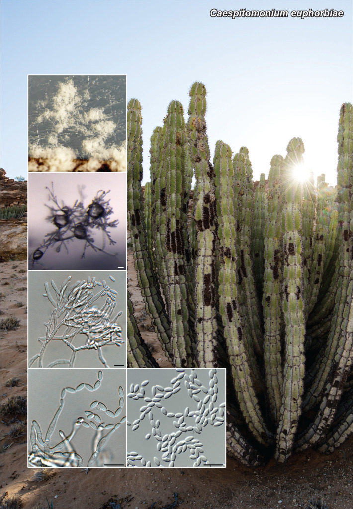
Conidiophores solitary to aggregated, penicillate, branching extensively, aggregating, becoming tuft-like, arising from superficial mycelium, subcylindrical, hyaline, smooth, 40–100 μm tall, 3–4 μm diam at base. Stipe hyaline, smooth, subcylindrical, septate, 15–30 × 3–4 μm. Primary and secondary branches subcylindrical, hyaline, smooth, aseptate, 8–12 × 2.5–3 μm. Conidiogenous cells phialidic, hyaline, smooth, terminal and intercalary, subcylindrical to narrowly ampulliform, with apical taper, 12–22 × 2.5–3 μm; apex with short, 1–1.5 μm long, non-flared collarette. Conidia in long unbranched chains, aseptate, hyaline, smooth, ellipsoid, apex subobtuse, base with truncate hilum, (5–)5.5–6(–6.5) × 2.5–3 μm.

Culture characteristics — Colonies flat, spreading, surface with concentric zone lines, with sparse to moderate aerial mycelium and smooth, lobate margin, reaching 40 mm diam after 2 wk at 25 °C. On malt extract agar (MEA), potato dextrose agar (PDA) and oatmeal agar (OA) surface and reverse buff.

Typus. NAMIBIA, Walvis Bay, on Euphorbia sp. (Euphorbiaceae), 20 Nov. 2019, P.W. Crous, HPC 3127 (holotype CBS H-24539, culture ex-type CPC 39083 = CBS 147075, ITS, LSU, actA, rpb2, tef1 (second part) and tub2 sequences GenBank OK664698.1, OK663737.1, OK651122.1, OK651157.1, OK651197.1 and OK651201.1, MycoBank MB 841777).

Caespitomonium squamicola (Berk. & Broome) Crous, comb. nov. MycoBank MB 841778

Basionym. Fusidium squamicola Berk. & Broome, as ‘squamicolum’, J. Linn. Soc., Bot. 14: 98. 1873 (1875).

Synonyms. Monosporium squamicola (Berk. & Broome) Petch, as ‘squamicolum’, Ann. R. bot. Gdns Peradeniya 9: 169. 1924.

Nalanthamala squamicola (Berk. & Broome) W. Gams, Trans. Br. Mycol. Soc. 64: 402. 1975.

Material examined. SRI LANKA, Peradeniya Gardens, from sooty mould and aphids on Clerodendron monahassa, collection date and collector unknown, CBS H-14970, culture CBS 701.73 = IMI 185382.

Caespitomonium hyalinulum (Sacc.) Crous, comb. nov.

MycoBank MB 841879

Basionym. Torula hyalinula Sacc., Michelia 1 (no. 2): 265. 1878.

Synonyms. Oospora hyalinula (Sacc.) Sacc., Fungi italica autogr. del. 17–28: t. 878. 1881.

Alysidium hyalinulum (Sacc.) Pound & Clem., Minn. bot. Stud. 1(Bulletin 9): 650. 1896.

Acremonium hyalinulum (Sacc.) W. Gams, Cephalosporium-artige Schimmelpilze (Stuttgart): 104. 1971.

Culture examined. USA, Maryland, Baltimore, air in hospital, date unknown, Walker No. 589, CBS H-8191, culture CBS 271.36.

Notes — Caespitomonium represents a new genus in Bionectriaceae that is acremonium-like in morphology but distinguished from Acremonium s.str. based on its extensively branched, tuft-like conidiophores (Gams 1971, 1975).

Based on a megablast search of NCBIs GenBank nucleotide database, the closest hits using the ITS sequence had highest similarity to Acremonium polychromum (strain BXT10-7, GenBank JX076941.1; Identities = 498/501 (99 %), no gaps), Hydropisphaera erubescens (strain 5141/HMAS 91779, GenBank FJ969800.1; Identities = 527/578 (91 %), 17 gaps (2 %)), and Paracylindrocarpon aloicola (strain CBS 141300, GenBank NR_154346.1; Identities = 530/583 (91 %), 17 gaps (2 %)). Closest hits using the LSU sequence are Nalanthamala squamicola (GenBank AF373281.1; Identities = 868/869 (99 %), no gaps), Acremonium hyalinulum (strain CBS 271.36, GenBank HQ232045.1; Identities = 846/848 (99 %), one gap (0 %)), and Acremonium persicinum (strain CBS 203.73, GenBank MH877816.1; Identities = 865/873 (99 %), four gaps (0 %)). Distant hits obtained using the actA sequence had highest similarity to Volutella consors (strain CBS 122767, GenBank KM231160.1; Identities = 384/400 (96 %), no gaps), Gliocladiopsis irregularis (strain CBS 755.97, GenBank KM231113.1; Identities = 384/400 (96 %), no gaps), and Gliocladiopsis pseudotenuis (strain CBS 116074, GenBank KM231112.1; Identities = 384/400 (96 %), no gaps). Distant hits obtained using the rpb2 sequence had highest similarity to Heleococcum aurantiacum (strain CBS 201.35, GenBank JX158463.1; Identities = 708/845 (84 %), six gaps (0 %)), Hydropisphaera peziza (strain CBS 102038, GenBank DQ522444.1; Identities = 682/823 (83 %), eight gaps (0 %)), and Gliomastix murorum (as Acremonium murorum; strain AFTOL-ID 1393, GenBank FJ238363.1; Identities = 602/731 (82 %), six gaps (0 %)).

(Notes continues on Supplementary material page FP1284)

Colour illustrations. Euphorbia virosa in Namibia (photo Oliver Halsey). Conidiophores on synthetic nutrient-poor agar (SNA); conidiophores and conidiogenous cells giving rise to conidia; conidia. Scale bars = 10 μm.

Supplementary material

FP1284

Phylogenetic tree.

Persoonia. 2021 Dec 24;47:184–185.

Fungal Planet 1285 – 24 December 2021

Roigiella syzygii Crous, sp. nov.

Pedro W Crous 1, Johannes Z Groenewald 1

Etymology. Name refers to the host genus Syzygium from which it was isolated.

Classification — Orbiliaceae, Orbiliales, Orbiliomycetes.

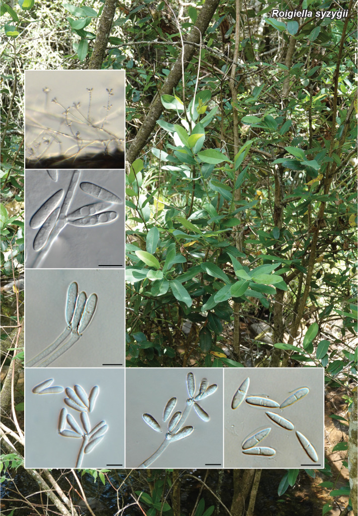
Mycelium consisting of hyaline, smooth, branched, septate, 2.5–3 μm diam hyphae. Conidiophores solitary, erect, arising from superficial hyphae, branched, septate, subcylindrical, up to 300 μm tall, 4–5 μm wide. Conidiogenous cells terminal and intercalary, subcylindrical, hyaline, smooth, 50–100 × 3–4 μm, with subdenticulate loci aggregated in dense, swollen clusters; denticles 1 μm diam, peg-like, not thickened nor darkened. Conidia in clusters, straight, obovoid, apex subobtuse, tapering to a truncate hilum, 1 μm diam, not constricted at median septum, enclosed in mucoid sheath, hyaline, smooth, granular, (16–)18–20(–22) × (4–)4.5–5 μm.

Culture characteristics — Colonies flat, spreading, with fluffy, moderate aerial mycelium and smooth, even margin, reaching 30 mm diam after 2 wk at 25 °C. On MEA, PDA and OA surface and reverse pale luteous.

Typus. SOUTH AFRICA, Northern Province, Nelspruit, Buffelskloof Nature Reserve, on twigs of Syzygium cordatum (Myrtaceae), Nov. 2018, P.W. Crous, HPC 3149 (holotype CBS H-24536, culture ex-type CPC 39044 = CBS 147072, ITS and LSU sequences GenBank OK664699.1 and OK663738.1, MycoBank MB 841779).

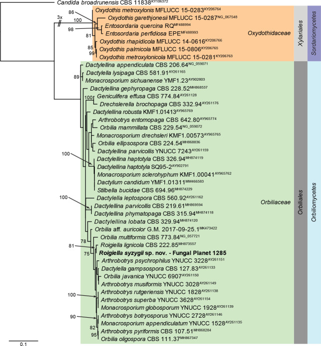
Notes — Roigiella syzygii was isolated from synnematal conidiophores on host tissue, but these structures did not develop in culture. Phylogenetically it clusters with Roigiella lignicola, which is a synnematous genus in the Arthobotrys complex (Castañeda-Ruiz 1984). Morphologically R. syzygii is distinct from R. lignicola (conidia 11–17 × 2–3 μm) in that it has larger conidia that are encased in a mucilaginous sheath.

Based on a megablast search of NCBIs GenBank nucleotide database, the closest hits using the ITS sequence had highest similarity to Roigiella lignicola (strain CBS 222.85, GenBank MH861869.1; Identities = 511/519 (98 %), three gaps (0 %)), Arthrobotrys dendroides (strain CBS 431.85, GenBank NR_159642.1; Identities = 545/568 (96 %), five gaps (0 %)), and Arthrobotrys ellipsospora (strain NBRC 31826, GenBank LC146721.1; Identities = 538/561 (96 %), nine gaps (1 %)). Closest hits using the LSU sequence are Roigiella lignicola (strain CBS 222.85, GenBank MH873557.1; Identities = 882/886 (99 %), no gaps), Arthrobotrys superba (strain YNUCC 3628, GenBank AY261154.1; Identities = 862/872 (99 %), no gaps), and Arthrobotrys botryosporus (strain YNUCC 2728, GenBank AY261146.1; Identities = 862/872 (99 %), no gaps).

graphic file with name per-2023-47-6-i001.jpg

Consensus phylogram (50 % majority rule) obtained from the maximum likelihood analysis with IQ-TREE v. 2.1.3 (Minh et al. 2020) of the Orbiliomycetes LSU nucleotide alignment. Bootstrap support values (> 74 % are shown) from 1 000 non-parametric bootstrap replicates are shown at the nodes. Culture collection numbers and GenBank accession numbers (superscript) are indicated for all species. The tree was rooted to Candida broadrunensis (culture CBS 11838; GenBank KY106372.1) and the species described here is highlighted with bold face. Families, orders and classes are shown in coloured blocks. Alignment statistics: 44 strains including the outgroup; 855 characters including alignment gaps analysed: 209 distinct patterns, 191 parsimony-informative, 59 singleton sites, 605 constant sites. The best model identified for the entire alignment in IQ-TREE using the TESTNEW option was: TN+F+I+G4. The alignment and tree were deposited in figshare.com (10.6084/m9.figshare.16918582).

Colour illustrations. Syzygium cordatum in Buffelskloof Nature Reserve. Conidiophores on SNA; conidiogenous cells giving rise to conidia; conidia with mucoid sheath. Scale bars = 10 μm

graphic file with name per-2023-47-6-g002.jpg

Roigiella syzygii

Persoonia. 2021 Dec 24;47:186–187.

Fungal Planet 1286 – 24 December 2021

Protocreopsis euphorbiae Crous, sp. nov.

Pedro W Crous 1, Lin Zhao 1, Johannes Z Groenewald 1

Etymology. Name refers to the host genus Euphorbia from which it was isolated.

Classification — Bionectriaceae, Hypocreales, Sordariomycetes.

Conidiophores solitary to penicillate, aggregated in clusters forming superficial sporodochia (up to 450 μm diam), with orange mucoid spore masses. Conidiophores branched, septate, subcylindrical, up to 150 μm tall, 2.5–3.5 μm diam. Conidiogenous cells integrated, terminal and intercalary, phialidic, hyaline, smooth, subcylindrical to aculiform, flexuous with slight apical taper towards truncate apex, 1–1.5 μm diam, with periclinal thickening lacking collarette, 20–35 × 2.5–3 μm. Conidia solitary, aseptate, hyaline, smooth, granular, ellipsoid, apex subobtuse, base truncate, 1–1.5 μm diam, (3.5–)6–7(–7.5) × (3–)4 μm.

Culture characteristics — Colonies flat, spreading, with sparse to moderate aerial mycelium and feathery, lobate margin, reaching 20 mm diam after 2 wk at 25 °C. On MEA, PDA and OA surface and reverse orange.

Typus. SOUTH AFRICA, Northern Province, Nelspruit, Buffelskloof Nature Reserve, on leaf litter of Euphorbia ingens (Euphorbiaceae), Nov. 2018, P.W. Crous, HPC 3140 (holotype CBS H-24484, culture ex-type CPC 38896 = CBS 146972, ITS, LSU, actA, tef1 (second part) and tub2 sequences GenBank OK664700.1, OK663739.1, OK651123.1, OK651198.1 and OK651202.1, MycoBank MB 841780).

Notes — Protocreopsis (based on P. musicola = syn. P. fusigera) is a nectria-like genus with a hyphal stroma and acremonium-like asexual morph that was treated by Rossman et al. (1999). The present collection, known only from its asexual morph, is best accommodated in this genus based on its phylogenetic relationship to other acremonium-like species of Protocreopsis.

Based on a megablast search of NCBIs GenBank nucleotide database, the closest hits using the ITS sequence had highest similarity to Acremonium rutilum (strain JCM 23088, GenBank NR_077124.1; Identities = 565/587 (96 %), 11 gaps (1 %)), Protocreopsis phormiicola (strain CBS 567.76, GenBank MH861001.1; Identities = 520/597 (87 %), 24 gaps (4 %)), and Beauveria bassiana (strain BbN06B02, GenBank MK952492.1; Identities = 484/558 (87 %), 21 gaps (3 %)). Closest hits using the LSU sequence are Acremonium rutilum (strain MY6, GenBank KY873378.1; Identities = 860/863 (99 %), no gaps), Acremonium persicinum (strain CBS 378.70C, GenBank HQ232080.1; Identities = 809/812 (99 %), one gap (0 %)), and Pronectria robergei (strain CBS 128021, GenBank MH876199.1; Identities = 855/869 (98 %), one gap (0 %)). Distant hits obtained using the actA sequence had highest similarity to Penicillifer bipapillatus (strain CBS 420.88, GenBank KM231105.1; Identities = 375/406 (92 %), no gaps), Dactylonectria novozelandica (strain CBS 113552, GenBank KM231157.1; Identities = 380/416 (91 %), two gaps (0 %)), and Dactylonectria alcacerensis (strain CBS 129087, GenBank KM231158.1; Identities = 380/416 (91 %), two gaps (0 %)). Distant hits obtained using the tef1 (second part) sequence had highest similarity to Podospora curvicolla (strain VLV, GenBank X96614.1; Identities = 439/478 (92 %), two gaps (0 %)), Clonostachys rosea (strain GJS 90-227, GenBank AY489611.1; Identities = 438/477 (92 %), no gaps), and Engyodontium parvisporum (strain IHEM 22910, GenBank LC425558.1; Identities = 426/464 (92 %), two gaps (0 %)). Distant hits obtained using the tub2 sequence had highest similarity to Septofusidium herbarum (strain CBS 265.58, GenBank KM232113.1; Identities = 310/367 (84 %), 19 gaps (5 %)), Atractium crassum (as Fusarium merismoides var. crassum; strain F-241,346, GenBank EU860033.1; Identities = 303/359 (84 %), 13 gaps (3 %)), and Allantonectria miltina (strain CBS 125499, GenBank KM232107.1; Identities = 303/359 (84 %), 15 gaps (4 %)).

Colour illustrations. Buffelskloof Nature Reserve. Colonies forming sporodochia on SNA; conidiogenous cells giving rise to conidia; conidia. Scale bars = 10 μm.

graphic file with name per-2023-47-6-g003.jpg

Protocreopsis euphorbiae

Supplementary material

See the phylogenetic tree provided with the supplementary material FP1284.

Persoonia. 2021 Dec 24;47:188–189.

Fungal Planet 1287 – 24 December 2021

Cryptometrion metrosideri Crous, sp. nov.

Pedro W Crous 1, Johannes Z Groenewald 1, Raja Thangavel 2

Etymology. Name refers to the host genus Metrosideros from which it was isolated.

Classification — Cryphonectriaceae, Diaporthales, Sordariomycetes.

Conidiomata on PNA erumpent, solitary, globose, pycnidial with central ostiole, punctiform, 100–150 μm diam, red-brown to orange, exuding a creamy conidial mass. Conidiophores lining the inner cavity, hyaline, smooth, subcylindrical, septate, 10–20 × 2–3.5 μm, intermingled among subcylindrical, hyaline, smooth, septate paraphyses with obtuse ends. Conidiogenous cells subcylindrical, integrated, terminal and intercalary, phialidic, hyaline, smooth, 3–7 × 2–3 μm. Conidia solitary, subcylindrical, hyaline, smooth, aseptate, (2.5–)3–4(–6) × 1.5 μm, exuding in orange droplets.

Culture characteristics — Colonies flat, spreading, with moderate aerial mycelium and smooth, even margin, covering dish after 2 wk at 25 °C. On MEA surface cinnamon to sepia, reverse sepia; on PDA surface and reverse orange; on OA surface sepia.

Typus. NEW ZEALAND, Auckland, Devonport, Lake Road, on Metrosideros sp. (Myrtaceae), 10 Apr. 2019, K. Hofer (holotype CBS H-24413, culture ex-type CPC 38512 = T19_03462C = CBS 146785, ITS, LSU, rpb2, tef1 (first part) and tub2 sequences GenBank OK664701.1, OK663740.1, OK651158.1, OK651182.1 and OK651203.1, MycoBank MB 841781).

Additional material examined. NEW ZEALAND, Auckland, Mangere, on Metrosideros excelsa, 20 Aug. 2013, N. Scott, culture T13_03336.

Notes — Cryptometrion metrosideri is closely related to C. aestuescens (CBS 124014, Eucalyptus grandis, Indonesia; Gryzenhout et al. 2010). Morphologically the asexual morph is similar to C. aestuescens, as are the cultures, which on MEA turn sienna to umber with age. The present collection is thus best accommodated in Cryptometrion, pending additional collections to better resolve this generic complex.

Based on a megablast search of NCBIs GenBank nucleotide database, the closest hits using the ITS sequence had highest similarity to Aurapex penicillata (strain CR1207B, GenBank JQ864529.1; Identities = 615/650 (95 %), 13 gaps (2 %)), Diversimorbus metrosideri (strain CMW 37322, GenBank NR_153977.1; Identities = 577/618 (93 %), 15 gaps (2 %)), and Corticimorbus sinomyrti (strain CERC3634, GenBank KT167171.1; Identities = 583/630 (93 %), 22 gaps (3 %)). Closest hits using the LSU sequence are Cryptometrion aestuescens (strain CBS 124014, GenBank MH874864.1; Identities = 804/805 (99 %), no gaps), Holocryphia eucalypti (strain CMW 7037, GenBank JQ862753.1; Identities = 770/772 (99 %), no gaps), and Diversimorbus metrosideri (strain CMW 37322, GenBank NG_070365.1; Identities = 770/772 (99 %), no gaps). Closest hits using the rpb2 sequence are Cryptometrion aestuescens (strain CBS 124007, GenBank MN271798.1; Identities = 649/662 (98 %), no gaps), Holocryphia eucalypti (strain CBS 115842, GenBank MN271804.1; Identities = 734/756 (97 %), no gaps), and Corticimorbus sinomyrti (strain CBS 140205, GenBank MN271794.1; Identities = 428/441 (97 %), no gaps). Closest hits using the tef1 (first part) sequence had highest similarity to Cryptometrion aestuescens (strain CBS 124007, GenBank MN271851.1; Identities = 509/563 (90 %), 17 gaps (3 %)), Ursicollum fallax (strain CBS 118663, GenBank MN271897.1; Identities = 512/576 (89 %), 19 gaps (3 %)), and Diversimorbus metrosiderotis (strain CBS 132866, GenBank MN271857.1; Identities = 515/580 (89 %), 30 gaps (5 %)). Distant hits using the tub2 sequence had highest similarity to Cryphonectria macrospora (voucher YMJ 94031513, GenBank EF025618.1; Identities = 699/811 (86 %), 41 gaps (5 %)), Diversimorbus metrosideri (as Cryphonectriaceae sp. SFC-2012a; strain CMW 37320, GenBank JQ862951.1; Identities = 414/444 (93 %), two gaps (0 %)), and Parvomorbus guangdongensis (strain CSF10738, GenBank MN258814.1; Identities = 388/424 (92 %), 11 gaps (2 %)).

Colour illustrations. Devonport, New Zealand. Conidioma on pine needle agar (PNA); conidiogenous cells giving rise to conidia; conidia. Scale bars = 150 μm (conidioma), 10 μm (all others).

graphic file with name per-2023-47-6-g004.jpg

Cryptometrion metrosideri

Supplementary material

FP1287

Phylogenetic tree.

per-2023-47-6-SF1287.jpg (643.7KB, jpg)
Persoonia. 2021 Dec 24;47:190–191.

Fungal Planet 1288 – 24 December 2021

Harzia combreti Crous, sp. nov.

Pedro W Crous 1, Johannes Z Groenewald 1

Etymology. Name refers to the host genus Combretum from which it was isolated.

Classification — Ceratostomataceae, Coronophorales, Sordariomycetes.

Mycelium consisting of hyaline to olivaceous, smooth, branched, septate, 3.5–4 μm diam hyphae. Conidiophores macronematous, hyaline, smooth, subcylindrical, multiseptate with terminal and intercalary conidiogenous cells; conidiogenous cells hyaline, smooth, subcylindrical with apical taper, 20–70 × 3–4.5 μm. Conidia pale brown, smooth to finely roughened, granular, aseptate, dry, ovoid, thick-walled, (18–)20–22(–23) × (13–)15–17(–18) μm, with marginal frill.

Culture characteristics — Colonies flat, spreading, with moderate aerial mycelium and feathery, lobate margin, reaching 50 mm diam after 2 wk at 25 °C. On MEA surface pale luteous, reverse luteous; on PDA surface and reverse saffron; on OA surface pale luteous.

Typus. SOUTH AFRICA, Northern Province, Nelspruit, Lowveld Botanical Garden, on leaf litter of Combretum collinum ssp. sulvense (Combretaceae), 24 Nov. 2018, P.W. Crous, HPC 3170 (holotype CBS H-24881, culture ex-type CPC 39059 = CBS 148439, ITS and LSU sequences GenBank OK664702.1 and OK663741.1, MycoBank MB 841782).

Notes — Harzia is characterised by sympodially branched, hyaline superficial mycelium, hyaline to brown ovoid conidia and a Proteophiala synasexual morph (Domsch et al. 2007, Schultes et al. 2017). Harzia combreti is closely related to an isolate identified as H. patula. However, H. patula has larger conidia ((16–)25–37.5(–50) × (12.5–)15–28(–37.5) μm; Holubová-Jechová 1974) and thus easily distinguished from H. combreti. Other similar species include H. acremonioides (conidia 20–30 × 15–20 μm; Domsch et al. 2007) and H. metrosideri (conidia (15–)16–18(–20) × (12–)15–16 μm; Crous et al. 2019d), from which it is also morphologically and phylogenetically distinct.

Based on a megablast search of NCBIs GenBank nucleotide database, the closest hits using the ITS sequence had highest similarity to ‘Harzia patula’ (as Olpitrichum patulum; strain CBS 121524, GenBank KY628686.1; Identities = 644/657 (98 %), two gaps (0 %)), Harzia patula (strain CBS 379.88, GenBank NR_161009.1; Identities = 636/658 (97 %), seven gaps (1 %)), and Harzia acremonioides (strain NRRL 54327, GenBank HQ698593.1; Identities = 594/615 (97 %), five gaps (0 %)). Closest hits using the LSU sequence are ‘Harzia patula’ (as Olpitrichum patulum; strain CBS 121524, GenBank KY628687.1; Identities = 829/832 (99 %), no gaps), Harzia acremonioides (strain NRRL 54327, GenBank HQ698593.1; Identities = 502/505 (99 %), one gap (0 %)), and Harzia macrospora (strain CBS 343.67, GenBank MH870687.1; Identities = 827/832 (99 %), one gap (0 %)).

graphic file with name per-2023-47-6-i002.jpg

The first of 61 equally most parsimonious trees obtained from a phylogenetic analysis of the Harzia ITS nucleotide alignment. The tree was rooted to Sphaerodes retispora var. retispora (culture CBS 994.72; GenBank KY628702.1) and the scale bar indicates the number of changes. Parsimony bootstrap support values from 1 000 replicates and > 79 % are shown at the nodes and the treated species is highlighted with a coloured block and bold text. GenBank accession (superscript) and/or culture collection/voucher numbers (in bold face when having a type status) are indicated for all species. Branches present in the strict consensus tree are thickened. Alignment statistics: 24 strains including the outgroup; 598 characters including alignment gaps analysed: 300 constant, 212 variable and parsimony-uninformative and 86 parsimony-informative. Tree statistics: Tree Length = 466, Consistency Index = 0.856, Retention Index = 0.740, Rescaled Consistency Index = 0.634. The alignment and tree were deposited in figshare.com (10.6084/m9.figshare.16918582).

Colour illustrations. Leaves of Combretum collinum ssp. sulvense. Conidiophores on PNA; conidiogenous cells giving rise to conidia; conidia. Scale bars = 10 μm.

graphic file with name per-2023-47-6-g005.jpg

Harzia combreti

Persoonia. 2021 Dec 24;46:192–193.

Fungal Planet 1289 – 24 December 2021

Phaeoisaria dalbergiae Crous, sp. nov.

Pedro W Crous 1, Johannes Z Groenewald 1

Etymology. Name refers to the host genus Dalbergia from which it was isolated.

Classification — Pleurotheciaceae, Pleurotheciales, Sordariomycetes.

Mycelium consisting of hyaline, smooth, branched, septate, 1.5–2 μm diam hyphae. Conidiophores erect, subcylindrical, hyaline, smooth, 0–2-septate, unbranched or branched at apex, 10–50 × 1.5–2.5 μm, giving rise to 1–4 apical conidiogenous cells; conidiogenous cells lateral, arising in apical clusters, 7–15 × 1.5–2.5 μm, with terminal whorl of denticles, subcylindrical, hyaline, smooth, 0.5–2 × 1 μm, not thickened nor darkened. Conidia solitary, hyaline, smooth, aseptate, thin-walled, guttulate, subcylindrical to obovoid, tapering towards both ends, apex subobtuse, base with truncate hilum, 0.5 μm diam, (5–)6–7 × (1.5–)2 μm.

Culture characteristics — Colonies flat, spreading, surface folded, with sparse aerial mycelium and smooth, lobate margin, reaching 6–12 mm diam after 2 wk at 25 °C. On MEA, PDA and OA surface and reverse isabelline.

Typus. SOUTH AFRICA, Northern Province, Nelspruit, Buffelskloof Nature Reserve, on bark of Dalbergia armata (Fabaceae), 23 Nov. 2018, P.W. Crous, HPC 3147 (holotype CBS H-24882, culture ex-type CPC 39540 = CBS 148440, ITS, LSU, rpb2 and SSU sequences GenBank OK664703.1, OK663742.1, OK663796.1 and OK651159.1, MycoBank MB 841783).

Notes — Phaeoisaria includes species with solitary conidiophores and synnemata, with polyblastic, denticulate conidiogenous cells (Seifert et al. 2011). Phaeoisaria dalbergiae was originally isolated as a synnematous mycophylic fungus on Dalbergia armata in South Africa. In culture however, only solitary conidiophores were observed. Phaeoisaria dalbergiae is closely related to P. clematidis (conidia 5–7 × 2–3 μm; Luo et al. 2018) and P. annesophieae (conidia 4.5–9 × 2–3.5 μm; Crous et al. 2017), from which it is phylogenetically distinct.

Based on a megablast search of NCBIs GenBank nucleotide database, the closest hits using the ITS sequence had highest similarity to Phaeoisaria clematidis (strain CBS 113340, GenBank EU552148.1; Identities = 554/570 (97 %), one gap (0 %)), Phaeoisaria annesophieae (voucher MFLU 19-0531, GenBank MT559109.1; Identities = 518/549 (94 %), 11 gaps (2 %)), and Phaeoisaria filiformis (voucher MFLU 18-1462, GenBank NR_168819.1; Identities = 488/525 (93 %), 12 gaps (2 %)). Closest hits using the LSU sequence are Phaeoisaria annesophieae (voucher MFLU 19-0531, GenBank MT559084.1; Iden-tities = 806/816 (99 %), no gaps), Phaeoisaria sp. JB-2020a (strain FCCUFG 03, GenBank MT375866.1; Identities = 805/815 (99 %), no gaps), and Phaeoisaria clematidis (strain MFLUCC 18-1017, GenBank MW132065.1; Identities = 783/793 (99 %), no gaps). Closest hits using the rpb2 sequence had highest similarity to Phaeoisaria clematidis (strain MFLUCC 17-1341, GenBank MF401400.1; Identities = 455/489 (93 %), no gaps), Phaeoisaria aquatica (as Phaeoisaria sp. ZLL-2017a; strain MFLUCC 16-1298, GenBank MF401406.1; Identities = 611/669 (91 %), no gaps), and Phaeoisaria microspora (strain M0104, GenBank MF167352.1; Identities = 666/757 (88 %), seven gaps (0 %)). Closest hits using the SSU sequence are Phaeoisaria clematidis (strain MFLUCC 17-1968, GenBank MG837027.1; Identities = 987/992 (99 %), no gaps), Phaeoisaria fasciculata (strain DAOM 230055, GenBank KT278694.1; Identities = 919/927 (99 %), no gaps), and Phaeoisaria guttulata (as Phaeoisaria sp. JY-2018; voucher MFLU 18-0139, GenBank MG837026.1; Identities = 983/992 (99 %), no gaps).

graphic file with name per-2023-47-6-i003.jpg

The first of two equally most parsimonious trees obtained from a phylogenetic analysis of the Phaeoisaria ITS nucleotide alignment. The tree was rooted to Pleurotheciella aquatica (voucher MFLU 17-0911; GenBank NR_160591.1) and the scale bar indicates the number of changes. Parsimony bootstrap support values from 1 000 replicates and > 79 % are shown at the nodes and the treated species is highlighted with a coloured block and bold text. GenBank accession (superscript) and/or culture collection/voucher numbers (in bold face when having a type status) are indicated for all species. Branches present in the strict consensus tree are thickened. Alignment statistics: 33 strains including the outgroup; 520 characters including alignment gaps analysed: 321 constant, 96 variable and parsimony-uninformative and 101 parsimony-informative. Tree statistics: Tree Length = 334, Consistency Index = 0.778, Retention Index = 0.894, Rescaled Consistency Index = 0.696. The alignment and tree were deposited in figshare.com (10.6084/m9.figshare.16918582).

Colour illustrations. Stem of Dalbergia armata. Conidiophores and conidiogenous cells giving rise to conidia; conidia. Scale bars = 10 μm.

graphic file with name per-2023-47-6-g006.jpg

Phaeoisaria dalbergiae

Persoonia. 2021 Dec 24;47:194–195.

Fungal Planet 1290 – 24 December 2021

Pseudosoloacrosporiella Crous, gen. nov.

Pedro W Crous 1, Johannes Z Groenewald 1

Etymology. Morphologically similar to Soloacrosporiella.

Classification — Incertae sedis, Microthyriales, Dothideomycetes.

Mycelium consisting of pale brown, smooth, branched, septate hyphae. Conidiophores solitary, erect, pale brown, smooth, subcylindrical with apical taper, 0–1-septate, mostly reduced to conidiogenous cells. Conidiogenous cells with 1–3 sympodial apical loci with rhexolytic conidiogenesis, leaving minute collarettes on denticulate loci. Ramoconidia fusoid-ellipsoid, tapering towards both ends, pale brown, smooth, guttulate, septate. Conidia in long unbranched chains, septate, fusoid-ellipsoid, pale brown, smooth; loci rhexolytic, with minute marginal frill.

Type species. Pseudosoloacrosporiella cryptomeriae Crous

MycoBank MB 841784.

graphic file with name per-2023-47-6-g007.jpg

Pseudosoloacrosporiella cryptomeriae

Persoonia. 2021 Dec 24;47:195.

Fungal Planet 1290 – 24 December 2021

Pseudosoloacrosporiella cryptomeriae Crous, sp. nov.


Etymology. Name refers to the host genus Cryptomeria from which it was isolated.

Mycelium consisting of pale brown, smooth, branched, septate, 1.5–2 μm diam hyphae. Conidiophores solitary, erect, pale brown, smooth, subcylindrical with apical taper, 0–1-septate, mostly reduced to conidiogenous cells. Conidiogenous cells 15–30 × 3–4 μm, with 1–3 sympodial apical loci with rhexolytic conidiogenesis, leaving minute collarettes on denticulate loci, 0.5 μm diam. Ramoconidia fusoid-ellipsoid, tapering towards both ends, pale brown, smooth, guttulate, 1–3-septate, 15–20 × 3–4 μm, with 1–3 apical loci. Conidia in long (–15) unbranched chains, 0–1-septate, fusoid-ellipsoid, pale brown, smooth, (7–)12–13(–15) × (2.5–)3(–3.5) μm; loci rhexolytic, 0.5 μm diam, with minute marginal frill.

Culture characteristics — Colonies flat, spreading, surface folded, with moderate aerial mycelium and smooth, lobate margin, reaching 8 mm diam after 7 d at 25 °C. On MEA, PDA and OA surface and reverse ochreous.

Typus. NETHERLANDS, Gelderland Province, Wageningen, Belmonte Botanical Garden, on leaves of Cryptomeria japonica (Cupressaceae), 28 July 2020, P.W. Crous, HPC 3301 (holotype CBS H-24883, culture ex-type CPC 39587 = CBS 148441, ITS, LSU and tef1 (first part) sequences GenBank OK664704.1, OK663743.1 and OK651183.1, MycoBank MB 841785).

Notes — Pseudoacrosporiella is phylogenetically closely related to Soloacrosporiella, but distinct in that it lacks setae, and its conidia do not have thickened and darkened hila, but rather have a characteristic marginal frill (also on conidiogenous loci), which also distinguishes it from Soloacrospora and Neocladophialophora (Castañeda Ruiz et al. 1997, Crous et al. 2014b).

Based on a megablast search of NCBIs GenBank nucleotide database, the closest hits using the ITS sequence had highest similarity to Neocladophialophora quercina (strain CBS 138874, GenBank NR_137948.1; Identities = 497/557 (89 %), 17 gaps (3 %)) and Soloacrosporiella acaciae (strain CBS 139894, GenBank NR_137986.1; Identities = 413/494 (84 %), 38 gaps (7 %)). Closest hits using the LSU sequence are Neocladophialophora quercina (strain CBS 138874, GenBank MH877670.1; Identities = 772/810 (95 %), one gap (0 %)), Soloacrosporiella acaciae (strain CBS 139894, GenBank NG_058164.1; Identities = 761/814 (93 %), two gaps (0 %)), and Neoscolecobasidium agapanthi (strain CPC 28778, GenBank NG_059748.1; Identities = 695/793 (88 %), 14 gaps (1 %)). No significant hits were obtained when the tef1 (first part) sequence was used in blastn and megablast searches.

Colour illustrations. Cryptomeria japonica. Conidiophores on SNA, giving rise to branched chains of conidia; conidia. Scale bars = 10 μm.

Supplementary material

FP1290

Phylogenetic tree.

per-2023-47-6-SF1290.jpg (974.6KB, jpg)
Persoonia. 2021 Dec 24;47:196–197.

Fungal Planet 1291 – 24 December 2021

Parasitella quercicola Crous & Denman, sp. nov.

Pedro W Crous 1, Johannes Z Groenewald 1, Sandra Denman 2

Etymology. Name refers to the host genus Quercus from which it was isolated.

Classification — Mucoraceae, Mucorales, Mucoromycetes.

Mycelium consisting of rhizoid-like hyphae, 4–7 μm diam, smooth, hyaline. Sporangiophores arising from aerial mycelium, simple or sympodially branched, erect, smooth-walled, hyaline, 5–7 μm diam. Sporangia olivaceous, globose to subglobose, 25–60 μm diam, shortly echinulate; wall evanescent, leaving columella with short collarettes. Columellae hyaline, smooth-walled, globose to subglobose, 11–17 μm diam. Sporangiospores hyaline, smooth-walled, aseptate, ellipsoid to reniform, (5–)6–7(–9) × (3–)3.5(–4) μm. Oidia and zygosporangia not observed.

Culture characteristics — Colonies erumpent, spreading, with abundant aerial mycelium, reaching 7 cm diam after 7 d at 25 °C. On MEA surface smoke grey, reverse luteous; on PDA surface and reverse dirty white; on OA surface white.

Typus. UK, E379015.3 N243233.1, on Quercus robur (Fabaceae), 29 July 2020, S. Denman, FPPH 459 (holotype CBS H-24884, culture ex-type CPC 39612 = CBS 148442, ITS and LSU sequences GenBank OK664705.1 and OK663744.1, MycoBank MB 841786).

Notes — Parasitella quercicola is closely related to P. parasitica (neotype CBS 412.66), a species known to be mycoparasitic on other fungi, including Mucorales (Schipper 1978, O’Donnell et al. 2001). Parasitella is presently seen as monotypic, with P. quercicola being phylogenetically distinct from P. parasitica.

Based on a megablast search of NCBIs GenBank nucleotide database, the closest hits using the ITS sequence had highest similarity to Parasitella parasitica (strain CBS 208.28, GenBank MH854982.1; Identities = 540/593 (91 %), 21 gaps (3 %)), Mucor plumbeus (strain LF9, GenBank KX011014.1; Identities = 489/603 (81 %), 54 gaps (8 %)), and Mucor racemosus f. chibinensis (strain LT4-3, GenBank MW850517.1; Identities = 488/603 (81 %), 52 gaps (8 %)). Closest hits using the LSU sequence are Parasitella parasitica (strain CBS 153.69, GenBank MH871012.1; Identities = 853/880 (97 %), nine gaps (1 %)), Mucor mousanensis (strain CBS 999.70, GenBank MH871807.1; Identities = 823/879 (94 %), 13 gaps (1 %)), and Mucor plumbeus (strain FMR 16013, GenBank LT984540.1; Identities = 821/879 (93 %), 13 gaps (1 %)).

graphic file with name per-2023-47-6-i004.jpg

The first of 11 equally most parsimonious trees obtained from a phylogenetic analysis of the Mucor ITS nucleotide alignment. The tree was rooted to Absidia montepascoalis (voucher URM 8218; GenBank NR_172995.1) and the scale bar indicates the number of changes. Parsimony bootstrap support values from 1 000 replicates and > 79 % are shown at the nodes and the treated species is highlighted with a coloured block and bold text. GenBank accession (superscript) and/or culture collection/voucher numbers (in bold face when having a type status) are indicated for all species. Branches present in the strict consensus tree are thickened. Alignment statistics: 62 strains including the outgroup; 809 characters including alignment gaps analysed: 153 constant, 164 variable and parsimony-uninformative and 492 parsimony-informative. Tree statistics: Tree Length = 3 931, Consistency Index = 0.364, Retention Index = 0.612, Rescaled Consistency Index = 0.223. The alignment and tree were deposited in figshare.com (10.6084/m9.figshare.16918582).

Colour illustrations. Quercus robur. Sporangiophores; sporangiophores with sporangia, sporangiospores, and columella; sporangiospores. Scale bars = 10 μm.

graphic file with name per-2023-47-6-g008.jpg

Parasitella quercicola

Persoonia. 2021 Dec 24;47:198–199.

Fungal Planet 1292 – 24 December 2021

Paramacroventuria Crous & Bulgakov, gen. nov.

Pedro W Crous 1, Johannes Z Groenewald 1, Timur S Bulgakov 2

Etymology. Name refers to the fact that it is closely related to Macroventuria.

Classification — Didymellaceae, Pleosporales, Dothideomycetes.

Conidiomata pycnidial, globose, brown, with central periphysate ostiole; wall of 3–6 layers of medium brown textura angularis. Conidiophores reduced to conidiogenous cells lining inner cavity, hyaline, smooth, ampulliform, proliferating percurrently at apex. Conidia solitary, brown, finely roughened, medianly septate, guttulate, ellipsoid, apex obtuse, base with truncate hilum.

Type species. Paramacroventuria ribis Crous & Bulgakov

MycoBank MB 841787.

graphic file with name per-2023-47-6-g009.jpg

Paramacroventuria ribis

Persoonia. 2021 Dec 24;47:199.

Fungal Planet 1292 – 24 December 2021

Paramacroventuria ribis Crous & Bulgakov, sp. nov.


Etymology. Name refers to the host genus Ribes from which it was isolated.

Leaf spots superfial, round, merging, 1–6 mm diam, firstly medium brown, later fair grey, with raised, dark brown-purple edge. Conidiomata pycnidial, globose, brown, 150–250 μm diam, with central periphysate ostiole; wall of 3–6 layers of medium brown textura angularis. Conidiophores reduced to conidiogenous cells lining inner cavity, hyaline, smooth, ampulliform, 5–8 × 4–7 μm, proliferating percurrently at apex. Conidia solitary, brown, finely roughened, medianly septate, guttulate, ellipsoid, apex obtuse, base with truncate hilum, 2–3 μm diam, (9–)10–12(–15) × 5(–6) μm.

Culture characteristics — Colonies flat, spreading, with sparse aerial mycelium and smooth, even margin, reaching 35 mm diam after 7 d at 25 °C. On MEA surface umber, reverse isabelline; on PDA surface and reverse isabelline; on OA surface isabelline.

Typus. RUSSIA, Rostov region, Shakhty city district, urban street greenery, on Ribes aureum (Grossulariaceae), 18 July 2020, T.S. Bulgakov, HPC 3326 = PC 107 (holotype CBS H-24885, isotype LE F-332405, culture ex-type CPC 39692 = CBS 148443, ITS, LSU, rpb2 and tub2 sequences GenBank OK664706.1, OK663745.1, OK651160.1 and OK651204.1, MycoBank MB 841788).

Notes — Paramacroventuria clusters close to Juxtiphoma and Macroventuria. Juxtiphoma was introduced to accommodate Phoma eupyrena reported on stems of Solanum tuberosum (Valenzuela-Lopez et al. 2018), while the sexual genus Macroventuria is based on M. anomochaeta, which was described from decayed canvas (Hou et al. 2020b). Paramacroventuria is an asexual phoma-like genus, distinct from Juxtiphoma in that it lacks chlamydospores, and has percurrently proliferating conidiogenous cells.

Based on a megablast search of NCBIs GenBank nucleotide database, the closest hits using the ITS sequence had highest similarity to Briansuttonomyces eucalypti (strain CBS 114879, GenBank NR_153618.1; Identities = 535/541 (99 %), no gaps), Macroventuria wentii (strain CBS 526.71, GenBank MH860250.1; Identities = 534/540 (99 %), no gaps), and Leptosphaerulina australis (strain CBS 297.54, GenBank MH857342.1; Identities = 536/543 (99 %), one gap (0 %)). Closest hits using the LSU sequence are Microsphaeropsis hellebori (strain CBS 569.82, GenBank MH873273.1; Identities = 820/821 (99 %), no gaps), Didymella aurea (strain CBS 269.93, GenBank NG_069042.1; Identities = 819/821 (99 %), no gaps), and Microsphaeropsis ononidicola (strain MFLUCC 15-0459, GenBank MG967668.1; Identities = 819/821 (99 %), no gaps). Closest hits using the rpb2 sequence had highest similarity to Macroventuria anomochaeta (strain CBS 525.71, GenBank GU456346.1; Identities = 691/751 (92 %), no gaps), Paraboeremia adianticola (strain CBS 187.83, GenBank KP330401.1; Identities = 693/760 (91 %), no gaps), and Juxtiphoma eupyrena (as Phoma eupyrena; strain CBS 374.91, GenBank LT623268.1; Identities = 692/760 (91 %), no gaps). Closest hits using the tub2 sequence had highest similarity to Ascochyta pilosella (strain CBS 583.97, GenBank MT005696.1; Identities = 316/333 (95 %), no gaps), Paramicrosphaeropsis ellipsoidea (strain CBS 197.97, GenBank MT005680.1; Identities = 314/333 (94 %), no gaps), and Stagonosporopsis helianthi (strain CBS 200.87, GenBank KT389848.1; Identities = 314/334 (94 %), three gaps (0 %)).

Colour illustrations. Ribes aureum. Conidiomata on PNA; colony sporulating on OA; conidioma; conidiogenous cells giving rise to conidia; conidia. Scale bars = 250 μm (conidioma), 10 μm (all others).

Supplementary material

FP1292

Phylogenetic tree.

per-2023-47-6-SF1292.jpg (729.3KB, jpg)
Persoonia. 2021 Dec 24;47:200–201.

Fungal Planet 1293 – 24 December 2021

Myrmecridium sambuci Crous, sp. nov.

Pedro W Crous 1, Arien L van Iperen 1, Mieke Starink-Willemse 1, Johannes Z Groenewald 1

Etymology. Name refers to the host genus Sambucus from which it was isolated.

Classification — Myrmecridiaceae, Myrmecridiales, Sordariomycetes.

On SNA. Mycelium consisting of hyaline, smooth, branched, septate, 1.5–2.5 μm diam hyphae. Conidiophores unbranched, erect, straight, medium brown, thick-walled, (1–)3–7-septate, up to 170 μm tall, 2.5–3 μm diam; basal cell 3–5 μm diam. Conidiogenous cells terminal, integrated, subcylindrical, 30–75 μm long, pale brown, forming a rachis with pimple-shaped denticles less than 1 μm long, 0.5 μm diam, slightly thickened. Conidia solitary, aseptate, pale brown, thin-walled, smooth, guttulate, with or without wing-like gelatinous sheath in middle, ellipsoid to fusoid, (7–)8–9(–10) × (2.5–)3(–3.5) μm; hilum unthickened, nor darkened, 0.5 μm diam.

Culture characteristics — Colonies flat, spreading, with sparse aerial mycelium and smooth, lobate margin, reaching 25 mm diam after 7 d at 25 °C. On MEA surface honey to isabelline, reverse isabelline; on PDA surface and reverse luteous; on OA surface umber.

Typus. NETHERLANDS, Zeeland Province, Oranjezon, on Sambucus nigra (Adoxaceae), 17 Sept. 2020, A.L. van Iperen, HPC 3510 (holotype CBS H-24886, culture ex-type CPC 39959 = CBS 148444, ITS and LSU sequences GenBank OK664707.1 and OK663746.1, MycoBank MB 841789).

Notes — Myrmecridium was established by Arzanlou et al. (2007) for ramichloridium-like fungi with solitary conidiophores having pimple-shaped denticles along the medium brown rachis, and subhyaline conidia that frequently have a wing-like gelatinous sheath. Myrmecridium sambuci is closely related to M. phragmiticola known from Phragmites australis in Ukraine (conidia ellipsoid to fusoid, (7–)8–9 × (2.5–)3 μm, conidiophores 2–4-septate, up to 70 μm tall, 3–3.5 μm diam; basal cell 4–6 μm diam; Crous et al. 2020b), but can be distinguished based on its taller and narrower conidiophores.

Based on a megablast search of NCBIs GenBank nucleotide database, the closest hits using the ITS sequence had highest similarity to Myrmecridium phragmiticola (strain CPC 36367, GenBank NR_170826.1; Identities = 541/551 (98 %), three gaps (0 %)), Myrmecridium schulzeri (strain NTOU 4421, GenBank MZ422994.1; Identities = 541/555 (97 %), three gaps (0 %)), and Myrmecridium fluviae (strain CNUFC-YR61-2, GenBank KX839679.1; Identities = 456/471 (97 %), four gaps (0 %)). Closest hits using the LSU sequence are Myrmecridium phragmiticola (strain CPC 36367, GenBank NG_074444.1; Identities = 799/802 (99 %), one gap (0 %)), Myrmecridium schulzeri (strain CBS 188.96, GenBank EU041829.1; Identities = 794/802 (99 %), one gap (0 %)), and Myrmecridium banksiae (strain CBS 132536, GenBank NG_042684.1; Identities = 793/802 (99 %), one gap (0 %)).

Myrmecridium is based on M. sambuci (basionym: Psilobotrys schulzeri, isolated from a trunk of Carpinus betulus, Croatia). Unfortunately, neither De Hoog (1977) nor Arzanlou et al. (2007) could locate the type specimen, which is presumably lost. Since its description, however, numerous isolates have been assigned to this species name, which cluster phylogenetically throughout the genus. To facilitate a more stable taxonomy, we therefore designate the reference strain used by Arzanlou et al. (2007) as neotype below.

Myrmecridium schulzeri (Sacc.) Arzanlou et al., Stud. Mycol. 58: 84. 2007

Basionym. Psilobotrys schulzeri Sacc., Hedwigia 23: 126. 1884.

For additional synonyms see Arzanlou et al. (2007).

Typus. NETHERLANDS, Lienden, isolated from Triticum aestivum root, C.L. de Graaff (CBS 325.74, preserved as metabolically inactive culture, designated as neotype here, MBT 10003688, culture ex-neotype CBS 325.74 = JCM 7234).

Myrmecridium aquaticum (conidia 3-septate, 14–16 × 4–6 μm, on submerged decaying wood, China; Luo et al. 2019), is morphologically similar to, and represents the older name for Neomyrmecridium guizhouense (conidia (2–)3-septate, 8.9–12.7 × 2.8–4.8 μm, on submerged decaying wood, China; Hyde et al. 2020), and phylogenetically, they also cluster in the same clade.

Neomyrmecridium aquaticum (Z.L. Luo et al.) Crous, comb. nov. MycoBank MB 841858

Basionym: Myrmecridium aquaticum Z.L. Luo et al., Fungal Diversity 99: 501. 2019.

Synonym: Neomyrmecridium guizhouense N.G. Liu et al., Fungal Diversity 100: 187. 2020.

Colour illustrations. Zeeland Province, the Netherlands. Conidiophores on SNA; conidiophores with conidiogenous cells giving rise to conidia; conidia with mucoid sheath. Scale bars = 10 μm.

graphic file with name per-2023-47-6-g010.jpg

Myrmecridium sambuci

Supplementary material

FP1293

Phylogenetic tree.

per-2023-47-6-SF1293.jpg (589.8KB, jpg)
Persoonia. 2021 Dec 24;47:202–203.

Fungal Planet 1294 – 24 December 2021

Neofabraea salicina Crous, sp. nov.

Pedro W Crous 1, Arien L van Iperen 1, Mieke Starink-Willemse 1, Johannes Z Groenewald 1

Etymology. Name refers to the host genus Salix from which it was isolated.

Classification — Dermateaceae, Helotiales, Leotiomycetes.

Sporulating poorly on SNA. Conidiomata poorly developed, stromatic, acervular. Conidiophores simple to branched, smooth, hyaline, septate. Conidiogenous cells integrated, phialidic, smooth, hyaline, cylindrical to narrowly ampulliform, giving rise to macro- and microconidia, 10–15 × 2.5–3.5 μm. Macroconidia cylindrical-fusoid to ellipsoid, aseptate, hyaline, smooth-walled, with basal scar, (10–)15–17(–25) × (4–)5–6(–6.5) μm. Microconidia cylindrical, apex obtuse, base truncate, aseptate, hyaline, smooth-walled, (5–)7–8 × (2–)2.5–3 μm.

Culture characteristics — Colonies erumpent, spreading, surface folded, with sparse aerial mycelium and smooth, lobate margin, reaching 10 mm diam after 7 d at 25 °C. On MEA surface dirty white, reverse ochreous; on PDA surface and reverse dirty white; on OA surface sienna.

Typus. NETHERLANDS, Gelderland Province, Beuningen, river Waal, on Salix sp. (Salicaceae), 17 Oct. 2020, A.L. van Iperen, HPC 3485 (holotype CBS H-24887, culture ex-type CPC 39973 = CBS 148445, ITS, LSU, rpb2, tef1 (first part) and tub2 sequences GenBank OK664708.1, OK663747.1, OK651161.1, OK651184.1 and OK651205.1, MycoBank MB 841790).

Notes — Although Phlyctema vagabunda (syn. Neofabraea alba) is a known pathogen of Salix spp., we are not aware of Neofabraea spp. on this host. Neofabraea salicina is closely related to species such as N. brasiliensis (macroconidia 12–22 × 2.5–3.7 μm; Crous et al. 2015), N. kienholzii (macroconidia 12–17.5 ×2.5–3.5 μm; Spotts et al. 2009) N. perennans (macroconidia 12–25 × 3–6 μm; Spotts et al. 2009) and N. malicorticis (macroconidia 16–31 × 4.5–6 μm; Chen et al. 2016), but can easily be distinguished based on its DNA phylogeny.

Based on a megablast search of NCBIs GenBank nucleotide database, the closest hits using the ITS sequence had highest similarity to Neofabraea perennans (strain CBS 453.64, GenBank KR859089.1; Identities = 536/541 (99 %), no gaps), Neofabraea malicorticis (strain CBS 102863, GenBank KR859085.1; Identities = 536/541 (99 %), no gaps), and Neofabraea inaequalis (strain CBS 326.75, GenBank NR_155470.1; Identities = 535/541 (99 %), no gaps). Closest hits using the LSU sequence are Neofabraea malicorticis (strain CBS 122030, GenBank NG_068983.1; Identities = 761/764 (99 %), two gaps (0 %)), Neofabraea kienholzii (strain KARE1951, GenBank MK160254.1; Identities = 761/764 (99 %), two gaps (0 %)), and Neofabraea perennans (strain PPO-45431, GenBank KX768551.1; Identities = 761/764 (99 %), two gaps (0 %)). Closest hits using the rpb2 sequence had highest similarity to Neofabraea malicorticis (strain CBS 102863, GenBank KR859325.1; Identities = 722/735 (98 %), no gaps), Neofabraea perennans (strain GC57, GenBank MN729470.1; Identities = 721/735 (98 %), no gaps), and Neofabraea inaequalis (strain CBS 326.75, GenBank KR859321.1; Identities = 711/735 (97 %), no gaps). Closest hits using the tef1 (first part) sequence had highest similarity to Neofabraea perennans (strain CBS 139.41, GenBank KX982710.1; Identities = 510/531 (96 %), seven gaps (1 %)), Neofabraea malicorticis (strain CBS 102863, GenBank KX982708.1; Identities = 494/534 (93 %), 23 gaps (4 %)), and Neofabraea kienholzii (strain CBS 355.72, GenBank KX982716.1; Identities = 248/269 (92 %), four gaps (1 %)). Closest hits using the tub2 sequence had highest similarity to Neofabraea eucalyptorum (strain CBS 146634, GenBank MT375121.1; Identities = 527/578 (91 %), six gaps (1 %)), Phlyctema phoenicis (strain CPC 29372, GenBank KY173611.1; Identities = 314/384 (82 %), 13 gaps (3 %)), and Coleophoma eucalyptorum (strain CBS 131314, GenBank KU728606.1; Identities = 453/589 (77 %), 45 gaps (7 %)).

graphic file with name per-2023-47-6-i005.jpg

The first of five equally most parsimonious trees obtained from a phylogenetic analysis of the Neofabrea rpb2 nucleotide alignment. The tree was rooted to Pezicula acericola (culture CBS 245.97; GenBank KF376213.1) and the scale bar indicates the number of changes. Parsimony bootstrap support values from 1 000 replicates and > 79 % are shown at the nodes and the treated species is highlighted with a coloured block and bold text. GenBank accession (superscript) and/or culture collection/voucher numbers (in bold face when having a type status) are indicated for all species. Branches present in the strict consensus tree are thickened. Alignment statistics: 17 strains including the outgroup; 736 characters including alignment gaps analysed: 546 constant, 99 variable and parsimony-uninformative and 91 parsimony-informative. Tree statistics: Tree Length = 268, Consistency Index = 0.832, Retention Index = 0.858, Rescaled Consistency Index = 0.714. The alignment and tree were deposited in figshare.com (10.6084/m9.figshare.16918582).

Colour illustrations. Gelderland Province, Beuningen, river Waal. Conidiophores and conidiogenous cells giving rise to conidia; conidia. Scale bars = 10 μm.

graphic file with name per-2023-47-6-g011.jpg

Neofabraea salicina

Persoonia. 2021 Dec 24;47:204–205.

Fungal Planet 1295 – 24 December 2021

Polyscytalum vaccinii Crous, sp. nov.

Pedro W Crous 1, Arien L van Iperen 1, Mieke Starink-Willemse 1, Johannes Z Groenewald 1

Etymology. Name refers to the host genus Vaccinium from which it was isolated.

Classification — Phlogicylindriaceae, Xylariales, Sordariomycetes.

Mycelium consisting of pale brown, smooth, septate, branched, 1.5–2 μm diam hyphae. Conidiophores erect, flexuous, dark brown, thick-walled, septate, up to 200 μm tall, 2.5–3 μm diam. Ramoconidia giving rise to branched chains of cylindrical conidia, pale brown, smooth-walled, subcylindrical, 0(–1)-septate, (12–)15–18(–20) × (2–)2.5 μm, with 1–4 flat-tipped scars, 1–1.5 μm diam. Conidia hyaline to pale olivaceous, cylindrical, 0(–1)-septate, guttulate, (10–)13–16(–20) × 2(–2.5) μm.

Culture characteristics — Colonies flat, spreading, with moderate aerial mycelium and feathery, lobate margin, reaching 10 mm diam after 7 d at 25 °C. On MEA surface and reverse umber, with diffuse sienna pigment; on PDA surface and reverse umber; on OA surface umber with diffuse umber pigment.

Typus. NETHERLANDS, Limburg Province, Weerterbos, on Vaccinium myrtillus (Ericaceae), 6 Sept. 2020, A.L. van Iperen, HPC 3463 (holotype CBS H-24888, culture ex-type CPC 39935 = CBS 148446, ITS and LSU sequences GenBank OK664709.1 and OK663748.1, MycoBank MB 841791).

Notes — Polyscytalum was treated by Crous et al. (2018b), with several species placed in Cylindrium. Polyscytalum vaccinii is phylogenetically distinct from taxa presently known in the genus, and morphologically distinguishable based on its conidial dimensions. It is similar but distinct from P. pinicola (conidiophores up to 80 μm tall, conidia (13–)14–15(–16) × 2 μm; Crous et al. 2020c) based on a combination of conidiophore and conidium characteristics. It also needs to be compared to P. pini-canariensis (conidia (18–)22–26(–48) × 3(–3.5) μm; Crous et al. 2020c), P. fecundissium (conidia 13–18 × 2 mm) and P. neofecundissimum (conidia (12–)14–17(–20) × 2(–3) μm) (Crous et al. 2018b), which have overlapping conidial dimensions, but are phylogenetically distinct.

Based on a megablast search of NCBIs GenBank nucleotide database, the closest hits using the ITS sequence had highest similarity to Anungitea eucalyptigena (strain CPC 28762, GenBank NR_164411.1; Identities = 404/417 (97 %), two gaps (0 %)), Polyscytalum neofecundissimum (strain CBS 143390, GenBank NR_158959.1; Identities = 567/589 (96 %), 11 gaps (1 %)), and Anungitea nullicana (strain CBS 143406, GenBank NR_156391.1; Identities = 401/417 (96 %), two gaps (0 %)). Closest hits using the LSU sequence are Polyscytalum neofecundissimum (strain CBS 143390, GenBank NG_066207.1; Identities = 812/817 (99 %), two gaps (0 %)), Polyscytalum pinicola (strain CPC 36759, GenBank NG_074425.1; Identities = 798/805 (99 %), one gap (1 %)), and Polyscytalum pini-canariensis (strain CBS 146819, GenBank NG_074496.1; Identities = 805/813 (99 %), one gap (0 %)).

graphic file with name per-2023-47-6-i006.jpg

The first of 16 equally most parsimonious trees obtained from a phylogenetic analysis of the Polyscytalum / Phlogicylindrium ITS nucleotide alignment. The tree was rooted to Sarcostroma grevilleae (culture CBS 101.71; GenBank MH553952.1) and the scale bar indicates the number of changes. Parsimony bootstrap support values from 1 000 replicates and > 79 % are shown at the nodes and the treated species is highlighted with a coloured block and bold text. GenBank accession (superscript) and/or culture collection/voucher numbers (in bold face when having a type status) are indicated for all species. Branches present in the strict consensus tree are thickened. Alignment statistics: 26 strains including the outgroup; 590 characters including alignment gaps analysed: 321 constant, 75 variable and parsimony-uninformative and 194 parsimony-informative. Tree statistics: Tree Length = 572, Consistency Index = 0.682, Retention Index = 0.772, Rescaled Consistency Index = 0.526. The alignment and tree were deposited in figshare.com (10.6084/m9.figshare.16918582).

Colour illustrations. Vaccinium myrtillus. Conidiophores and conidiogenous cells giving rise to conidial chains; conidia. Scale bars = 10 μm.

graphic file with name per-2023-47-6-g012.jpg

Polyscytalum vaccinii

Persoonia. 2021 Dec 24;47:206–207.

Fungal Planet 1296 – 24 December 2021

Ramularia pararhabdospora Crous, sp. nov.

Pedro W Crous 1, Arien L van Iperen 1, Mieke Starink-Willemse 1, Johannes Z Groenewald 1

Etymology. Name refers to its morphological similarity to Ramularia rhabdospora.

Classification — Mycosphaerellaceae, Mycosphaerellales, Dothideomycetes.

Leaf spots amphigenous, subcircular, medium brown with raised margin, 3–7 mm diam. Conidiomata fasciculate, amphigenous, stroma absent, arising from stomata in moderately dense fascicles. Conidiophores 0–2-septate, subcylindrical, hyaline, smooth-walled, 15–55 × 4–5 μm. Conidiogenous cells integrated, subcylindrical, straight to geniculate-sinuous, hyaline, smooth-walled, 15–25 × 3–4 μm; scars thickened, darkened, refractive, 1.5–2 μm diam. Conidia solitary, hyaline, smooth-walled, subcylindrical, tapering from middle to subobtuse apex, base obconically rounded, with truncate hilum, thickened, darkened and refractive, 1.5–2 μm diam, 3(–5)-septate, (38–)45–55(–75) × (3–)3.5–4 μm. In culture conidia are up to 100 μm long, flexuous, and undergo microcyclic conidiation.

Culture characteristics — Colonies erumpent, spreading, with sparse to moderate aerial mycelium and smooth, lobate margin, reaching 5 mm diam after 7 d at 25 °C. On MEA surface saffron, reverse ochrous; on PDA surface and reverse grey olivaceous in centre, pale luteous in outer region; on OA surface smoke grey.

Typus. NETHERLANDS, Gelderland Province, Beuningen, river Waal, on Plantago lanceolata (Plantaginaceae), 17 Oct. 2020, A.L. van Iperen, HPC 3473 (holotype CBS H-24889, culture ex-type CPC 39983 = CBS 148447, ITS, LSU, actA, gapdh, his3, rpb2 and tef1 (first part) sequences GenBank OK664710.1, OK663749.1, OK651124.1, OK651147.1, OK651148.1, OK651162.1 and OK651185.1, MycoBank MB 841792).

Notes — Two species are known from this host in Europe. Ramularia rhabdospora is common on Plantago lanceolata, and has catenate conidia that are (10–)15–40(–50) × 3–7 μm, 0–3(–4)-septate. Ramularia plantaginis has catenate, ellipsoid-ovoid to cylindrical conidia, 10–35 × 3–6 μm, 0–3-septate (Braun 1998), thus different from those of R. pararhabdospora. Ramularia rhabdospora and R. pararhabdospora are phylogenetically related and it is possible that more cryptic species exist within R. rhabdospora.

Based on a megablast search of NCBIs GenBank nucleotide database, the closest hits using the ITS sequence had highest similarity to Ramularia hieracii-umbellati (strain CPC 10691 GenBank KX287429.1; Identities = 518/522 (99 %), no gaps), Ramularia rhabdospora (strain CBS 118415, GenBank KX287492.1; Identities = 518/525 (99 %), no gaps), and Ramularia kriegeriana (strain CPC 10827, GenBank KX287461.1; Identities = 509/516 (99 %), one gap (0 %)). Closest hits using the LSU sequence are Ramularia veronicicola (strain CBS 113981, GenBank KX287241.1; Identities = 804/804 (100 %), no gaps), Ramularia rhabdospora (strain CBS 312.92, GenBank KX287193.1; Identities = 804/804 (100 %), no gaps), and Ramularia bosniaca (strain CBS 123880, GenBank KX287045.1; Identities = 804/804 (100 %), no gaps). Closest hits using the actA sequence had highest similarity to Ramularia rhabdospora (strain CBS 118415, GenBank KX287774.1; Identities = 552/568 (97 %), one gap (0 %)), Ramularia inaequalis (strain CPC 25742, GenBank KP894336.1; Identities = 538/582 (92 %), 12 gaps (2 %)), and Ramularia armoraciae (strain CBS 241.90, GenBank KX287613.1; Identities = 528/573 (92 %), six gaps (1 %)). Closest hits using the gapdh sequence had highest similarity to Ramularia rhabdospora (strain CBS 118415, GenBank KX288332.1; Identities = 500/522 (96 %), no gaps), Ramularia digitalis-ambiguae (strain CBS 434.67, GenBank KX288247.1; Identities = 424/462 (92 %), five gaps (1 %)), and Ramularia hieracii-umbellati (strain CPC 10789, GenBank KX288272.1; Identities = 411/455 (90 %), five gaps (1 %)). Closest hits using the his3 sequence had highest similarity to Ramularia rhabdospora (strain CBS 312.92, GenBank KX288940.1; Identities = 330/342 (96 %), no gaps), Ramularia gaultheriae (strain CBS 299.80, GenBank KX288859.1; Identities = 324/343 (94 %), two gaps (0 %)), and Ramularia tricherae (strain CBS 108995, GenBank KX288968.1; Identities = 322/342 (94 %), no gaps). Closest hits using the rpb2 sequence had highest similarity to Ramularia rhabdospora (strain CBS 312.92, GenBank KX288651.1; Identities = 758/781 (97 %), no gaps), Ramularia digitalis-ambiguae (strain CBS 297.37, GenBank KX288567.1; Identities = 721/781 (92 %), no gaps), and Ramularia geraniicola (strain CPC 25912, GenBank KX288574.1; Identities = 716/778 (92 %), no gaps). Closest hits using the tef1 (first part) sequence had highest similarity to Ramularia rhabdospora (strain CBS 118415, GenBank KX288052.1; Identities = 294/310 (95 %), one gap (0 %)), Ramularia abscondita (strain CBS 114727, GenBank KX287864.1; Identities = 325/357 (91 %), seven gaps (1 %)), and Ramularia hieracii-umbellati (strain CPC 10789, GenBank KX287993.1; Identities = 323/356 (91 %), four gaps (1 %)).

Colour illustrations. Gelderland Province, Beuningen, river Waal. Leaf spot; conidiophores and conidiogenous cells giving rise to conidia; conidia. Scale bars = 10 μm.

graphic file with name per-2023-47-6-g013.jpg

Ramularia pararhabdospora

Supplementary material

FP1296

Phylogenetic tree.

per-2023-47-6-SF1296.jpg (704.1KB, jpg)
Persoonia. 2021 Dec 24;47:208–209.

Fungal Planet 1297 – 24 December 2021

Cyphellophora sambuci Crous, sp. nov.

Pedro W Crous 1, Arien L van Iperen 1, Mieke Starink-Willemse 1, Johannes Z Groenewald 1

Etymology. Name refers to the host genus Sambucus from which it was isolated.

Classification — Cyphellophoraceae, Chaetothyriales, Eurotiomycetes.

Mycelium consisting of pale brown, smooth, guttulate, branched, septate, 1.5–2 μm diam hyphae. Conidiophores reduced to solitary conidiogenous cells on hyphae. Conidiogenous cells phialidic, flared, pale brown, 1–3 × 1–3 μm, giving rise to dimorphic conidia. Macroconidia subcylindrical to narrowly obclavate, guttulate, straight to slightly bent, apex subobtuse, base obconically truncate, (1–)3-septate, constricted at septa, hilum truncate, 1 μm diam, not darkened nor thickened, (15–)18–22(–28) × (1.5–)2 μm. Microconidia solitary, aseptate, pale brown, obovoid to ellipsoid, 2–5 × 1.5–2.5 μm.

Culture characteristics — Colonies erumpent, sectored, with moderate aerial mycelium and smooth, lobate margin, reaching 7 mm diam after 7 d at 25 °C. On MEA and PDA surface pale olivaceous grey, margin iron grey, reverse iron grey; on OA surface iron grey.

Typus. NETHERLANDS, South Holland Province, Goeree-Overflakkee, De Kwade Hoek, on Sambucus nigra (Adoxaceae), 13 Sept. 2020, A.L. van Iperen, HPC 3507 (holotype CBS H-24890, culture ex-type CPC 39957 = CBS 148448, ITS, LSU and tub2 sequences GenBank OK664711.1, OK663750.1 and OK651206.1, MycoBank MB 841793).

Notes — Cyphellophora is characterised by having elongated, curved, one- to multiseptate conidia, and well-developed, flared phialides, which frequently develop directly on fungal hyphae (Crous et al. 2009). Species have been isolated from human skin and nail infections (De Hoog et al. 2000, Réblová et al. 2013), soil, plants, water and other substrates (Feng et al. 2014, Crous et al. 2019b, 2020a). Cyphellophora sambuci is phylogenetically closely related to C. phyllostachydis (conidia 0–3 septate, 25–30(–45) × 1.5–2 μm; Gao et al. 2015) and C. europaea (conidia aseptate, 1.8–2.5 μm diam; De Hoog et al. 2000), but can be distinguished based on its conidial dimensions.

Based on a megablast search of NCBIs GenBank nucleotide database, the closest hits using the ITS sequence had highest similarity to Cyphellophora europaea (strain CF27, GenBank KY038650.1; Identities = 469/493 (95 %), seven gaps (1 %)), Cyphellophora oxyspora (strain CBS 416.89, GenBank MH862181.1; Identities = 491/533 (92 %), ten gaps (1 %)), and Cyphellophora phyllostachydis (strain HMAS 245769, GenBank NR_158393.1; Identities = 563/613 (92 %), 25 gaps (4 %)). Closest hits using the LSU sequence are Cyphellophora europaea (strain CBS 101466, GenBank KC455259.1; Identities = 807/814 (99 %), one gap (0 %)), Cyphellophora phyllostachydis (strain HLHNZWYZZ08, GenBank KP122933.1; Identities = 806/814 (99 %), one gap (0 %)), and Cyphellophora musae (strain GLMMZZ4, GenBank KP122934.1; Identities = 754/763 (99 %), one gap (0 %)). Closest hits using the tub2 sequence had highest similarity to Cyphellophora phyllostachydis (strain HLHNZWYZZ08, GenBank KP122929.1; Identities = 284/343 (83 %), seven gaps (2 %)), Cyphellophora europaea (strain CBS 101466, GenBank KC455229.1; Identities = 317/393 (81 %), 12 gaps (3 %)), and Cyphellophora ludoviensis (as Cyphellophora sp. RG-2016; strain CMRP1317, GenBank KX583749.1; Identities = 283/357 (79 %), 24 gaps (6 %)).

graphic file with name per-2023-47-6-i007.jpg

The first of 10 equally most parsimonious trees obtained from a phylogenetic analysis of the Cyphellophora ITS nucleotide alignment. The tree was rooted to Exophiala salmonis (culture CBS 157.67; GenBank MH858932.1) and the scale bar indicates the number of changes. Parsimony bootstrap support values from 1 000 replicates and > 79 % are shown at the nodes and the treated species is highlighted with a coloured block and bold text. GenBank accession (superscript) and/or culture collection/voucher numbers (in bold face when having a type status) are indicated for all species. Branches present in the strict consensus tree are thickened. Alignment statistics: 31 strains including the outgroup; 681 characters including alignment gaps analysed: 328 constant, 99 variable and parsimony-uninformative and 254 parsimony-informative. Tree statistics: Tree Length = 1 379, Consistency Index = 0.520, Retention Index = 0.623, Rescaled Consistency Index = 0.324. The alignment and tree were deposited in figshare.com (10.6084/m9.figshare.16918582).

Colour illustrations. Sambucus nigra at De Kwade Hoek, South Holland Province. Conidiogenous cells giving rise to conidia; conidia. Scale bars = 10 μm.

graphic file with name per-2023-47-6-g014.jpg

Cyphellophora sambuci

Persoonia. 2021 Dec 24;47:210–211.

Fungal Planet 1298 – 24 December 2021

Paradissoconium Crous & Boers, gen. nov.

Pedro W Crous 1, Johannes Z Groenewald 1, Jannes Boers 2

Etymology. Name refers to its morphological similarity to Dissoconium.

Classification — Dissoconiaceae, Mycosphaerellales, Dothideomycetes.

Mycelium consisting of hyaline, smooth, branched, septate hyphae. Conidiophores solitary, unbranched, erect, subcylindrical to subulate, with truncate apex, straight to gently curved, smooth, pale brown, septate. Conidiogenous cells terminal, integrated, loci slightly darkened, proliferating sympodially, and rejuvenating percurrently. Primary conidia solitary, straight, constricted at median septum, smooth, hyaline, ellipsoid, apex obtuse, base obconically truncate, hilum pronounced, darkened. Secondary conidia adjacent to primary conidia, hyaline, smooth, aseptate, ellipsoid, tapering to truncate hilum; anastomosing with primary conidia after active discharge.

Type species. Paradissoconium narthecii Crous & Boers

MycoBank MB 841794.

graphic file with name per-2023-47-6-g015.jpg

Paradissoconium narthecii

Persoonia. 2021 Dec 24;47:211.

Fungal Planet 1298 – 24 December 2021

Paradissoconium narthecii Crous & Boers, sp. nov.


Etymology. Name refers to the host genus Narthecium from which it was isolated.

Ascospores could not be linked to specific ascomata on host tissue, as several taxa were involved, including species with pseudothecia and thyrothecia. Ascospores smooth-walled, fusoid-ellipsoid, widest just above septum, germinating with flexuous germ tubes from both polar ends, spore becoming brown and verruculose, 6 μm diam, constricted at septum, not distorted. Mycelium consisting of hyaline, smooth, branched, septate, 2–3 μm diam hyphae. Conidiophores solitary, unbranched, erect, subcylindrical to subulate, with truncate apex, straight to gently curved, smooth, pale brown, 0–2-septate, 15–100 × 3.5–4.5 μm. Conidiogenous cells terminal, integrated, 15–45 × 3–4 μm, loci slightly darkened, 1–2 μm diam, cicatrized, at times subdenticulate, proliferating sympodially, and rejuvenating percurrently. Primary conidia (12–)14–16(–18) × (5–)6–7 μm, solitary, straight, constricted at median septum, smooth, hyaline, ellipsoid, apex obtuse, base obconically truncate, hilum pronounced, darkened, 2–2.5 μm diam. Secondary conidia 6–9 × 5–6 μm, adjacent to primary conidia, hyaline, smooth, aseptate, ellipsoid, tapering to truncate hilum, 2–2.5 μm; anastomosing with primary conidia after active discharge.

Culture characteristics — Colonies erumpent, spreading, with moderate aerial mycelium and feathery, lobate margin, reaching 6 mm diam after 7 d at 25 °C. On MEA, PDA and OA surface and reverse olivaceous grey.

Typus. NETHERLANDS, Drenthe Province, Dwingelderveld National Park, 52.829188, 6.432495, on dead leaves of Narthecium ossifragum (Nartheciaceae), 4 July 2021, J. Boers, HPC 3653 (holotype CBS H-24891, cultures ex-type CPC 41970, 41971 = CBS 148449, ITS, LSU, actA and rpb1 sequences GenBank OK664712.1, OK663751.1, OK651125.1 and OK651151.1, MycoBank MB 841795).

Additional materials examined. NETHERLANDS, Drenthe Province, Dwingelderveld National Park, 52.829188, 6.432495, on dead leaves of N. ossifragum, 4 July 2021, J. Boers, HPC 3653, single ascospore cultures CPC 41976, 41977.

Notes — The distinction between Dissoconium and Uwebraunia was discussed by Li et al. (2012). Both genera have solitary conidiophores with actively discharging aseptate secondary and 1-septate primary conidia that anastomose upon discharge onto the agar surface. Uwebraunia and Dissoconium have pseudothecial sexual morphs (Crous et al. 2004, Hyde et al. 2020). Paradissoconium has a typical dissoconium-like asexual morph that develops in culture, and is morphologically similar to Dissoconium s.str. Thus far Paradissoconium is monotypic but appears commonly on dead leaves of Narthecium ossifragum, where it is associated with thyrothecia. Although we tried on several locations to link the asexual and sexual morph, all attempts were unsuccessful, as too many diverse sexual morphs were involved. Paradissoconium is located basal in Dissoconiaceae.

Based on a megablast search of NCBIs GenBank nucleotide database, the closest hits using the ITS sequence had highest similarity to ‘Dissoconium sp. GS-2011’ (strain MYQWGNL19, GenBank JF701607.1; Identities = 483/491 (98 %), four gaps (0 %)), Ramichloridium punctatum (strain NTOU 4892, GenBank MK448255.1; Identities = 465/509 (91 %), 14 gaps (2 %)), and Dissoconium proteae (strain CBS 122900, GenBank NR_156213.1; Identities = 469/514 (91 %), 17 gaps (3 %)). Closest hits using the LSU sequence are Ramichloridium eucleae (strain CBS 138000, GenBank NG_058086.1; Identities = 814/828 (98 %), two gaps (0 %)), Ramichloridium indicum (strain CBS 171.96, GenBank EU041856.1; Identities = 811/827 (98 %), no gaps), and Ramichloridium apiculatum (strain CPC 12310, GenBank GU214687.1; Identities = 809/828 (98 %), two gaps (0 %)). Closest hits using the actA sequence had highest similarity to Uwebraunia communis (as Mycosphaerella communis; strain X1023, GenBank EU514299.1; Identities = 490/569 (86 %), 29 gaps (5 %)), Pseudophaeophleospora phormii (strain CBS 144606, GenBank MK442643.1; Identities = 427/480 (89 %), 16 gaps (3 %)), and Zasmidium commune (strain CBS 142528, GenBank KY979857.1; Identities = 366/392 (93 %), one gap (0 %)). Distant hits obtained using the rpb1 sequence had highest similarity to Nothophaeocryptopus gaeumannii (strain CBS 267.37, GenBank GU357770.1; Identities = 513/723 (71 %), 34 gaps (4 %)), Zymoseptoria tritici (strain ST99CH_3D1, GenBank LT854276.1; Identities = 493/717 (69 %), 27 gaps (3 %)), and Rasutoria tsugae (strain ratstk, GenBank GU371809.1; Identities = 489/720 (68 %), 31 gaps (4 %)).

Colour illustrations. Narthecium ossifragum. Conidiophores on SNA with primary and secondary conidia; primary and secondary conidia that discharge together and then anastomose. Scale bars = 10 μm.

Supplementary material

FP1298

Phylogenetic tree.

Persoonia. 2021 Dec 24;47:212–213.

Fungal Planet 1299 – 24 December 2021

Paramyrothecium lathyri Crous & Bulgakov, sp. nov.

Pedro W Crous 1, Johannes Z Groenewald 1, Timur S Bulgakov 2

Etymology. Name refers to the host genus Lathyrus from which it was isolated.

Classification — Stachybotryaceae, Hypocreales, Sordariomycetes.

Spots on living leaves and stems amphigenous, subcircular, 5–14 mm diam, fair brown to grey, with clear border. Conidiomata sporodochial, stromatic, cupulate, superficial, scattered, oval, 200–300 μm diam, with white, setose fringe, surrounding an olivaceous green mucoid conidial mass. Stroma well developed, of hyaline to subhyaline textura angularis. Setae arising from stroma, thick-walled, 3–10-septate, flexuous, up to 300 μm long, 3–4 μm diam, tapering to rounded apex. Conidiophores arising from basal stroma, consisting of penicillate, branched conidiophores; stipes unbranched, hyaline, septate, smooth-walled, 5–10 × 2–3.5 μm; primary branches aseptate, smooth-walled, 5–10 × 2–3 μm; secondary branches aseptate, smooth-walled, 5–8 × 2–3 μm; terminal whorl of 3–6 conidiogenous cells cylindrical to subcylindrical, phialidic, hyaline, smooth-walled, straight to slightly curved, 5–10 × 2–3 μm, with periclinal thickening and minute collarettes. Conidia aseptate, subhyaline, smooth-walled, cylindrical, ends rounded, guttulate, (8–)9(–10) × 2(–2.5) μm.

Culture characteristics — Colonies erumpent, spreading, surface folded, with moderate aerial mycelium and smooth, lobate margin, reaching 50 mm diam after 7 d at 25 °C. On MEA surface pale luteous, reverse ochreous; on PDA surface and reverse pale luteous; on OA surface saffron.

Typus. RUSSIA, Rostov region, Krasnosulinsky district, state natural wildlife area ‘Gornensky’, edge of artificial forest, on Lathyrus tuberosus (Fabaceae), 10 July 2020, T.S. Bulgakov, HPC 3370 = PC 084 (holotype CBS H-24793, isotype LE F-332408, culture ex-type CPC 39712 = CBS 148248, ITS, LSU, cmdA, rpb2 and tef1 (first part) sequences GenBank OK664713.1, OK663752.1, OK651141.1, OK651163.1 and OK651186.1, MycoBank MB 841796).

Notes — Paramyrothecium is distinguished from Myrothecium by having septate, thin-walled setae surrounding its sporodochia. Morphologically P. lathyri is distinct from the other species presently known, based on a combination of its conidial morphology and multiseptate setae (Lombard et al. 2016, Crous et al. 2021a). Paramyrothecium lathyri is located basal in Paramyrothecium.

Based on a megablast search of NCBIs GenBank nucleotide database, the closest hits using the ITS sequence had highest similarity to Myrothecium lachastrae (strain 13-1913, GenBank MH001945.1; Identities = 538/563 (96 %), eight gaps (1 %)), Paramyrothecium parvum (strain CBS 257.35, GenBank NR_145076.1; Identities = 556/582 (96 %), seven gaps (1 %)), and Paramyrothecium roridum (as Myrothecium roridum; strain BBA 62764, GenBank AJ301993.1; Identities = 563/590 (95 %), seven gaps (1 %)). Closest hits using the LSU sequence are Paramyrothecium roridum (strain CBS 357.89, GenBank NG_069343.1; Identities = 816/820 (99 %), one gap (0 %)), Paramyrothecium breviseta (strain CBS 544.75, GenBank NG_069340.1; Identities = 816/820 (99 %), one gap (0 %)), and Paramyrothecium foliicola (strain Bas4_m2, GenBank MH807539.1; Identities = 816/820 (99 %), one gap (0 %)). Closest hits using the cmdA sequence had highest similarity to Paramyrothecium pituitipietianum (strain CPC 38688, GenBank MW173100.1; Identities = 528/631 (84 %), 23 gaps (3 %)), Paramyrothecium foliicola (strain QB3, GenBank MT415358.1; Identities = 500/601 (83 %), 20 gaps (3 %)), and Paramyrothecium tellicola (strain CBS 478.91, GenBank KU846272.1; Identities = 497/602 (83 %), 24 gaps (3 %)). Closest hits using the rpb2 sequence had highest similarity to Paramyrothecium sinense (strain ZSY21, GenBank MH818825.1; Identities = 526/593 (89 %), no gaps), Paramyrothecium roridum (as Myrothecium roridum; strain UAMH 1369, GenBank DQ676588.1; Identities = 568/642 (88 %), no gaps), and Paramyrothecium nigrum (strain CBS 116537, GenBank KU846357.1; Identities = 637/721 (88 %), no gaps). Closest hits using the tef1 (first part) sequence had highest similarity to Neomyrothecium humicola (strain CBS 310.96, GenBank KU846527.1; Identities = 211/233 (91 %), six gaps (2 %)), Gregatothecium humicola (strain CBS 205.96, GenBank KU846402.1; Identities = 168/174 (97 %), two gaps (1 %)), and Albifimbria verrucaria (strain 207C9, GenBank KU845942.1; Identities = 201/224 (90 %), seven gaps (3 %)).

Colour illustrations. State natural wildlife area ‘Gornensky’, Russia. Symptomatic leaf of Lathyrus tuberosus. Conidiomata with setae on PNA; conidioma with setae on SNA; conidiogenous cells giving rise to conidia; conidia. Scale bars = 300 μm (conidiomata), 10 μm (all others).

graphic file with name per-2023-47-6-g016.jpg

Paramyrothecium lathyri

Supplementary material

FP1299

Phylogenetic tree.

per-2023-47-6-SF1299.jpg (588.8KB, jpg)
Persoonia. 2021 Dec 24;47:214–215.

Fungal Planet 1300 – 24 December 2021

Colpoma junipericola Crous & Bulgakov, sp. nov.

Pedro W Crous 1, Johannes Z Groenewald 1, Timur S Bulgakov 2

Etymology. Name refers to the host genus Juniperus from which it was isolated.

Classification — Rhytismataceae, Rhytismatales, Leotiomycetes.

Ascomata amphigenous on leaf litter, lens-shaped, immersed in epidermis, black, up to 1 mm long, 300–400 μm diam, opening by longitudinal slit. Asci cylindrical-clavate, short-stalked, 8-spored, not staining in Melzer’s reagent, 70–95 × 8–9 μm. Ascospores filiform, tapering more prominently to base, hyaline, aseptate, guttulate, at times with gelatinous sheath, (25–)30–35(–40) × 1.5–2 μm. Paraphyses filiform, extending above asci, hyaline, septate, guttulate, apex clavate, 2–2.5 μm diam, up to 100 μm tall, 1.5–2 μm wide, covered in mucoid layer. Conidiomata developing in culture, acervular, 150–200 μm diam, pale brown. Conidiophores reduced to conidiogenous cells lining inner cavity, ampulliform to subcylindrical, straight, hyaline, smooth-walled, 6–15 × 2.5–3 μm, phialidic with periclinal thickening. Conidia aseptate, hyaline, smooth-walled, subcylindrical, straight with obtuse ends, 5–7 × 1.5–2 μm.

Culture characteristics — Colonies flat, spreading, with sparse aerial mycelium and feathery, lobate margin, reaching 40 mm diam after 7 d at 25 °C. On MEA surface saffron, reverse peach; on PDA surface and reverse saffron; on OA surface pale luteous..

Typus. RUSSIA, Rostov region, Shakhty, private garden, on needles of Juniperus sabina (Cupressaceae), 22 May 2020, T.S. Bulgakov, HPC 3350 = PC 118 (holotype CBS H-24792, isotype LE F-332409, culture ex-type CPC 39702 = CBS 148247, ITS and LSU sequences GenBank OK664714.1 and OK663753.1, MycoBank MB 841797).

Notes — The Rhytismataceae occurring on Juniperus was reviewed by Hou et al. (2005). Colpoma junipericola is related to taxa that have been described in Colpoma and Coccomyces. Both genera are highly polyphyletic and thus we tentatively describe the new taxon on Colpoma. It is distinct from Colpoma juniperi which has ascomata that open via irregular splits, ascospores filiform, aseptate, 40–50 × 1–2 μm, enclosed in mucoid sheath, and paraphyses that are curled or coiled (but not swollen) (Minter 1996), and Coccomyces juniperi, which has ascospores 40–50 × 1–2 μm, filiform, often with branched paraphyses, and intracortical apothecia, opening via irregular splits (Sherwood 1980).

Based on a megablast search of NCBIs GenBank nucleotide database, the closest hits using the ITS sequence had highest similarity to Colpoma quercinum (strain C, GenBank AJ293879.1; Identities = 446/479 (93 %), five gaps (1 %)), Coccomyces strobi (strain NB-645C, GenBank MH457134.1; Identities = 470/511 (92 %), 10 gaps (1 %)), and Tryblidiopsis magnesii (strain DAOMC 252096, GenBank NR_164611.1; Identities = 503/559 (90 %), 13 gaps (2 %)). Closest hits using the LSU sequence are Colpoma quercinum (strain COL, GenBank MT707243.1; Identities = 740/759 (97 %), no gaps), Sporomega degenerans (voucher Lantz 367 (UPS), GenBank HM140567.1; Identities = 737/759 (97 %), no gaps), and Colpoma ledi (voucher Lantz 379 (UPS), GenBank HM140512.1; Identities = 734/759 (97 %), no gaps).

Colour illustrations. Juniperus sabina. Needles with ascomata; conidiomata on PNA; asci with paraphyses; ascus; conidiogenous cells giving rise to conidia; conidia. Scale bars = 10 μm.

graphic file with name per-2023-47-6-g017.jpg

Colpoma junipericola

Supplementary material

FP1300

Phylogenetic tree.

per-2023-47-6-SF1300.jpg (1,022.3KB, jpg)
Persoonia. 2021 Dec 24;47:216–217.

Fungal Planet 1301 & 1302 – 24 December 2021

Neochalara Crous, gen. nov.

Pedro W Crous 1, Johannes Z Groenewald 1, Ondřej Koukol 2

Etymology. Name refers to the genus Chalara, to which it is morphologically similar.

Classification — Pezizellaceae, Rhytismatales, Leotiomycetes.

Mycelium consisting of hyaline, smooth-walled, branched, septate hyphae. Conidiophores arising from bulbous base, or as extension of hyphae, frequently aggregated, cylindrical, brown, erect, thick-walled, unbranched, septate, with terminal conidiogenous cells. Conidiogenous cells phialidic, brown, smooth-walled, consisting of a venter that gradually merges into a cylindrical collarette that is pale brown. Conidia in long, unbranched chains, cylindrical with both ends truncate with marginal frill, medianly 1-septate, hyaline, guttulate, smooth-walled.

Type species. Neochalara spiraeae Crous

MycoBank MB 841798.

graphic file with name per-2023-47-6-g018.jpg

Neochalara spiraeae & Sporidesmium spiraeae

Persoonia. 2021 Dec 24;47:217.

Fungal Planet 1301 & 1302 – 24 December 2021

Neochalara spiraeae Crous, sp. nov.


Etymology. Name refers to the host genus Spiraea from which it was isolated.

Mycelium consisting of hyaline, smooth-walled, branched, septate, 3–4 μm diam hyphae. Conidiophores arising from bulbous base, or as extension of hyphae, frequently aggregated, cylindrical, brown, erect, thick-walled, unbranched, straight to slightly bent, 1–4-septate, sometimes with the basal cell inflated, with terminal conidiogenous cell, 100–170 × 6–8 μm. Conidiogenous cells phialidic, brown, smooth-walled, consisting of a subcylindrical venter that gradually merges into a pale brown cylindrical collarette; venter 30–40 μm long, 7–10 μm wide, neck collarette 30–50 μm long, 4–5 μm wide, ratio of mean lengths of collarette and venter 1 : 1. Conidia in long, unbranched chains, cylindrical with both ends truncate with marginal frill, medianly 1-septate, hyaline, guttulate, smooth-walled, (11–)12–14(–16) × (3–)4(–4.5) μm, av. 13 × 4.

Culture characteristics — Colonies flat, spreading, with sparse aerial mycelium and smooth, lobate margin, reaching 30 mm diam after 7 d at 25 °C. On MEA surface luteous, reverse ochreous; on PDA surface and reverse ochreous; on OA surface ochreous.

Typus. NETHERLANDS, Gelderland Province, Wageningen, Belmonte Botanical Garden, on Spiraea japonica (Rosaceae), 28 July 2020, P.W. Crous, HPC 3291 (holotype CBS H-24865, culture ex-type CPC 39565 = CBS 148332, ITS and LSU sequences GenBank OK664715.1 and OK663754.1, MycoBank MB 841799).

Notes — Although morphologically resembling members of the genus Chalara, the newly described species cannot be accommodated in any of the known Chalara species. Members of the genus Chalara with 1-septate conidia either have conidia with rounded ends, or a rounded apex and truncate base (Nag Raj & Kendrick 1976, McKenzie et al. 2002). However, N. spiraeae is distinct in having conidia with both ends being truncate. Furthermore, the distinct phylogenetic position also supports the taxonomical novelty. The present genus, Neochalara, appears to be allied to orbiliopsis-like taxa, although data to support this potential link presently remains inconclusive.

(notes Neochalara spiraeae continues on Supplementary material page FP1301 & 1302)

Supplementary material

See the phylogenetic tree provided with the supplementary material FP1300.

per-2023-47-6-SF1300.jpg (1,022.3KB, jpg)
Persoonia. 2021 Dec 24;47:217.

Fungal Planet 1301 & 1302 – 24 December 2021

Sporidesmium spiraeae Crous, sp. nov.


Etymology. Name refers to the host genus Spiraea from which it was isolated.

Classification — Sporidesmiaceae, Sporidesmiales, Sordariomycetes.

Mycelium consisting of hyaline, smooth, branched, septate, 1.5–2 μm diam hyphae. Conidiophores solitary or aggregated in clusters, erect, dark brown, subcylindrical, unbranched, 1–4-septate, thick-walled, 15–40 × 5–7 μm. Conidiogenous cells terminal, integrated, subcylindrical, blastic, medium brown, smooth-walled, 10–20 × 5–6 μm. Conidia solitary, obclavate, straight to flexuous, apex subobtuse, base obconically truncate, (4–)6–10-distoseptate, septa with central pore, wall smooth, pale brown, basal cell dark brown, hilum truncate, 4–5 μm diam, (45–)70–85(–100) × (8–)9(–10) μm.

Culture characteristics — Colonies flat, spreading, with sparse aerial mycelium and smooth, lobate margin, reaching 8 mm diam after 7 d at 25 °C. On MEA surface dirty white, reverse pale luteous; on PDA surface and reverse dirty white; OA surface dirty white.

Typus. NETHERLANDS, Gelderland Province, Wageningen, Belmonte Botanical Garden, on Spiraea japonica (Rosaceae), 28 July 2020, P.W. Crous, HPC 3291 (holotype CBS H-24837, culture ex-type CPC 39766 = CBS 148298, ITS, LSU and rpb2 sequences GenBank OK664716.1, OK663755.1 and OK651164.1, MycoBank MB 841800).

Notes — Although the type species of Sporidesmium (S. atrum) is not known from DNA data, Sporidesmium has been shown to be polyphyletic (Liu et al. 2019). No species of Sporidesmium (syn. Ellisembia) is known from Spiraea, and based on its phylogeny, as well as the morphological key provided by Wu & Zhuang (2005), S. spiraeae appears distinct.

(notes Sporidesmium spiraeae continues on Supplementary material page FP1301 & 1302)

Colour illustrations. Spiraea japonica at Belmonte Botanical Garden. Left column: Neochalara spiraeae. Conidiophores on SNA; conidiogenous cells giving rise to conidia; conidial chains. Right column: Sporidesmium spiraeae. Conidiophores on SNA; conidiogenous cells giving rise to conidia; conidia. Scale bars = 10 μm.

Supplementary material

FP1302

Phylogenetic tree.

per-2023-47-6-SF1302.jpg (909.1KB, jpg)
Persoonia. 2021 Dec 24;47:218–219.

Fungal Planet 1303 – 24 December 2021

Toxicocladosporium glendoranum Crous & Jurjević, sp. nov.

Pedro W Crous 1, Johannes Z Groenewald 1, Želko Jurjević 2, Sergey Balashov 2

Etymology. Name refers to the city where this fungus was collected, Glendora, California.

Classification — Cladosporiaceae, Cladosporiales, Dothideomycetes.

Conidiophores erect, arising from globose cells in superficial mycelium, 1–8-septate, up to 80 μm tall, 2.5–3.5 μm diam, brown, smooth-walled, mostly unbranched. Conidiogenous cells terminal and lateral, integrated, brown, smooth-walled, subcylindrical, 7–11 × 2–2.5 μm, with 1–3 apical loci, darkened, thickened, not refractive, 0.5 μm diam. Conidia occurring in branched chains, pale brown, smooth-walled, fusoid-ellipsoid; primary ramoconidia fusoid to subcylindrical, 0–1-septate, 7–20 × 2.5–3 μm; hila thickened and darkened, not refractive, 1 μm diam. Secondary ramoconidia aseptate, 5–10 × 2–2.5 μm; intercalary conidia in long chains (–12), fusoid-ellipsoid, (4–)5–6 × 1.5–2 μm; terminal conidia fusoid-ellipsoid, 3–4 × 1.5–2 μm; hila darkened, thickened, not refractive, 0.5 μm diam.

Culture characteristics — Colonies flat, spreading, with sparse aerial mycelium and smooth, lobate margin, reaching 7–10 mm diam after 7 d at 25 °C. On MEA, PDA, Czapek yeast agar (CYA) and OA surface greenish olivaceous, reverse iron grey.

Typus. USA, California, Glendora, from air in a brick room, Oct. 2019, Z. Jurjević, 5421 (holotype CBS H-24802, culture ex-type CPC 40763 = CBS 148256, ITS, LSU, actA and tef1 (first part) sequences GenBank OK664717.1, OK663756.1, OK651126.1 and OK651187.1, MycoBank MB 841801).

Notes — Toxicocladosporium glendoranum clusters basal to Toxicocladosporium but is also closely related to several Cladosporium spp. (Crous et al. 2016, Bezerra et al. 2017, Bensch et al. 2018). Morphologically it is characterised by having rather short conidiophores, and branched conidial chains with smooth, primarily aseptate conidia. Phylogenetically it is distinct from all species presently known in Toxicocladosporium.

Based on a megablast search of NCBIs GenBank nucleotide database, the closest hits using the ITS sequence had highest similarity to Cladosporium halotolerans (strain CBS 127014, GenBank MH864391.1; Identities = 516/550 (94 %), eight gaps (1 %)), Cladosporium endophyticum (strain MFLUCC 17-0599, GenBank NR_158360.1; Identities = 516/550 (94 %), eight gaps (1 %)), and Cladosporium parahalotolerans (strain CPC 22376, GenBank MF473155.1; Identities = 516/550 (94 %), eight gaps (1 %)). Closest hits using the LSU sequence are Toxicocladosporium ficiniae (strain CBS 136406, GenBank NG_058054.1; Identities = 792/810 (98 %), no gaps), Toxicocladosporium hominis (strain CBS 140694, GenBank MH878169.1; Identities = 773/794 (97 %), two gaps (0 %)), and Toxicocladosporium irritans (strain CBS 185.58, GenBank MH869283.1; Identities = 789/811 (97 %), two gaps (0 %)). Closest hits using the actA sequence had highest similarity to Cladosporium velox (strain CPC 18450, GenBank KT600654.1; Identities = 536/606 (88 %), 32 gaps (5 %)) and Cladosporium inversicolor (strain CPC 18238, GenBank KT600588.1; Identities = 530/602 (88 %), 26 gaps (4 %)). No significant hits were obtained when the tef1 sequence was used in blastn and megablast searches.

Colour illustrations. Brick wall in Glendora, California. Conidiophores and conidiogenous cells giving rise to conidia; conidial chains. Scale bars = 10 μm.

graphic file with name per-2023-47-6-g019.jpg

Toxicocladosporium glendoranum

Supplementary material

FP1303

Phylogenetic tree.

per-2023-47-6-SF1303.jpg (518.6KB, jpg)
Persoonia. 2021 Dec 24;47:220–221.

Fungal Planet 1304 – 24 December 2021

Sporidesmiella juncicola Crous & Osieck, sp. nov.

Pedro W Crous 1, Johannes Z Groenewald 1, Eduard R Osieck 2

Etymology. Name refers to the host genus Juncus from which it was isolated.

Classification — Junewangiaceae, Incertae sedis, Sordariomycetes.

Mycelium consisting of hyaline, smooth-walled, branched, septate, 1.5–2 μm diam hyphae. Conidiophores solitary, erect, macronematous, straight to somewhat flexuous, brown, smooth- and thick-walled, base swollen, 8–12 μm diam, 2–3-septate, 70–100 × 4–5 μm. Conidiogenous cells integrated, terminal, subcylindrical, scars inconspicuous, proliferating percurrently with delayed succession, conidia remaining attached via a marginal frill, rejuvenating sympodially, and appearing as polyblastic (sensu Luo et al. 2019; but in fact its percurrent, as can be seen from the marginal frill attached to the conidial hila), pale brown, smooth-walled, 20–45 × 4–5 μm. Conidia solitary, dry, subcylindrical to obovoid, medium brown, smooth-walled, guttulate, apex obtuse, base 2.5–3 μm diam, (3–)4-distoseptate, septa with central pore, (20–)28–32(–35) × (8–)9–10 μm.

Culture characteristics — Colonies erumpent, spreading, with sparse aerial mycelium and smooth, lobate margin, reaching 7 mm diam after 7 d at 25 °C. On MEA, PDA and OA surface saffron, reverse ochreous.

Typus. NETHERLANDS, Utrecht Province, Bornia, near Driebergen, 8 m a.s.l., N52°02’23" E05°17’56", on dead culm of Juncus effusus (Juncaceae), 25 Jan. 2021, E.R. Osieck, HPC 3583 = WI-15/#4211 (holotype CBS H-24850, culture ex-type CPC 41075 = CBS 148311, ITS, LSU and rpb2 sequences GenBank OK664718.1, OK663757.1 and OK651165.1, MycoBank MB 841802).

Additional material examined. NETHERLANDS, Utrecht Province, Nieuw Wulven, near Houten, 1.5 m a.s.l., N52°02’44" E05°09’40", on dead culm of Juncus inflexus, 4 Feb. 2021, E.R. Osieck, HPC 3590 = WI-21/#4205, CBS H-24824, culture CPC 41109 = CBS 148278, ITS, LSU, rpb2 and tef1 (first part) sequences GenBank OK664719.1, OK663758.1, OK651166.1 and OK651188.1.

Notes — The most common species of Sporidesmiella occurring in Europe (on various substrates) is S. hyalosperma (conidia brown, smooth, thick-walled, 4-distoseptate, (17–)22–25(–29) × 9–12(–13) μm, Kirk 1982; (19–)20–22(–24) × (9–)10(–11) μm, Crous et al. 2020c). Phylogenetically S. juncicola is closely related to S. aquatica (conidia 51–59 × 8–22, 3–4-distoseptate, on submerged wood, Luo et al. 2019), from which it is morphologically distinct based on its conidium dimensions.

Based on a megablast search of NCBIs GenBank nucleotide database, the closest hits using the ITS sequence of CPC 41075 had highest similarity to Sporidesmiella aquatica (voucher MFLU 18-1602, GenBank NR_168811.1; Identities = 483/496 (97 %), five gaps (1 %)), Sporidesmiella hyalosperma (strain CPC 37552, GenBank MT223845.1; Identities = 507/573 (88 %), 24 gaps (4 %)), and Sporidesmiella obovoidia (strain MFLUCC 17-2372, GenBank NR_172446.1; Identities = 504/579 (87 %), 37 gaps (6 %)). The ITS sequences of CPC 41075 and CPC 41109 differ with a single indel (500/501 (99 %), one gap (0 %)). Closest hits using the LSU sequence of CPC 41075 are Sporidesmiella aquatica (as Sporidesmiella sp. ZLL-2019a; strain S-777, GenBank MK849843.1; Identities = 763/763 (100 %), no gaps), Sporidesmiella obovoidia (strain MFLUCC 17-2372, GenBank NG_075412.1; Identities = 780/803 (97 %), one gap (0 %)), and Sporidesmiella hyalosperma (strain S-1518, GenBank MK849842.1; Identities = 773/797 (97 %), one gap (0 %)). The LSU sequences of CPC 41075 and CPC 41109 differ with a single indel (799/800 (99 %), one gap (0 %)). Closest hits using the rpb2 sequence of CPC 41075 had highest similarity to Sporidesmiella aquatica (voucher MFLU 18-2331, GenBank MN124524.1; Identities = 638/671 (95 %), no gaps), Sporidesmiella novae-zelandiae (voucher MFLU 18-1604, GenBank MN124526.1; Identities = 591/674 (88 %), no gaps), and Sporidesmiella hyalosperma (strain MFLUCC 18-1013, GenBank MW504070.1; Identities = 584/674 (87 %), no gaps). The rpb2 sequences of CPC 41075 and CPC 41109 are identical (674/674 nucleotides). No significant hits were obtained when the tef1 (first part) sequence of CPC 41109 was used in blastn and megablast searches.

Colour illustrations. Juncus effusus at Nieuw Wulven, near Houten. Conidiophores on SNA; conidiogenous cells giving rise to conidia; conidia. Scale bars = 10 μm.

graphic file with name per-2023-47-6-g020.jpg

Sporidesmiella juncicola

Supplementary material

FP1304

Phylogenetic tree.

per-2023-47-6-SF1304.jpg (419.8KB, jpg)
Persoonia. 2021 Dec 24;47:222–223.

Fungal Planet 1305 & 1306 – 24 December 2021

Xenopyricularia junci Crous & Osieck, sp. nov.

Pedro W Crous 1, Johannes Z Groenewald 1, Eduard R Osieck 2

Etymology. Name refers to the host genus Juncus from which it was isolated.

Classification — Pyriculariaceae, Magnaporthales, Sordariomycetes.

Mycelium consisting of hyaline to subhyaline, smooth-walled, branched, septate, 1.5–2 μm diam hyphae. Conidiophores arising from superficial mycelium, erect, flexuous, dark brown, thick-walled, up to 25-septate, base somewhat swollen, 5–7 μm diam, pale brown, unbranched, up to 800 μm tall, 3.5–4.5 μm wide. Conidiogenous cells integrated, terminal and intercalary, forming a rachis of pimple-like denticles, 0.5–1 × 0.5–1 μm, slightly darkened, up to 100 μm long, pale brown, smooth-walled, 4.5–6 μm wide. Conidia in dry clusters, solitary, pale brown, guttulate, 2-septate, with middle cell darker brown, (18–)27–30(–35) × (5–)6(–6.5) μm, pyriform to narrowly fusoid-ellipsoid, apex subobtuse, tapering toward truncate hilum, 1.5–2 μm diam, somewhat darkened.

Culture characteristics — Colonies flat, spreading, with sparse aerial mycelium and smooth, even margin, reaching 25 mm diam after 2 wk at 25 °C. On MEA, PDA and OA surface and reverse dirty white.

Typus. NETHERLANDS, Utrecht Province, Nieuw Wulven, near Houten, 1.5 m a.s.l., N52°02’45" E05°10’34", on dead culm of Juncus effusus (Juncaceae), 8 Jan. 2021, E.R. Osieck, HPC 3563 = WI-09/#4195 (holotype CBS H-24847, culture ex-type CPC 40968 = CBS 148308, ITS, LSU, actA, cmdA and rpb1 sequences GenBank OK664720.1, OK663759.1, OK651127.1, OK651142.1 and OK651152.1, MycoBank MB 841803).

(notes Xenopyricularia junci on Supplementary material page FP1305 & 1306)

graphic file with name per-2023-47-6-g021.jpg

Xenopyricularia junci & Zaanenomyces quadripartis

Supplementary material

FP1305

Phylogenetic tree.

per-2023-47-6-SF1306.jpg (903.4KB, jpg)
Persoonia. 2021 Dec 24;47:223.

Fungal Planet 1305 & 1306 – 24 December 2021

Zaanenomyces Crous & Osieck, gen. nov.


Etymology. Named for Mieke Zaanen LL.M., who was the Director General of the Royal Dutch Academy of Arts and Sciences (Feb. 2015 – July 2021), and overall responsible for the day-to-day management of the Academy. During this period, she also oversaw the expansion of the research infrastructure and rebranding of the Centraalbureau voor Schimmelcultures to the present Westerdijk Fungal Biodiversity Institute (WI).

Classification — Tubeufiaceae, Tubeufiales, Dothideomycetes.

Mycelium consisting of hyaline, smooth-walled, branched, septate hyphae. Conidiophores solitary, arising from superficial mycelium, erect, subcylindrical, unbranched, brown, smooth-walled, septate; base swollen or with rhizoids, conidiophore with or without brown nodulose swellings, thick-walled. Conidiogenous cells terminal, integrated, forming a rachis with pimple-like denticles, slightly thickened and darkened, not refractive. Conidia solitary, forming a dry mass at apex, narrowly obclavate, straight to flexuous, apex subobtuse, base obconically truncate, septate, hyaline, smooth-walled, guttulate, hilum thickened and darkened.

Type species. Zaanenomyces quadripartis Crous & Osieck

MycoBank MB 841804.

For Zaanenomyces (see also FP 1307 & 1313)

Persoonia. 2021 Dec 24;47:223.

Fungal Planet 1305 & 1306 – 24 December 2021

Zaanenomyces quadripartis Crous & Osieck, sp. nov.


Etymology. Name refers to the expansion of the WI research infrastructure (D-wing; quadripartis (‘the fourth part’), during the Westerdijk year (2017), celebrating Johanna Westerdijk, who was the first female professor in the Netherlands (10 February 1917).

Mycelium consisting of hyaline, smooth-walled, branched, sep-tate, 1.5–2 μm diam hyphae. Conidiophores solitary, arising from superficial mycelium, erect, subcylindrical, unbranched, brown, smooth-walled, 1–4-septate; base swollen or with rhizoids, conidiophore with or without brown nodulose swellings, thick-walled, 15–100 × 2.5–3.5 μm. Conidiogenous cells terminal, integrated, forming a rachis with pimple-like denticles, slightly thickened and darkened, not refractive, 0.5 μm diam, 10–35 × 2.5–3.5 μm. Conidia solitary, forming a dry mass at apex, narrowly obclavate, straight to flexuous, apex subobtuse, base obconically truncate, (1–)4-septate, hyaline, smooth-walled, guttulate, hilum thickened and darkened, 0.5–1 μm diam, (12–)23–30(–35) × (2.5–)3 μm; conidia becoming prominently constricted at septa with age.

Culture characteristics — Colonies erumpent, spreading, with sparse aerial mycelium and smooth, lobate margin, reaching 3 mm diam after 7 d at 25 °C. On MEA surface and reverse sienna; on PDA surface and reverse pale luteous; on OA surface pale luteous.

Typus. NETHERLANDS, Utrecht Province, Nieuw Wulven, near Houten, 1.5 m a.s.l., N52°02’45" E05°10’34", on dead culm of Juncus effusus (Juncaceae), 8 Jan. 2021, E.R. Osieck, HPC 3563 = WI-09/#4195 (holotype CBS H-24849, culture ex-type CPC 41073 = CBS 148310, ITS and LSU sequences GenBank OK664721.1 and OK663760.1, MycoBank MB 841805).

Additional material examined. NETHERLANDS, Utrecht Province, Nieuw Wulven, near Houten, on dead culm of Juncus effusus (Juncaceae), 8 Jan. 2021, E.R. Osieck, HPC 3565 = WI-11, CBS H-24818, culture CPC 41013 = CBS 148272, ITS and LSU sequences GenBank OK664722.1 and OK663761.1.

(notes Zaanenomyces quadripartis on Supplementary material page FP1305 & 1306)

Colour illustrations. Juncus effusus at Nieuw Wulven, near Houten. Left column: Xenopyricularia junci. Conidiophores on SNA; conidiogenous cells giving rise to conidia; conidia. Right column: Zaanenomyces quadripartis. Conidiophores developing on SNA; conidiogenous cells giving rise to conidia. Scale bars = 10 μm.

Supplementary material

See the phylogenetic tree provided with the supplementary material FP1290.

per-2023-47-6-SF1290.jpg (974.6KB, jpg)
Persoonia. 2021 Dec 24;47:224–225.

Fungal Planet 1307 – 24 December 2021

Zaanenomyces moderatricis-academiae Crous & Osieck, sp. nov.

Pedro W Crous 1, Johannes Z Groenewald 1, Eduard R Osieck 2

Etymology. Name refers to the role Mieke Zaanen LL.M. occupied in the Netherlands (Feb. 2015 – July 2021); ‘moderatrix’ = female director, and ‘academia’ = academy.

Classification — Tubeufiaceae, Tubeufiales, Dothideomycetes.

Mycelium consisting of branched, septate, pale brown, smooth-walled, 1.5–2 μm diam hyphae. Conidiophores solitary, erect, subcylindrical, pale brown, smooth-walled, straight, unbranched, 1–2-septate, 7–30 × 2–2.5 μm. Conidiogenous cells integrated, terminal, pale brown, smooth-walled, holoblastic, at times sympodial, 5–15 × 2–2.5 μm; scars somewhat darkened, non-refractive. Conidia solitary, straight to slightly curved, narrowly obclavate to subcylindrical, widest in lowest third, pale brown, smooth-walled, guttulate, apex subobtuse, base truncate, 1.5–2 μm diam, not darkened, 4–10-septate, (32–)40–52(–57) × 2.5–3 μm.

Culture characteristics — Colonies erumpent, spreading, with sparse aerial mycelium and feathery, lobate margin, reaching 7 mm diam after 7 d at 25 °C. On MEA, PDA and OA surface and reverse olivaceous grey.

Typus. NETHERLANDS, Utrecht Province, Nieuw Wulven, near Houten, 1.5 m a.s.l., N52°02’44" E05°09’40", on dead culm of Juncus inflexus (Juncaceae), 4 Feb. 2021, E.R. Osieck, HPC 3590 = WI-21/#4205 (holotype CBS H-24854, culture ex-type CPC 41273 = CBS 148315, ITS, LSU and rpb2 sequences GenBank OK664723.1, OK663762.1 and OK651167.1, MycoBank MB 841806).

Notes — Zaanenomyces moderatricis-academiae represents a second species of the genus Zaanenomyces, also collected from dead culms of Juncus spp. in the Netherlands (see FP 1306). The ex-type strain was cultured from minute, dark, setose ascomata (material too depauperate for description), suggesting that it has a sexual morph. Unfortunately, only the asexual morph developed in culture. Zaanenomyces moderatricis-academiae is distinguished from Z. quadripartis based on its conidiophore and conidium morphology; it is also phylogenetically distinct.

Based on a megablast search of NCBIs GenBank nucleotide database, the closest hits using the ITS sequence had highest similarity to Acanthostigma perpusillum (strain UAMH 7237, GenBank AY916492.1; Identities = 442/506 (87 %), 16 gaps (3 %)), Camporesiomyces vaccinii (strain CBS 216.90, GenBank NR_156202.1; Identities = 432/505 (86 %), 13 gaps (2 %)), and Helicosporium flavisporum (strain MFLUCC 17-2020, GenBank NR_160372.1; Identities = 409/472 (87 %), 28 gaps (5 %)). Closest hits using the LSU sequence are Helicosporium luteosporum (strain MFLUCC 16-0226, GenBank NG_059773.1; Identities = 765/793 (96 %), no gaps), Helicosporium setiferum (as Helicosporium sp. YZL-2018c; strain MFLUCC 17-2007, GenBank MH558863.1; Identities = 763/793 (96 %), no gaps), and Helicosporium flavisporum (strain MFLUCC 17-2020, GenBank NG_066235.1; Identities = 761/793 (96 %), no gaps). No significant hits were obtained when the rpb2 was used in blastn and megablast searches.

Colour illustrations. Juncus inflexus growing at Nieuw Wulven, near Houten. Conidiogenous cells giving rise to conidia; conidia. Scale bars = 10 μm.

graphic file with name per-2023-47-6-g022.jpg

Zaanenomyces moderatricis-academiae

Supplementary material

See the phylogenetic tree provided with the supplementary material FP1290.

per-2023-47-6-SF1290.jpg (974.6KB, jpg)
Persoonia. 2021 Dec 24;47:226–227.

Fungal Planet 1308 – 24 December 2021

Tetracoccosporium pseudoaerium Crous & Jurjević, sp. nov.

Pedro W Crous 1, Johannes Z Groenewald 1, Želko Jurjević 2

Etymology. Name refers to its morphological similarity to Tetracoccosporium aerium.

Classification — Sympoventuriaceae, Venturiales, Dothideomycetes.

Mycelium consisting of pale brown, smooth-walled, septate, branched, 2.5–3 μm diam hyphae. Conidiophores terminal and lateral on hyphae, erect, subcylindrical, pale brown, smooth-walled, 1–2-septate, with 1–3 terminal conidiogenous cells, 7–25 × 2.5–3 μm. Conidiogenous cells pale brown, smooth, doliiform to subcylindrical, monoblastic, tapering toward apex, 1 μm diam, 3–10 × 2–3 μm. Conidia solitary, dry, acrogenous, spherical, medium brown, verruculose, becoming warty, divided by septa at right angles into four sections, becoming swollen with age and constricted at septa, 10–13 μm wide, 7–8 μm high.

Culture characteristics — Colonies flat, spreading, with sparse aerial mycelium and smooth, lobate margin, reaching 10 mm diam after 7 d at 25 °C. On MEA, PDA, Czapek yeast agar (CYA) and OA surface and reverse umber. On CYA reaching 14 mm diam after 7 d at 37 °C.

Typus. USA, Delaware, Wilmington, from air sample in house, Jan. 2010, Ž. Jurjević 5594 (holotype CBS H-24822, culture ex-type CPC 41065 = CBS 148276, ITS and LSU sequences GenBank OK664724.1 and OK663763.1, MycoBank MB 841807).

Notes — Tetracoccosporium aerium (ex-type CBS 493.92) was described to have conidia that are dark brown, 4-celled, 8–12 × 6–8 μm, constricted at septa. As is the case with CBS 148276, it was also isolated from air, but collected in India (Misra & Srivastava 1976). Morphologically the two isolates have overlapping conidial dimensions, but are phylogenetically distinct, and thus the isolate from the USA is herewith described as a new species.

Based on a megablast search of NCBIs GenBank nucleotide database, the closest hits using the ITS sequence had highest similarity to Tetracoccosporium aerium (strain CBS 493.92, GenBank NR_160189.1; Identities = 484/526 (92 %), 19 gaps (3 %)), Ochroconis bacilliformis (strain CBS 100442, GenBank NR_155566.1; Identities = 269/295 (91 %), 18 gaps (6 %)), and Ochroconis musae (strain CBS 145061, GenBank MK442605.1; Identities = 271/301 (90 %), 12 gaps (3 %)). Closest hits using the LSU sequence are Tetracoccosporium aerium (strain CBS 493.92, GenBank MH874035.1; Identities = 796/812 (98 %), three gaps (0 %)), Melnikomyces vietnamensis (strain CBS 136209, GenBank NG_058087.1; Identities = 780/813 (96 %), one gap (0 %)), and Melnikomyces thailandicus (strain CBS 145767, GenBank NG_068685.1; Identities = 741/774 (96 %), one gap (0 %)).

Colour illustrations. House at Wilmington, Delaware, USA. Conidiophores on SNA; conidiogenous cells giving rise to conidia; conidia. Scale bars = 10 μm.

graphic file with name per-2023-47-6-g023.jpg

Tetracoccosporium pseudoaerium

Supplementary material

See the phylogenetic tree provided with the supplementary material FP1290.

per-2023-47-6-SF1290.jpg (974.6KB, jpg)
Persoonia. 2021 Dec 24;47:228–229.

Fungal Planet 1309 – 24 December 2021

Myrmecridium junci Crous & Osieck, sp. nov.

Pedro W Crous 1, Johannes Z Groenewald 1, Eduard R Osieck 2

Etymology. Name refers to the host genus Juncus from which it was isolated.

Classification — Myrmecridiaceae, Myrmecridiales, Sordariomycetes.

Mycelium consisting of hyaline, smooth-walled, branched, septate, 2–3 μm diam hyphae. Conidiophores solitary, erect, subcylindrical, medium brown, smooth-walled, unbranched, straight to flexuous, septate, 45–200 × 3–4 μm. Conidiogenous cells integrated, terminal, and intercalary, subcylindrical, pale brown, smooth-walled, forming a rachis in upper part with numerous pimple-like denticles, 0.5–1 × 0.5 μm, somewhat refractive; conidiophore has whorls of conidia along its length, 7–80 × 3–3.5 μm. Conidia aseptate, solitary, obovoid, hyaline, pale olivaceous with age, apex obtuse, tapering to truncate hilum, 1 μm diam, aseptate, with mucoid sheath surrounding median region, 1–2 μm diam, (8–)9–10(–11) × 2.5–3 μm.

Culture characteristics — Colonies flat, spreading, with sparse aerial mycelium and smooth, lobate margin, reaching 15 mm diam after 7 d at 25 °C. On MEA surface orange, reverse scarlet; on PDA surface and reverse pale luteous; on OA surface orange.

Typus. NETHERLANDS, Utrecht Province, Bornia, near Driebergen, 8 m a.s.l., N52°02’23" E05°17’56", on dead culm of Juncus effusus (Juncaceae), 25 Jan. 2021, E.R. Osieck, HPC 3583 = WI-15 (holotype CBS H-24820, culture ex-type CPC 41032 = CBS 148274, ITS and LSU sequences GenBank OK664725.1 and OK663764.1, MycoBank MB 841808).

Notes — Myrmecridium junci has conidia of similar dimensions to those of M. sambuci ((7–)8–9(–10) × (2.5–)3(–3.5) μm), but is distinct in that conidia are arranged in distinct whorls along the length of the conidiophore, which is not the case with M. sambuci. The species is distinct from all other Myrmecridium species known from DNA data.

Based on a megablast search of NCBIs GenBank nucleotide database, the closest hits using the ITS sequence had highest similarity to Myrmecridium schulzeri (strain CSB_F134, GenBank KU574706.1; Identities = 522/531 (98 %), four gaps (0 %)), Myrmecridium flexuosum (strain CBS 398.76, GenBank NR_146238.1; Identities = 511/522 (98 %), three gaps (0 %)), and Myrmecridium fluviae (strain CNUFC YR61-1, GenBank NR_164555.1; Identities = 461/471 (98 %), one gap (0 %)). Closest hits using the LSU sequence are Myrmecridium schulzeri (strain CBS 114996, GenBank EU041835.1; Identities = 780/790 (99 %), no gaps), Myrmecridium banksiae (strain CBS 132536, GenBank NG_042684.1; Identities = 779/790 (99 %), no gaps), and Myrmecridium spartii (strain CPC 24953, GenBank KR611902.1; Identities = 778/790 (98 %), no gaps).

Colour illustrations. Juncus effusus at Bornia, near Driebergen. Conidiophores on SNA; conidiophores and conidiogenous cells giving rise to conidia; conidia. Scale bars = 10 μm.

graphic file with name per-2023-47-6-g024.jpg

Myrmecridium junci

Supplementary material

See the phylogenetic tree provided with the supplementary material FP1293.

per-2023-47-6-SF1293.jpg (589.8KB, jpg)
Persoonia. 2021 Dec 24;47:230–231.

Fungal Planet 1310 – 24 December 2021

Caeliomyces Crous & Jurjević, gen. nov.

Pedro W Crous 1, Johannes Z Groenewald 1, Želko Jurjević 2

Etymology. Name refers to the fact that it was isolated from air ‘caeli’.

Classification — Teratosphaeriaceae, Mycosphaerellales, Dothideomycetes.

Conidiomata solitary, pycnidial, brown, globose, with central ostiole; wall of 3–6 layers of brown textura angularis. Conidiophores reduced to conidiogenous cells lining the inner cavity, hyaline, smooth-walled, ampulliform, phialidic. Conidia solitary, aseptate, ellipsoid, ends obtuse, green-brown, smooth-walled.

Type species. Caeliomyces tampanus Crous & Jurjević

MycoBank MB 841809.

graphic file with name per-2023-47-6-g025.jpg

Caeliomyces tampanus

Persoonia. 2021 Dec 24;47:231.

Fungal Planet 1310 – 24 December 2021

Caeliomyces tampanus Crous & Jurjević, sp. nov.


Etymology. Name refers to the collection site, Tampa, Florida.

Conidiomata solitary, pycnidial, brown, globose, 20–80 μm diam, with central ostiole, 3–6 μm diam; wall of 3–6 layers of brown textura angularis. Conidiophores reduced to conidiogenous cells lining the inner cavity, hyaline, smooth-walled, ampulliform, 3–6 × 2–3 μm, phialidic. Conidia solitary, aseptate, ellipsoid, ends obtuse, green-brown, smooth-walled, 2–4 × 1.5–2 μm.

Culture characteristics — Colonies flat, spreading, with sparse to moderate aerial mycelium and smooth, lobate margin, reaching 10–20 mm diam after 2 wk at 25 °C. On MEA surface and reverse olivaceous grey; on PDA and OA surface and reverse iron grey.

Typus. USA, Florida, Tampa, from office dust, Oct. 2018, Ž. Jurjević 5589 (holotype CBS H-24821, culture ex-type CPC 41060 = CBS 148275, ITS, LSU, cmdA, tef1 (first part) and tub2 sequences GenBank OK664726.1, OK663765.1, OK651143.1, OK651189.1 and OK651207.1, MycoBank MB 841810).

Notes — Caeliomyces is phylogenetically closely related to coelomycetous genera such as Caatingomyces (Hyde et al. 2019) and Readeriella (Crous et al. 2019c). All three genera have pycnidial conidiomata, phialidic conidiogenous cells, and aseptate, pigmented conidia. However, Readeriella has conidia that tend to vary in shape, and have a thicker wall, darker pigmentation, and percurrently proliferating conidiogenous cells, while Caatingomyces has septate, branched conidiophores with terminal and intercalary conidiogenous cells.

Based on a megablast search of NCBIs GenBank nucleotide database, the closest hits using the ITS sequence had highest similarity to Readeriella deanei (strain CBS 134746, GenBank MH866077.1; Identities = 416/448 (93 %), ten gaps (2 %)), Caatingomyces brasiliensis (as Capnodiales sp. JB-2019a; strain URM 7916, GenBank MH929437.1; Identities = 460/497 (93 %), 11 gaps (2 %)), and Readeriella angustia (strain CBS 124997, GenBank NR_156531.1; Identities = 491/533 (92 %), 14 gaps (2 %)). Closest hits using the LSU sequence are Readeriella angustia (strain CBS 124998, GenBank NG_057843.1; Identities = 801/817 (98 %), four gaps (0 %)), Caatingomyces brasiliensis (as Capnodiales sp. JB-2019a; strain URM 7916, GenBank MH929439.1; Identities = 799/815 (98 %), five gaps (0 %)), and Readeriella dimorphospora (strain CPC 25379, GenBank KX306792.1; Identities = 763/779 (98 %), four gaps (0 %)). Closest hits using the cmdA sequence had highest similarity to Readeriella tasmanica (strain CPC 13631, GenBank KF902686.1; Identities = 283/311 (91 %), no gaps), Readeriella dendritica (strain CBS 120032, GenBank KF902674.1; Identities = 282/310 (91 %), no gaps), and Readeriella novaezelandiae (strain CBS 114357, GenBank KF902683.1; Identities = 280/309 (91 %), no gaps). No significant hits were obtained when the tef1 sequence was used in blastn and megablast searches. Closest hits using the tub2 sequence had highest similarity to Readeriella ellipsoidea (strain CBS 142525, GenBank KY979925.1; Identities = 302/362 (83 %), 24 gaps (6 %)), Readeriella lehmannii (strain CPC 28935, GenBank KY173617.1; Identities = 301/361 (83 %), 22 gaps (6 %)), and Readeriella menaiensis (strain CBS 125003, GenBank KF442490.1; Identities = 291/352 (83 %), 17 gaps (4 %)).

Colour illustrations. Office at Tampa, Florida. Conidiomata on SNA; conidiogenous cells giving rise to conidia; conidia. Scale bars = 80 μm (conidiomata), 10 μm (all others).

Supplementary material

See the phylogenetic tree provided with the supplementary material FP1298.

Persoonia. 2021 Dec 24;47:232–233.

Fungal Planet 1311 – 24 December 2021

Microcera physciae Crous & Boers, sp. nov.

Pedro W Crous 1, Johannes Z Groenewald 1, Jannes Boers 2

Etymology. Name refers to the host genus Physcia from which it was isolated.

Classification — Nectriaceae, Hypocreales, Sordariomycetes.

Associated with discoloured thalli of Physcia tenella. Mycelium consisting of hyaline, smooth-walled, branched, septate, 2–3 μm diam hyphae. Conidiomata sporodochial, pale cream on SNA, consisting of tightly aggregated conidiophores that are reduced to conidiogenous cells or with a short stipe, giving rise to 1–4 conidiogenous cells. Conidiogenous cells subulate, tapering towards apex, flexuous, monophialidic with periclinal thickening and minute non-flared collarette, 10–20 × 2–3 μm. Conidia hyaline, smooth-walled, guttulate, (1–)3-septate, distinctly curved and twisted, apex attenuated, subobtuse, dorsiventrally curved, basal cell attenuated to subobtuse, non foot-shaped to slightly notched basal cell, (20–)24–26(–33) × 3(–3.5) μm.

Culture characteristics — Colonies flat, spreading, with sparse aerial mycelium and smooth, lobate margin, reaching 15 mm diam after 7 d at 25 °C. On MEA surface orange, reverse orange with patches of ochreous; on PDA surface and reverse orange; on OA surface ochreous with orange to creamy sporodochia.

Typus. NETHERLANDS, Drenthe Province, Dwingeloo graveyard, 52.831376, 6.365404, on Physcia tenella (Physciaceae), 10 Mar. 2021, J. Boers, HPC 3602 (holotype CBS H-24829, culture ex-type CPC 41284 = CBS 148283, ITS, LSU, rpb1, rpb2, tef1 (first part) and tub2 sequences GenBank OK664727.1, OK663766.1, OK651153.1, OK651168.1, OK651190.1 and OK651208.1, MycoBank MB 841811).

Additional material examined. NETHERLANDS, Drenthe Province, Dwingeloo, 52.835821, 6.364449, on Physcia tenella, 10 Feb. 2021, J. Boers, HPC 3585, CBS H-24834, culture CPC 41038 = CBS 148288, ITS, LSU, rpb1, rpb2, tef1 (first part) and tub2 sequences GenBank OK664728.1, OK663767.1, OK651154.1, OK651169.1, OK651191.1 and OK651209.1.

Notes — Species of Microcera are usually pathogens of scale insects (Gräfenhan et al. 2011, Crous et al. 2021b). It is thus surprising to find a species associated with discoloured thalli of Physcia tenella. Although no insects were observed at the time of collection, it could well be that this element was overlooked, and further observations would be needed to resolve the ecology of M. physciae. The present collections should be compared with Fusarium sampaioi, which was described from various lichens including Physcialeptalea’. The latter species was discussed by Hawksworth (1979), who concluded that the Fusarium name should be rejected since the studied material was based on discordant elements. Microcera physciae is related to sequences identified as M. coccophila.

Based on a megablast search of NCBIs GenBank nucleotide database, the closest hits using the ITS sequence of CPC 41284 had highest similarity to Microcera larvarum (strain ICMP 5444, GenBank MT107902.1; Identities = 486/498 (98 %), three gaps (0 %)), Cosmospora aurantiicola (strain F-251,305, GenBank EU860061.1; Identities = 486/498 (98 %), three gaps (0 %)), and Microcera rubra (strain CBS 638.76, GenBank MH861019.1; Identities = 485/498 (97 %), three gaps (0 %)). The ITS sequence of CPC 41038 differs with two nucleotides from that of CPC 41284 (495/497 (99 %), no gaps). Closest hits using the LSU sequence of CPC 41284 are Microcera rubra (strain CBS 638.76, GenBank NG_058100.1; Identities = 803/814 (99 %), no gaps), Microcera larvarum (strain CBS 738.79, GenBank KM231701.1; Identities = 803/814 (99 %), no gaps), and Microcera coccophila (strain MAFF 241482, GenBank KC291787.1; Identities = 780/793 (98 %), no gaps). The LSU sequence of CPC 41038 is identical to that of CPC 41284 (813/813 (100 %), no gaps). Closest hits using the rpb1 sequence of CPC 41284 had highest similarity to Microcera coccophila (strain MAFF 241482, GenBank KC291895.1; Identities = 629/688 (91 %), one gap (0 %)), Microcera larvarum (strain A.R. 4580, GenBank KC291894.1; Identities = 627/688 (91 %), one gap (0 %)), and Microcera rubra (strain CBS 638.76, GenBank KM232253.1; Identities = 658/730 (90 %), one gap (0 %)). The rpb1 sequences of CPC 41038 and 41230 differ with two and 29 nucleotides from that of CPC 41284 (740/742 (99 %), one gap, and 709/738 (96 %), one gap, respectively). Closest hits using the rpb2 sequence of CPC 41284 had highest similarity to Microcera sp. (strain NRRL 26790, GenBank JX171636.1; Identities = 843/850 (99 %), no gaps), Microcera larvarum (strain NRRL 20473, GenBank JX171587.1; Identities = 764/848 (90 %), no gaps), and Microcera rubra (strain CBS 638.76, GenBank KM232388.1; Identities = 761/845 (90 %), no gaps). The rpb2 sequence of CPC 41038 differs with five nucleotides from that of CPC 41284 (870/875 (99 %), no gaps). Closest hits using the tef1 (first part) sequence of CPC 41284 had highest similarity to ‘Fusarium sp.’ (strain NRRL 20473, GenBank JF740695.1; Identities = 397/479 (83 %), 19 gaps (3 %)), Microcera larvarum (strain CBS 738.79, GenBank KM231957.1; Identities = 388/479 (81 %), 26 gaps (5 %)), and Microcera coccophila (strain ZJUP0179, GenBank MN614420.1; Identities = 346/426 (81 %), 21 gaps (4 %)). The tef1 (first part) sequence of CPC 41038 differs with 15 nucleotides from that of CPC 41284 (455/470 (97 %), one gap). Closest hits using the tub2 sequence of CPC 41284 had highest similarity to ‘Fusarium sp.’ (strain F-267,620, GenBank EU860029.1; Identities = 521/526 (99 %), one gap (0 %)), Microcera larvarum (as Fusarium larvarum var. larvarum; strain F-266,784, GenBank EU860024.1; Identities = 455/522 (87 %), five gaps (0 %)), and Microcera rubra (as Fusarium larvarum var. rubrum; strain F-257,517, GenBank EU860021.1; Identities = 454/522 (87 %), five gaps (0 %)). The tub2 sequence of CPC 41038 differs with three nucleotides from that of CPC 41284 (343/346 (99 %), no gaps).

Colour illustrations. Physcia tenella. Sporodochia on SNA; conidiogenous cells giving rise to conidia; conidia. Scale bars = 10 μm.

graphic file with name per-2023-47-6-g026.jpg

Microcera physciae

Supplementary material

FP1311

Phylogenetic tree.

per-2023-47-6-SF1311.jpg (474.4KB, jpg)
Persoonia. 2021 Dec 24;47:234–235.

Fungal Planet 1312 & 1313 – 24 December 2021

Myrmecridium dactylidis Crous & Osieck, sp. nov.

Pedro W Crous 1, Johannes Z Groenewald 1, Eduard R Osieck 2

Etymology. Name refers to the host genus Dactylis from which it was isolated.

Classification — Myrmecridiaceae, Myrmecridiales, Sordariomycetes.

Mycelium consisting of branched, septate, hyaline, smooth-walled, 1–2 μm diam hyphae. Conidiophores arising from aerial hyphae, unbranched, straight, medium brown, thick-walled, 1–4-septate, up to 100 μm tall, 3–4 μm diam; basal cell not inflated. Conidiogenous cells integrated, cylindrical, 20–35 × 3.5–4 μm, pale brown, forming a rachis with scattered pimple-shaped denticles less than 1 μm long and ± 0.5 μm wide, apically pointed, pigmented, slightly thickened. Conidia solitary, 0–1(–2)-septate, subhyaline, thin-walled, smooth, guttulate, surrounded by a wing-like gelatinous sheath, ± 0.5 μm thick, ellipsoid to obovoid or fusoid, (6–)8–10(–11.5) × (3–)3.5(–4) μm, tapering to a subtruncate hilum; hilum un-pigmented, not darkened.

Culture characteristics — Colonies flat, spreading, with sparse to moderate aerial mycelium and smooth, lobate margin, reaching 12 mm diam after 2 wk at 25 °C. On MEA, PDA and OA surface and reverse orange.

Typus. NETHERLANDS, Utrecht Province, Nieuw Wulven, near Houten, 1.5 m a.s.l., N52°03’04" E05°09’48", on dead culms of Dactylis glomerata (Poaceae), 4 Feb. 2021, E.R. Osieck, HPC 3601 = WI-27/#4222 (holotype CBS H-24827, culture ex-type CPC 41247 = CBS 148281, ITS and LSU sequences GenBank OK664729.1 and OK663768.1, MycoBank MB 841812).

Notes — Myrmecridium dactylidis is closely related to M. phragmites (conidia 0–1-septate, (6.5–)7–8(–9) × (2.5–)3(–3.5) μm; Crous et al. 2011), but distinct in that it has larger conidia that can be up to 2-septate.

Based on a megablast search of NCBIs GenBank nucleotide database, the closest hits using the ITS sequence had highest similarity to Myrmecridium phragmitis (strain CBS 131311, GenBank NR_137782.1; Identities = 542/552 (98 %), four gaps (0 %)), Myrmecridium schulzeri (strain 9Y-G52, GenBank MT138583.1; Identities = 514/527 (98 %), two gaps (0 %)), and Myrmecridium phragmiticola (strain CPC 36367, GenBank NR_170826.1; Identities = 527/549 (96 %), four gaps (0 %)). Closest hits using the LSU sequence are Myrmecridium phragmitis (strain CBS 131311, GenBank NG_057948.1; Identities = 796/798 (99 %), no gaps), Myrmecridium schulzeri (strain DUCC4137, GenBank MH819185.1; Identities = 784/798 (98 %), no gaps), and Myrmecridium banksiae (strain CBS 132536, GenBank NG_042684.1; Identities = 784/798 (98 %), no gaps).

graphic file with name per-2023-47-6-g027.jpg

Myrmecridium dactylidis & Zaanenomyces versatilis

Supplementary material

See the phylogenetic tree provided with the supplementary material FP1293.

per-2023-47-6-SF1293.jpg (589.8KB, jpg)
Persoonia. 2021 Dec 24;47:235.

Fungal Planet 1312 & 1313 – 24 December 2021

Zaanenomyces versatilis Crous & Osieck, sp. nov.


Etymology. Name refers to the versatile role Mieke Zaanen LL.M. occupied as Director General of the Royal Dutch Academy of Arts and Sciences (Feb. 2015 – July 2021).

Classification — Tubeufiaceae, Tubeufiales, Dothideomycetes.

Mycelium consisting of pale brown, smooth-walled, branched, septate, 2–2.5 μm diam hyphae. Conidiophores solitary, erect, subcylindrical, straight to geniculous-sinuous, unbranched, 0–5-septate, 5–60 × 2.5–3.5 μm. Conidiogenous cells terminal, integrated, pale brown, smooth-walled, subcylindrical with apical rachis of denticles, cylindrical, 1–2 × 1–1.5 μm; scars not thickened nor darkened. Conidia solitary, in dry clusters, subcylindrical, widest in middle, tapering to subobtuse apex and truncate hilum, 1.5–2 μm diam, pale brown, smooth, guttulate, straight to slightly flexuous, (3–)7–10(–12)-septate, (16–)43–50(–55) × (2.5–)3(–3.5) μm.

Culture characteristics — Colonies erumpent, spreading, surface folded, with moderate aerial mycelium and smooth, lobate margin, reaching 6 mm diam after 7 d at 25 °C. On MEA, PDA and OA surface umber, reverse chestnut.

Typus. NETHERLANDS, Utrecht Province, Nieuw Wulven, near Houten, 1.5 m a.s.l., N52°02’44" E05°09’40", on dead culm of Juncus inflexus (Juncaceae), 4 Feb. 2021, E.R. Osieck, HPC 3590 = WI-21/#4205 (holotype CBS H-24851, culture ex-type CPC 41224 = CBS 148312, ITS and LSU sequences GenBank OK664730.1 and OK663769.1, MycoBank MB 841813).

Notes — Zaanenomyces versatilis represents the third species in the genus (see FP 1306 & 1307) collected on Juncus in the Netherlands, distinct from Z. quadripartis (conidia (1–)4-septate, (12–)23–30(–35) × (2.5–)3 μm), and Z. moderatricis-academiae (conidia 4–10-septate, (32–)40–52(–57) × 2.5–3 μm).

Based on a megablast search of NCBIs GenBank nucleotide database, the closest hits using the ITS sequence had highest similarity to Acanthostigma perpusillum (strain UAMH 7237, GenBank AY916492.1; Identities = 504/568 (89 %), 16 gaps (2 %)), Camporesiomyces vaccinii (strain CBS 216.90, GenBank NR_156202.1; Identities = 498/568 (88 %), 15 gaps (2 %)), and Tubeufia cerea (strain BCC 8125, GenBank AY916491.1; Identities = 493/568 (87 %), 30 gaps (5 %)). The ITS sequences of CPC 41273 (Zaanenomyces juncicola) and CPC 41224 are dissimilar (484/505 (96 %), six gaps (1 %)). Closest hits using the LSU sequence are Helicosporium luteosporum (strain MFLU 16-2871, GenBank NG_059773.1; Identities = 794/818 (97 %), no gaps), Helicosporium flavisporum (voucher MFLU 17-1105, GenBank NG_066235.1; Identities = 790/818 (97 %), no gaps), and Helicosporium vesicarium (as Helicosporium sp. YZL-2018e; strain MFLUCC 17-1795, GenBank MH558864.1; Identities = 790/818 (97 %), no gaps). The LSU sequences of CPC 41273 (Zaanenomyces juncicola) and CPC 41224 are dissimilar (789/793 (99 %), no gaps).

Colour illustrations. Juncus inflexus at Nieuw Wulven, near Houten. Left column: Myrmecridium dactylidis. Conidiophores on SNA; conidiogenous cells giving rise to conidia; conidia. Right column: Zaanenomyces versatilis. Conidiophores on SNA; conidiogenous cells giving rise to conidia; conidia. Scale bars = 10 μm.

Supplementary material

See the phylogenetic tree provided with the supplementary material FP1290.

per-2023-47-6-SF1290.jpg (974.6KB, jpg)
Persoonia. 2021 Dec 24;47:236–237.

Fungal Planet 1314 – 24 December 2021

Myrmecridium juncicola Crous & Osieck, sp. nov.

Pedro W Crous 1, Johannes Z Groenewald 1, Eduard R Osieck 2

Etymology. Name refers to the host genus Juncus from which it was isolated.

Classification — Myrmecridiaceae, Myrmecridiales, Sordariomycetes.

Mycelium consisting of pale brown, smooth-walled, branched, septate, 2.5–3 μm diam hyphae. Conidiophores solitary, erect, unbranched, subcylindrical, flexuous, brown, smooth- and thick-walled, 1–10-septate, up to 170 μm tall, 4–5 μm wide, arising from superficial mycelium. Conidiogenous cells integrated, terminal, subcylindrical, pale brown, smooth-walled, 20–35 × 3.5–4 μm, forming a rachis with numerous pimple-like denticles, 0.5–1 × 0.5 μm, slightly darkened, not reflective. Conidia solitary, fusoid, pale brown, smooth-walled, guttulate with mucoid sheath in mid region (up to 2 μm diam), apex subobtuse, tapering to truncate hilum, 0.5 μm diam, slightly darkened, 0–1-septate, (12–)14–16(–18) × (2.5–)3(–3.5) μm.

Culture characteristics — Colonies flat, spreading, with sparse to moderate aerial mycelium and smooth, lobate margin, reaching 20 mm diam after 7 d at 25 °C. On MEA surface and reverse ochreous; on PDA surface and reverse saffron; on OA surface saffron with patches of umber.

Typus. NETHERLANDS, Overijssel Province, Engbertsdijksvenen, near Vriezenveen, 13 m a.s.l., N52°26’53" E06°40’02", on dead culm of Juncus effusus (Juncaceae), 9 Mar. 2021, E.R. Osieck, HPC 3612 = WI-31/#4227 (holotype CBS H-24855, culture ex-type CPC 41313 = CBS 148316, ITS and LSU sequences GenBank OK664731.1 and OK663770.1, MycoBank MB 841814).

Additional materials examined. NETHERLANDS, Utrecht Province, Nieuw Wulven, near Houten, 1.5 m a.s.l., N52°02’45" E05°10’34", on dead culm of J. effusus, 8 Jan. 2021, E.R. Osieck, HPC 3565 = WI-11/#4195, CBS H-24813, culture CPC 40831 = CBS 148267, ITS and LSU sequences GenBank OK664733.1 and OK663772.1; ibid., CBS H-24812, culture CPC 40829 = CBS 148266, ITS and LSU sequences GenBank OK664732.1 and OK663771.1.

Notes — Myrmecridium juncicola needs to be compared to M. junci (conidia (8–)9–10(–11) × 2.5–3 μm, arranged in whorls; described elsewhere in this paper), but has larger conidia, also when compared to other species treated here, like M. dactylidis, and M. sambuci. Phylogenetically, the species is related to M. dactylidis, and M. phragmitis.

Based on a megablast search of NCBIs GenBank nucleotide database, the closest hits using the ITS sequence of CPC 41313 had highest similarity to Myrmecridium schulzeri (strain CSB_F134, GenBank KU574706.1; Identities = 484/505 (96 %), seven gaps (1 %)), Myrmecridium phragmiticola (strain CPC 36367, GenBank NR_170826.1; Identities = 480/505 (95 %), seven gaps (1 %)), and Myrmecridium flexuosum (strain CBS 398.76, GenBank NR_146238.1; Identities = 473/501 (94 %), ten gaps (1 %)). The ITS sequences of CPC 40829 and 40831 differ with one nucleotide each from that of CPC 41313 (499/500 (99 %), no gaps, and 499/500 (99 %), no gaps, respectively). Closest hits using the LSU sequence of CPC 41313 are Myrmecridium banksiae (strain CBS 132536, GenBank NG_042684.1; Identities = 804/813 (99 %), two gaps (0 %)), Myrmecridium spartii (strain CPC 24953, GenBank KR611902.1; Identities = 803/813 (99 %), two gaps (0 %)), and Myrmecridium schulzeri (strain CBS 114996, GenBank EU041835.1; Identities = 803/813 (99 %), two gaps (0 %)). The LSU sequences of CPC 40829 and 40831 are identical to that of CPC 41313 (811/811 (100 %), no gaps, and 788/788 (100 %), no gaps, respectively).

Colour illustrations. Juncus effusus at Engbertsdijksvenen, near Vriezenveen, Overijssel Province. Conidiophores on SNA; conidiogenous cells giving rise to conidia; conidia. Scale bars = 10 μm.

graphic file with name per-2023-47-6-g028.jpg

Myrmecridium juncicola

Supplementary material

See the phylogenetic tree provided with the supplementary material FP1293.

per-2023-47-6-SF1293.jpg (589.8KB, jpg)
Persoonia. 2021 Dec 24;47:238–239.

Fungal Planet 1315 – 24 December 2021

Sarocladium junci Crous & Osieck, sp. nov.

Pedro W Crous 1, Johannes Z Groenewald 1, Eduard R Osieck 2

Etymology. Name refers to the host genus Juncus from which it was isolated.

Classification — Sarocladiaceae, Hypocreales, Sordariomycetes.

Mycelium consisting of hyaline, smooth-walled, branched, septate, 1.5–2 μm diam hyphae. Conidiophores arising from superficial mycelium, erect, flexuous, rarely branched, subcylindrical with apical taper, hyaline, smooth-walled, 1–3-septate, 15–60 × 2–3 μm. Conidiogenous cells integrated, terminal rarely intercalary, 15–30 × 2–3 μm, monophialidic, with periclinal thickening; conidia aggregating in mucoid mass. Conidia solitary, hyaline, smooth-walled, aseptate, subcylindrical, tapering to subobtuse ends, (3.5–)4–5 × 1.5–2 μm. Chlamydospores or sexual morph not observed.

Culture characteristics — Colonies flat, spreading, surface folded, with sparse aerial mycelium and smooth, lobate margin, reaching 35 mm diam after 7 d at 25 °C. On MEA surface dirty white, reverse pale luteous; on PDA and OA surface and reverse dirty white.

Typus. NETHERLANDS, Utrecht Province, Nieuw Wulven, near Houten, 1.5 m a.s.l., N52°02’44" E05°09’40", on dead culm of Juncus inflexus (Juncaceae), 4 Feb. 2021, E.R. Osieck, HPC 3590 = WI-21/#4205 (holotype CBS H-24823, culture ex-type CPC 41107 = CBS 148277, ITS, LSU, actA, rpb2 and tub2 sequences GenBank OK664734.1, OK663773.1, OK651128.1, OK651170.1 and OK651210.1, MycoBank MB 841815).

Notes — Sarocladium is an acremonium-like genus that contains several important plant and human pathogens (Summerbell et al. 2011, Giraldo et al. 2015). Sarocladium junci is phylogenetically and morphologically distinct from known species in the genus, including its close relative S. spirale (conidia 2.5–4 × 1–2 μm, colonies pale yellowish on MEA and PDA; Ou et al. 2020).

Based on a megablast search of NCBIs GenBank nucleotide database, the closest hits using the ITS sequence had highest similarity to Sarocladium spirale (strain BCRC FU31117, GenBank NR_165864.1; Identities = 510/517 (99 %), one gap (0 %)), Sarocladium hominis (strain UTHSC 04-1034, GenBank NR_155779.1; Identities = 544/560 (97 %), five gaps (0 %)), and Sarocladium kiliense (strain R3PS(A), GenBank MK752433.1; Identities = 550/572(96 %), four gaps (0 %)). Closest hits using the LSU sequence are Sarocladium oryzae (strain CBS 180.74, GenBank NG_057866.1; Identities = 801/814 (98 %), no gaps), Sarocladium kiliense (strain CGMCC3.19018, GenBank MH380001.1; Identities = 802/816(98 %), two gaps (0 %)), and Sarocladium strictum (strain CBS 376.70I, GenBank MH871473.1; Identities = 802/816 (98 %), two gaps (0 %)). Closest hits using the actA sequence had highest similarity to Sarocladium kiliense (strain CBS 400.52, GenBank KM231258.1; Identities = 594/658 (90 %), seven gaps (1 %)), Sarocladium sasijaorum (strain NL19100007, GenBank MW890032.1; Identities = 584/655 (89 %), 13 gaps (1 %)), and Sarocladium dejongiae (strain CBS 144929, GenBank MK069412.1; Identities = 578/647 (89 %), 12 gaps (1 %)). Closest hits using the rpb2 sequence had highest similarity to Sarocladium kiliense (strain CBS 400.52, GenBank KM232425.1; Identities = 717/842 (85 %), six gaps (0 %)), Furcasterigmium furcatum (as Acremonium furcatum; strain CBS 145322, GenBank LR594790.1; Identities = 344/437 (79 %), ten gaps (2 %)), and Flavocillium bifurcatum (strain YFCC 6101, GenBank MN576897.1; Identities = 408/528 (77 %), 20 gaps (3 %)). Closest hits using the tub2 sequence had highest similarity to Sarocladium spirale (strain 3-22, GenBank LC464483.1; Identities = 562/598 (94 %), four gaps (0 %)), Sarocladium kiliense (strain CBS 400.52, GenBank KM232119.1; Identities = 522/607 (86 %), 32 gaps (5 %)), and Sarocladium strictum (strain AS5144, GenBank AY435440.1; Identities = 514/608 (85 %), 32 gaps (5 %)).

graphic file with name per-2023-47-6-i008.jpg

The first of two equally most parsimonious trees obtained from a phylogenetic analysis of the Sarocladium tub2 nucleotide alignment. The tree was rooted to Parasarocladium wereldwijsianum (culture NL_19095011; GenBank MW890143.1) and the scale bar indicates the number of changes. Parsimony bootstrap support values from 1 000 replicates and > 79 % are shown at the nodes and the treated species is highlighted with a coloured block and bold text. GenBank accession (superscript) and/or culture collection/voucher numbers (in bold face when having a type status) are indicated for all species. Branches present in the strict consensus tree are thickened. Alignment statistics: 17 strains including the outgroup; 647 characters including alignment gaps analysed: 293 constant, 147 variable and parsimony-uninformative and 207 parsimony-informative. Tree statistics: Tree Length = 620, Consistency Index = 0.831, Retention Index = 0.865, Rescaled Consistency Index = 0.718. The alignment and tree were deposited in figshare.com (10.6084/m9.figshare.16918582).

Colour illustrations. Juncus inflexus at Nieuw Wulven, near Houten. Conidiophores on SNA; conidiophores giving rise to conidia; conidia. Scale bars = 10 μm.

graphic file with name per-2023-47-6-g029.jpg

Sarocladium junci

Persoonia. 2021 Dec 24;47:240–241.

Fungal Planet 1316 – 24 December 2021

Myrmecridium juncigenum Crous & Osieck, sp. nov.

Pedro W Crous 1, Johannes Z Groenewald 1, Eduard R Osieck 2

Etymology. Name refers to the host genus Juncus from which it was isolated.

Classification — Myrmecridiaceae, Myrmecridiales, Sordariomycetes.

Mycelium consisting of hyaline, smooth-walled, branched, septate, 1.5–2.5 μm diam hyphae, frequently forming hyphal coils. Conidiophores solitary, erect, arising from superficial mycelium, subcylindrical, straight to geniculate-sinuous, brown, smooth-walled, unbranched, 1–2-septate, 25–60 × 3–4 μm. Conidiogenous cells integrated, terminal, pale brown, smooth-walled, subcylindrical, forming a rachis of pimple-like subdenticulate loci, 0.5 μm diam, 25–45 × 3–4 μm. Conidia solitary, in dry clusters, subcylindrical, pale brown, guttulate, tapering towards subobtuse apex and truncate hilum, 0.5 μm diam, slightly darkened, middle region with mucoid sheath, 1–2 μm diam, 3-septate, (17–)20–22(–24) × (2.5–)3 μm.

Culture characteristics — Colonies flat, spreading, with sparse aerial mycelium and smooth, lobate margin, reaching 15 mm diam after 7 d at 25 °C. On MEA, PDA and OA surface and reverse orange.

Typus. NETHERLANDS, Utrecht Province, Nieuw Wulven, near Houten, 1.5 m a.s.l., N52°02’45" E05°10’34", on dead culm of Juncus effusus (Juncaceae), 8 Jan. 2021, E.R. Osieck, HPC 3564 = WI-10/#4195 (holotype CBS H-24814, culture ex-type CPC 40970 = CBS 148268, ITS and LSU sequences GenBank OK664735.1 and OK663774.1, MycoBank MB 841816).

Notes — Myrmecridium juncigenum (conidia 3-septate, (17–)20–22(–24) × (2.5–)3 μm) is morphologically distinct from other species known from this host like M. juncicola (conidia 0–1-septate, (12–)14–16(–18) × (2.5–)3(–3.5) μm) and M. junci (conidia aseptate, (8–)9–10(–11) × 2.5–3 μm, arranged in whorls). Furthermore, it is also phylogenetically distinct from other species known in the genus.

Based on a megablast search of NCBIs GenBank nucleotide database, the closest hits using the ITS sequence had highest similarity to Myrmecridium hiemale (strain A64, GenBank MF614998.1; Identities = 506/515 (98 %), one gap (0 %)), Myrmecridium spartii (strain CBS 140006, GenBank NR_155376.1; Identities = 533/544 (98 %), four gaps (0 %)), and Myrmecridium schulzeri (strain CK1477, GenBank MH474102.1; Identities = 517/528 (98 %), four gaps (0 %)). Closest hits using the LSU sequence are Myrmecridium spartii (strain CPC 24953, GenBank KR611902.1; Identities = 811/813 (99 %), no gaps), Myrmecridium banksiae (strain CBS 132536, GenBank NG_042684.1; Identities = 814/817 (99 %), one gap (0 %)), and Myrmecridium schulzeri (strain CBS 114996, GenBank EU041835.1; Identities = 813/817 (99 %), one gap (0 %)).

Colour illustrations. Juncus effusus at Nieuw Wulven, near Houten. Conidiophores on SNA; conidiogenous cells giving rise to conidia; conidia with mucoid sheath. Scale bars = 10 μm.

graphic file with name per-2023-47-6-g030.jpg

Myrmecridium juncigenum

Supplementary material

See the phylogenetic tree provided with the supplementary material FP1293.

per-2023-47-6-SF1293.jpg (589.8KB, jpg)
Persoonia. 2021 Dec 24;47:242–243.

Fungal Planet 1317 & 1318 – 24 December 2021

Cylindromonium everniae Crous & Boers, sp. nov.

Pedro W Crous 1, Johannes Z Groenewald 1, Jannes Boers 2

Etymology. Name refers to the host genus Evernia from which it was isolated.

Classification — Nectriaceae, Hypocreales, Sordariomycetes.

Mycelium consisting of hyaline, smooth-walled, branched, septate, 1.5–2 μm diam hyphae. Conidiophores solitary, erect, flexuous, hyaline, smooth-walled, subcylindrical, unbranched, 1(–2)-septate, 40–100 × 2.5–3 μm. Conidiogenous cells integrated, terminal, subcylindrical with apical taper, hyaline, smooth-walled, 35–55 × 2.5–3 μm, monophialidic with periclinal thickening and non-flared collarette, 1.5–2 μm diam. Conidia solitary, aseptate, aggregating in mucoid mass, hyaline, smooth-walled, guttulate, subcylindrical, tapering to subobtuse apex and truncate, protruding hilum, 1 μm diam, (8–)12–14(–19) × (3–)3.5(–4) μm.

Culture characteristics — Colonies flat, spreading, surface folded, with sparse aerial mycelium and smooth, lobate margin, reaching 15 mm diam after 2 wk at 25 °C. On MEA surface pale luteous, reverse ochreous; on PDA surface pale luteous, reverse plate luteous with diffuse luteous pigment; on OA surface pale luteous.

Typus. NETHERLANDS, Drenthe Province, Dwingeloo graveyard, on Evernia prunastri (Parmeliaceae), Mar. 2021, J. Boers, HPC 3557 (holotype CBS H-24801, culture ex-type CPC 40760 = CBS 148255, ITS, LSU, actA, his3, rpb2 and tef1 (first part) sequences GenBank OK664736.1, OK663775.1, OK651129.1, OK651149.1, OK651171.1 and OK651192.1, MycoBank MB 841817).

(notes Cylindromonium everniae on Supplementary material page FP1317 & 1318)

graphic file with name per-2023-47-6-i009.jpg

The first of two equally most parsimonious trees obtained from a phylogenetic analysis of the Cylindromonium ITS nucleotide alignment. The tree was rooted to Neocosmospora citricola (culture CBS 146513; GenBank NR_172994.1) and the scale bar indicates the number of changes. Parsimony bootstrap support values from 1 000 replicates and > 79 % are shown at the nodes and the treated species is highlighted with a coloured block and bold text. GenBank accession (superscript) and/or culture collection/voucher numbers (in bold face when having a type status) are indicated for all species. Branches present in the strict consensus tree are thickened. Alignment statistics: eight strains including the outgroup; 527 characters including alignment gaps analysed: 361 constant, 109 variable and parsimony-uninformative and 57 parsimony-informative. Tree statistics: Tree Length = 240, Consistency Index = 0.896, Retention Index = 0.725, Rescaled Consistency Index = 0.650. The alignment and tree were deposited in figshare.com (10.6084/m9.figshare.16918582).

graphic file with name per-2023-47-6-g031.jpg

Cylindromonium everniae & Rhodoveronaea everniae

Supplementary material

Also see the phylogenetic tree provided with the supplementary material FP1284.

Persoonia. 2021 Dec 24;47:242–243.

Fungal Planet 1317 & 1318 – 24 December 2021

Rhodoveronaea everniae Crous & Boers, sp. nov.


Etymology. Name refers to the host genus Evernia from which it was isolated.

Classification — Rhamphoriaceae, Rhamphoriales, Sordariomycetes.

Mycelium consisting of hyaline, smooth-walled, branched, septate, 1.5–2 μm diam hyphae. Conidiophores erect, arising from superficial mycelium, subcylindrical, branched or not, red-brown, thick-walled, warty, 1–6-septate, straight to geniculate-sinuous, 25–100 × 3–4 μm. Conidiogenous cells integrated, terminal and intercalary, pale brown, smooth to warty, 15–40 × 3–4 μm, forming a rachis of minute pimple-like subdenticulate loci, 0.5 μm diam. Conidia solitary, pale brown, thin- and smooth-walled, ellipsoid to obovoid, 0–3-septate, (6.5–)9–11(–12) × (3–)4(–4.5) μm, with a protruding hilum, 1 μm diam, and minute marginal frill.

Culture characteristics — Colonies erumpent, spreading, with moderate aerial mycelium and smooth, lobate margin, reaching 4 mm diam after 7 d at 25 °C. On MEA surface saffron, reverse pale luteous; on PDA surface and reverse isabelline; on OA surface ochreous.

Typus. NETHERLANDS, Drenthe Province, Dwingeloo graveyard, 52.831376, 6.365404, on Evernia prunastri (Parmeliaceae), 6 Jan. 2021, J. Boers, HPC 3557 (holotype CBS H-24848, culture ex-type CPC 41005 = CBS 148309, ITS, LSU and rpb2 sequences GenBank OK664737.1, OK663776.1 and OK651172.1, MycoBank MB 841818).

(notes Rhodoveronaea everniae on Supplementary material page FP1317 & 1318)

Colour illustrations. Evernia prunastri at Dwingeloo graveyard, Drenthe Province. Left column: Cylindromonium everniae. Conidiophores on SNA; conidiogenous cells giving rise to conidia; conidia. Right column: Rhodoveronaea everniae. Conidiophores on SNA; conidiogenous cells giving rise to conidia; conidia. Scale bars = 10 μm.

Supplementary material

FP1318

Phylogenetic tree.

per-2023-47-6-SF1318.jpg (974.5KB, jpg)
Persoonia. 2021 Dec 24;47:244–245.

Fungal Planet 1319 – 24 December 2021

Kiflimonium junci Crous & Osieck, sp. nov.

Pedro W Crous 1, Johannes Z Groenewald 1, Eduard R Osieck 2

Etymology. Name refers to the host genus Juncus from which it was isolated.

Classification — Hypocreaceae, Hypocreales, Sordariomycetes.

Mycelium consisting of hyaline, smooth, branched, septate, 1.5–2 μm diam hyphae. Conidiophores erect, hyaline, smooth-walled, subcylindrical, arising from superficial mycelium, base slightly swollen, frequently branched, giving rise to 2–3 conidiogenous cells, 1–3-septate, 60–110 × 2.5–4 μm. Conidiogenous cells hyaline, smooth-walled, flexuous, subcylindrical with apical taper, monophialidic, with non-flared apical collarette, 1–1.5 μm diam, 50–100 × 2–3 μm. Conidia solitary, aggregating in mucoid mass, hyaline, smooth-walled, guttulate, reniform, apex obtuse to subobtuse, tapering to truncate hilum, 0.5 μm diam, (3.5–)5–6(–7) × (2–)2.5(–3) μm.

Culture characteristics — Colonies flat, spreading, surface folded, with sparse to moderate aerial mycelium and smooth, lobate margin, reaching 20 mm diam after 2 wk at 25 °C. On MEA surface ochreous, reverse umber; on PDA surface and reverse pale luteous; on OA surface pale luteous.

Typus. NETHERLANDS, Utrecht Province, Nieuw Wulven, near Houten, 1.5 m a.s.l., N52°02’44" E05°09’40", on dead culm of Juncus inflexus (Juncaceae), 4 Feb. 2021, E.R. Osieck, HPC 3590 = WI-21/#4205 (holotype CBS H-24825, cultures ex-type CPC 41222 = CBS 148279, CPC 41223= CBS 148280, ITS, LSU and actA sequences GenBank OK664738.1, OK663777.1 and OK651130.1, MycoBank MB 841819).

Notes — Kiflimonium (based on K. curvulum) was established by Summerbell et al. (2018) to accommodate a species with 0–1-septate, curved conidia (4–6.7 × 1.4–2.1 μm) (Gams 1971). Although the genus was monotypic, Summerbell et al. (2018) referred to several undescribed species deposited in the CBS collection, which will be named in a future study. Kiflimonium junci is closely related to K. curvulum.

Based on a megablast search of NCBIs GenBank nucleotide database, the closest hits using the ITS sequence of CPC 41222 had highest similarity to Kiflimonium curvulum (strain CBS 825.70, GenBank MH859964.1; Identities = 492/549 (90 %), 21 gaps (3 %)), Achroiostachys aurantispora (strain CBS 187.73, GenBank KU845803.1; Identities = 479/568 (84 %), 39 gaps (6 %)), and Neonectria ditissima (strain CBS 226.31, GenBank JF735309.1; Identities = 481/571 (84 %), 39 gaps (6 %)). The ITS sequences of CPC 41222 and CPC 41223 are identical (544/544 nucleotides, no gaps). Closest hits using the LSU sequence of CPC 41222 are Kiflimonium curvulum (strain CBS 384.70C, GenBank MH871504.1; Identities = 801/814 (98 %), no gaps), Paramyrothecium roridum (as Myrothecium roridum; strain ATCC 16297, GenBank AY489708.1; Identities = 770/815 (94 %), six gaps (0 %)), and Lasionectria martinicensis (strain CBS 129746, GenBank KP899121.1; Identities = 770/816 (94 %), five gaps (0 %)). Closest hits using the actA sequence of CPC 41222 had highest similarity to Sarocladium dejongiae (strain CBS 144929, GenBank MK069412.1; Identities = 469/514 (91 %), seven gaps (1 %)), Sarocladium sasijaorum (strain NL19100007, GenBank MW890032.1; Identities = 556/643 (86 %), 21 gaps (3 %)), and Sarocladium kiliense (strain CBS 400.52, GenBank KM231258.1; Identities = 555/651 (85 %), 31 gaps (4 %)).

Colour illustrations. Juncus inflexus at Nieuw Wulven, near Houten. Conidiophores on SNA; conidiophores and conidiogenous cells giving rise to conidia; conidia. Scale bars = 10 μm.

graphic file with name per-2023-47-6-g032.jpg

Kiflimonium junci

Supplementary material

See the phylogenetic tree provided with the supplementary material FP1284.

Persoonia. 2021 Dec 24;47:246–247.

Fungal Planet 1320 – 24 December 2021

Varicosporellopsis americana Crous & Jurjević, sp. nov.

Pedro W Crous 1, Johannes Z Groenewald 1, Želko Jurjević 2, Amy Erhard 2

Etymology. Name refers to the country where it was collected, USA.

Classification — Nectriaceae, Hypocreales, Sordariomycetes.

Mycelium consisting of hyaline, branched, septate, smooth, 3–5 μm diam hyphae, lacking chlamydospores, and frequently forming hyphal coils. Conidiophores solitary, erect, branched at base, 0–2-septate, or reduced to conidiogenous cells, 40–70 × 3–4 μm, giving rise to lateral branches or conidiogenous cells. Conidiogenous cells subcylindrical with slight apical taper, hyaline, smooth, 30–50 × 3–4 mm, apex phialidic with flared collarette, 2–3 mm diam, giving rise to clusters of slimy conidia. Conidia solitary, hyaline, smooth, guttulate, ellipsoid, aseptate, straight to curved, apex subobtuse, base tapered to a truncate hilum, 1–1.5 mm diam, (10–)12–16(–24) × (3–)4(–4.5) mm.

Culture characteristics — Colonies flat, spreading, with sparse to moderate aerial mycelium and smooth, lobate margin, reaching 30 mm diam after 2 wk at 25 °C. On MEA, PDA and OA surface and reverse saffron.

Typus. USA, California, San Francisco, from sludge in water reservoir, Apr. 2020, Ž. Jurjević 5496 (holotype CBS H-24803, culture ex-type CPC 40767 = CBS 148257, ITS, LSU, actA and tub2 sequences GenBank OK664739.1, OK663778.1, OK651131.1, and OK651211.1, MycoBank MB 841820); ibid., CBS H-24804, culture CPC 40768 = CBS 148258, ITS, LSU, actA and tub2 sequences GenBank OK664740.1, OK663779.1, OK651132.1, and OK651212.1.

Notes — The genus Varicosporellopsis is monotypic, based on V. aquatilis, which was described from submerged wood collected in freshwater in southwestern France (conidia 6–11 × 2.8–3.2 μm; Lechat & Fournier 2016), and later also reported from soil in the Netherlands (conidia (6–)11–13(–15) × (3–)4(–4.5) μm; Crous et al. 2018b). Varicosporellopsis americana is distinct in that it has larger conidia. Varicosporellopsis americana forms a lineage sister to V. aquatilis.

Based on a megablast search of NCBIs GenBank nucleotide database, the closest hits using the ITS sequence of CPC 40767 had highest similarity to Varicosporellopsis aquatilis (strain CBS 140158, GenBank NR_171219.1; Identities = 506/530 (95 %), five gaps (0 %)), Paracremonium variiforme (strain LC5832, GenBank KU746693.1; Identities = 473/498 (95 %), eight gaps (1 %)), and Paracremonium binnewijzendii (strain CBS 143277, GenBank NR_157491.1; Identities = 533/564 (95 %), 13 gaps (2 %)). The ITS sequences of CPC 40767 and 40768 are identical (551/551 nucleotides, no gaps). Closest hits using the LSU sequence of CPC 40767 are Varicosporellopsis aquatilis (strain CBS 140158, GenBank NG_058207.1; Identities = 802/815 (98 %), no gaps), Paracremonium pembeum (strain P169, GenBank MT252038.1; Identities = 797/814 (98 %), no gaps), and Paracremonium variiforme (strain LC5832, GenBank KU746739.1; Identities = 796/813 (98 %), one gap (0 %)). The LSU sequences of CPC 40767 and 40768 are identical (807/807 nucleotides, no gaps). Closest hits using the actA sequence of CPC 40767 had highest similarity to Paracremonium contagium (strain CBS 110348, GenBank KM231240.1; Identities = 502/567 (89 %), 18 gaps (3 %)), Paracremonium inflatum (strain CBS 485.77, GenBank KM231238.1; Identities = 487/548 (89 %), 11 gaps (2 %)), and Xenoacremonium recifei (strain CBS 137.35, GenBank KM231241.1; Identities = 425/470 (90 %), five gaps (1 %)). The actA sequences of CPC 40767 and 40768 are identical (565/565 nucleotides, no gaps). Closest hits using the tub2 sequence of CPC 40767 had highest similarity to Varicosporellopsis aquatilis (strain CBS 143509, GenBank MH108052.1; Identities = 509/542 (94 %), 12 gaps (2 %)), Paracremonium inflatum (strain CBS 485.77, GenBank KM232101.1; Identities = 480/536 (90 %), 21 gaps (3 %)), and Paracremonium bendijkiorum (strain NL19_24005, GenBank MW890139.1; Identities = 484/545 (89 %), 22 gaps (4 %)). The tub2 sequences of CPC 40767 and 40768 are identical (535/535 nucleotides, no gaps).

Colour illustrations. Water reservoir with sludge. Conidiophores on SNA; conidiogenous cells giving rise to conidia; conidia. Scale bars = 10 μm.

graphic file with name per-2023-47-6-g033.jpg

Varicosporellopsis americana

Supplementary material

See the phylogenetic tree provided with the supplementary material FP1284.

Persoonia. 2021 Dec 24;47:248–249.

Fungal Planet 1321 – 24 December 2021

Cylindrium desperesense Crous & Jurjević, sp. nov.

Pedro W Crous 1, Johannes Z Groenewald 1, Želko Jurjević 2, Sergey Balashov 2

Etymology. Name refers to the collection site, Des Peres, Missouri, USA.

Classification — Cylindriaceae, Hypocreales, Sordariomycetes.

Conidiomata sporodochial, crystalline to saffron, 50–300 μm diam, lacking chlamydospores. Conidiophores subcylindrical, smooth-walled, hyaline, branched, septate, up to 40 μm tall, 3–4 μm wide. Conidiogenous cells hyaline, smooth-walled, terminal and lateral, 5–10 × 1.5–2.5 μm; proliferating sympodially, scars flattened, not thickened nor darkened. Conidia hyaline, smooth-walled, aseptate, thick-walled, with two large guttules, granular, fusoid-ellipsoid, straight to slightly dorsiventrally curved, tapering towards both ends, apex bluntly rounded, somewhat truncate, 1–1.5 μm diam; hilum truncate, 1.5–2 μm diam, (12–)14–16(–22) × 4(–5) μm.

Culture characteristics — Colonies flat, spreading, with sparse aerial mycelium and smooth, lobate margin, reaching 40 mm diam after 2 wk at 25 °C. On MEA surface and reverse orange; on PDA and OA surface and reverse saffron.

Typus. USA, Missouri, Des Peres, from air in a store, May 2020, Ž. Jurjević 5505 (holotype CBS H-24805, culture ex-type CPC 40769 = CBS 148259, ITS, LSU, actA, cmdA, rpb2, tef1 (part 1) and tub2 sequences GenBank OK664741.1, OK663780.1, OK651133.1, OK651144.1, OK651173.1, OK651193.1 and OK651213.1, MycoBank MB 841821).

Notes — Cylindrium accommodates species with sporodochia, hyaline to pale brown, subcylindrical conidiophores, conidiogenous cells with sympodial proliferation, and aseptate, hyaline conidia (usually in chains) (Lombard et al. 2015, Crous et al. 2018b). Cylindrium desperesense represents a new, distinct species based on its conidial morphology and phylogeny.

Based on a megablast search of NCBIs GenBank nucleotide database, the closest hits using the ITS sequence had highest similarity to Cylindrium corymbiae (strain CPC 35637, GenBank MT223792.1; Identities = 518/560 (93 %), 23 gaps (4 %)), Cylindrium grande (strain CBS 145655, GenBank NR_165557.1; Identities = 520/563 (92 %), 24 gaps (4 %)), and Cylindrium elongatum (strain CBS 685.83A, GenBank KM231852.1; Identities = 514/561 (92 %), 23 gaps (4 %)). Closest hits using the LSU sequence are Pseudoidriella syzygii (strain CBS 131307, GenBank JQ044441.1; Identities = 813/817 (99 %), no gaps), Cylindrium grande (strain CBS 145655, GenBank NG_068656.1; Identities = 799/805 (99 %), no gaps), and Tristratiperidium microsporum (strain MFLUCC 15-0413, GenBank KT696539.1; Identities = 730/736 (99 %), no gaps). Closest hits using the actA sequence had highest similarity to Cylindrium grande (strain CBS 145578, GenBank MK876456.1; Identities = 599/693 (86 %), 30 gaps (4 %)), Cylindrium corymbiae (strain CBS 146087, GenBank MT223750.1; Identities = 481/526 (91 %), seven gaps (1 %)), and Cylindrium aeruginosum (strain CBS 693.83, GenBank KM231265.1; Identities = 497/550 (90 %), 13 gaps (2 %)). Closest hits using the cmdA sequence had highest similarity to Cylindrium grande (strain CBS 145578, GenBank MK876468.1; Identities = 483/620 (78 %), 38 gaps (6 %)), Cylindrium corymbiae (strain CBS 146087, GenBank MT223763.1; Identities = 484/622 (78 %), 41 gaps (6 %)), and Cylindrium elongatum (strain CBS 115974, GenBank KM231449.1; Identities = 350/444 (79 %), 30 gaps (6 %)). Closest hits using the rpb2 sequence had highest similarity to Cylindrium grande (strain CPC 35403, GenBank MK876481.1; Identities = 668/764 (87 %), no gaps), and Cylindrium corymbiae (strain CBS 146087, GenBank MT223679.1; Identities = 661/764 (87 %), no gaps). Closest hits using the tef1 (first part) sequence had highest similarity to Cylindrium corymbiae (strain CBS 146087, GenBank MT223712.1; Identities = 269/304 (88 %), 16 gaps (5 %)), Cylindrium elongatum (strain CBS 685.83A, GenBank KM231988.1; Identities = 258/288 (90 %), 15 gaps (5 %)), and Cylindrium grande (strain CPC 35403, GenBank MK876495.1; Identities = 255/286 (89 %), ten gaps (3 %)). Closest hits using the tub2 sequence had highest similarity to Cylindrium elongatum (strain CBS 685.83A, GenBank KM232122.1; Identities = 534/683 (78 %), 48 gaps (7 %)), and Cylindrium corymbiae (strain CBS 146087, GenBank MT223732.1; Identities = 519/674 (77 %), 41 gaps (6 %)).

graphic file with name per-2023-47-6-i010.jpg

The first of 36 equally most parsimonious trees obtained from a phylogenetic analysis of the Cylindrium ITS nucleotide alignment. The tree was rooted to Phlogicylindrium tereticornis (culture CBS 143168; GenBank NR_156660.1) and the scale bar indicates the number of changes. Parsimony bootstrap support values from 1 000 replicates and > 79 % are shown at the nodes and the treated species is highlighted with a coloured block and bold text. GenBank accession (superscript) and/or culture collection/voucher numbers (in bold face when having a type status) are indicated for all species. Branches present in the strict consensus tree are thickened. Alignment statistics: 18 strains including the outgroup; 533 characters including alignment gaps analysed: 314 constant, 130 variable and parsimony-uninformative and 89 parsimony-informative. Tree statistics: Tree Length = 366, Consistency Index = 0.790, Retention Index = 0.709, Rescaled Consistency Index = 0.560. The alignment and tree were deposited in figshare.com (10.6084/m9.figshare.16918582).

Colour illustrations. Store in Des Peres, Missouri, USA. Conidiophores on SNA; conidiogenous cells giving rise to conidia; conidia. Scale bars = 10 μm.

graphic file with name per-2023-47-6-g034.jpg

Cylindrium desperesense

Persoonia. 2021 Dec 24;47:250–251.

Fungal Planet 1322 – 24 December 2021

Valsonectria portsmouthensis Crous & Jurjević, sp. nov.

Pedro W Crous 1, Johannes Z Groenewald 1, Želko Jurjević 2, Sergey Balashov 2

Etymology. Name refers to the location where the fungus was collected, Portsmouth, New Hampshire, USA.

Classification — Incertae sedis, Hypocreales, Sordariomycetes.

Myceliumconsisting of hyaline, smooth-walled, branched, septate, 1.5–2 μm diam hyphae. Conidiophores reduced to conidiogenous cells, solitary, arising from superficial hyphae, flexuous, subulate with apical taper, hyaline, smooth-walled, 15–45 × 2.5–3 μm, monophialidic, apex without flared collarette, 1 μm diam. Conidia solitary, aggregating in mucoid mass, hyaline, smooth-walled, guttulate, 0–1-septate, subcylindrical, apex subobtuse, tapering at base to truncate hilum, 1 μm diam, (10–)14–16(–22) × 3.5–4 μm.

Culture characteristics — Colonies flat, spreading, with moderate aerial mycelium and feathery, lobate margin, reaching 30 mm diam after 2 wk at 25 °C. On MEA surface dirty white, reverse pale luteous; on PDA and OA surface and reverse dirty white.

Typus. USA, New Hampshire, Portsmouth, from air in men’s locker room, Aug. 2020, Ž. Jurjević 5534 (holotype CBS H-24806, culture ex-type CPC 40779 = CBS 148260, ITS, LSU, actA, rpb2 and tub2 sequences GenBank OK664742.1, OK663781.1, OK651134.1, OK651174.1 and OK651214.1, MycoBank MB 841822).

Notes — Valsonectria portsmouthensis clusters in a clade with V. pulchella, the type species of Valsonectria, which is known to have acremonium-like and stilbella-like asexual morphs (Rossman 2000). It thus seems prudent to name the present acremonium-like isolate in Valsonectria, where it is distinct from species presently known from culture (Summerbell et al. 2011) .

Based on a megablast search of NCBIs GenBank nucleotide database, the closest hits using the ITS sequence had highest similarity to Acremonium roseolum (strain CBS 289.62, GenBank MH858153.1; Identities = 530/575 (92 %), 15 gaps (2 %)), Acremonium hyalinulum (strain FMR 10351, GenBank KP131521.1; Identities = 526/574 (92 %), 17 gaps (2 %)), and Nectria bactridioides (strain IMI 063286, GenBank AY254161.1; Identities = 518/570 (91 %), 21 gaps (3 %)). Closest hits using the LSU sequence are Acremonium roseolum (strain CBS 289.62, GenBank MH869748.1; Identities = 799/810 (99 %), no gaps), Scopinella solani (strain CBS 770.84, GenBank AY015632.1; Identities = 797/808 (99 %), no gaps), and Valsonectria pulchella (strain MUT<ITA> 4890, GenBank KP671718.1; Identities = 800/812 (99 %), two gaps (0 %)). Closest hits using the actA sequence had highest similarity to Aquilomyces patris (strain CBS 135760, GenBank KP184120.1; Identities = 556/627 (89 %), 17 gaps (2 %)), Darksidea alpha (strain CBS 135630, GenBank KP184090.1; Identities = 531/616 (86 %), 18 gaps (2 %)), and Roussoella euonymi (strain CBS 143426, GenBank MH107986.1; Identities = 448/496 (90 %), three gaps (0 %)). No significant hits were obtained when the rpb2 and tub2 sequences were used in blastn and megablast searches.

Colour illustrations. Men’s locker room, Portsmouth, New Hampshire, USA. Conidiogenous cells on SNA; conidiogenous cells giving rise to conidia; conidia. Scale bars = 10 μm.

graphic file with name per-2023-47-6-g035.jpg

Valsonectria portsmouthensis

Supplementary material

See the phylogenetic tree provided with the supplementary material FP1284.

Persoonia. 2021 Dec 24;47:252–253.

Fungal Planet 1323 – 24 December 2021

Toxicocladosporium losalamitosense Crous & Jurjević, sp. nov.

Pedro W Crous 1, Johannes Z Groenewald 1, Želko Jurjević 2, Sergey Balashov 2

Etymology. Name refers to the place where the present fungus was collected, Los Alamitos, California.

Classification — Cladosporiaceae, Cladosporiales, Dothideomycetes.

Mycelium consisting of pale brown, smooth to roughened, septate, branched, 2–3 μm diam hyphae. Conidiophores reduced to individual conidiogenous loci on hyphae. Conidiogenous cells integrated, 1–2 × 1.5–2 μm; scars thickened, darkened and somewhat refractive. Ramoconidia 0–1-septate, pale brown, smooth-walled, subcylindrical, 8–17 × 2.5–3 μm; conidia in branched chains; intercalary conidia pale brown, smooth, fusoid-ellipsoid, aseptate, tapering toward both ends, (6–)9–11 × 2–2.5 μm; small terminal conidia aseptate, pale brown, smooth, fusoid-ellipsoid, 6–9 × 2(–2.5) μm; scars thickened, darkened, and somewhat refractive, 1–1.5 μm diam. Submerged hyphae forming chains of chlamydospores, becoming dark brown, smooth- and thick-walled, muriformly septate, 6–15 μm diam.

Culture characteristics — Colonies erumpent, spreading, surface folded, with sparse aerial mycelium and feathery, lobate margin, reaching 5 mm diam after 7 d at 25 °C. On MEA surface isabelline, reverse sepia; on PDA surface and reverse sepia; on OA surface sepia.

Typus. USA, California, Los Alamitos, from air in a classroom, Oct. 2020, Ž. Jurjević 5558 (holotype CBS H-24807, culture ex-type CPC 40792 = CBS 148261, ITS, LSU, actA, rpb2 and tub2 sequences GenBank OK664743.1, OK663782.1, OK651135.1, OK651175.1 and OK651215.1, MycoBank MB 841823).

Notes — Toxicocladosporium losalamitosense is related to T. ficiniae. It is morphologically and phylogenetically distinct from all known species (Bezerra et al. 2017).

Based on a megablast search of NCBIs GenBank nucleotide database, the closest hits using the ITS sequence had highest similarity to Toxicocladosporium hominis (as Toxicocladosporium sp.; strain UTHSC DI-13-172, GenBank LN834444.1; Identities = 467/481 (97 %), one gap (0 %)), Toxicocladosporium banksiae (strain CBS 128215, GenBank NR_152322.1; Identities = 532/550 (97 %), four gaps (0 %)), and Toxicocladosporium irritans (voucher KoLRI_EL006197, GenBank MN844802.1; Identities = 466/482 (97 %), three gaps (0 %)). Closest hits using the LSU sequence are Toxicocladosporium ficiniae (strain CBS 136406, GenBank NG_058054.1; Identities = 785/797 (98 %), two gaps (0 %)), Toxicocladosporium irritans (strain CBS 185.58, GenBank MH869283.1; Identities = 781/797 (98 %), two gaps (0 %)), and Toxicocladosporium strelitziae (strain CBS 132535, GenBank NG_042687.1; Identities = 781/797 (98 %), two gaps (0 %)). Closest hits using the actA sequence had highest similarity to Cladosporium inversicolor (strain CPC 18238, GenBank KT600588.1; Identities = 390/412 (95 %), no gaps), Cladosporium sinuosum (strain CPC 18365, GenBank KT600643.1; Identities = 403/433 (93 %), no gaps), and Cladosporium velox (strain CPC 18450, GenBank KT600654.1; Identities = 387/410 (94 %), no gaps). Closest hits using the rpb2 sequence had highest similarity to Toxicocladosporium strelitziae (strain CPC 19762, GenBank LT799788.1; Identities = 712/858 (83 %), 18 gaps (2 %)), Toxicocladosporium ficiniae (strain CPC 21283, GenBank LT799780.1; Identities = 683/834 (82 %), 14 gaps (1 %)), and Toxicocladosporium hominis (strain FMR 13297, GenBank LT799781.1; Identities = 677/831 (81 %), 18 gaps (2 %)). Closest hits using the tub2 sequence had highest similarity to Toxicocladosporium velox (strain CBS 124159, GenBank KY706609.1; Identities = 282/345 (82 %), 12 gaps (3 %)), Toxicocladosporium chlamydosporum (strain CBS 124157, GenBank KY706598.1; Identities = 279/341 (82 %), 12 gaps (3 %)), and Toxicocladosporium irritans (strain CBS 185.58, GenBank KY706601.1; Identities = 281/346 (81 %), 16 gaps (4 %)).

Colour illustrations. Classroom at Los Alamitos, California. Chlamydospores on SNA; conidiogenous cell giving rise to conidia; conidial chains. Scale bars = 10 μm.

graphic file with name per-2023-47-6-g036.jpg

Toxicocladosporium losalamitosense

Supplementary material

See the phylogenetic tree provided with the supplementary material FP1303.

per-2023-47-6-SF1303.jpg (518.6KB, jpg)
Persoonia. 2021 Dec 24;47:254–255.

Fungal Planet 1324 & 1325 – 24 December 2021

Phialoseptomonium junci Crous & Osieck, sp. nov.

Pedro W Crous 1, Johannes Z Groenewald 1, Eduard R Osieck 2

Etymology. Name refers to the host genus Juncus from which it was isolated.

Classification — Nectriaceae, Hypocreales, Sordariomycetes.

Mycelium consisting of hyaline, smooth-walled, branched, septate, 1.5–2 μm diam hyphae. Conidiophores erect, flexuous, unbranched, arising directly from superficial mycelium, cylindrical, 2–3-septate, 200–300 × 2.5–3.5 μm, base pale brown, smooth-walled, becoming hyaline toward apex. Conidiogenous cells apical, integrated, subcylindrical, phialidic, with minute non-flared collarette, 60–120 × 1.5–2 μm. Conidia solitary, aggregating in a mucoid mass, hyaline, smooth-walled, granular, guttulate, fusoid, straight, 3-septate, apex obtuse, base truncate, 1–1.5 μm diam, (20–)25–28(–32) × (3.5–)4–4.5(–5) μm.

Culture characteristics — Colonies flat, spreading, with moderate aerial mycelium and feathery, lobate margin, reaching 30 mm diam after 2 wk at 25 °C. On MEA surface saffron, reverse ochrous; on PDA and OA surface and reverse saffron.

Typus. NETHERLANDS, Utrecht Province, Nieuw Wulven, near Houten, 1.5 m a.s.l., N52°02’45" E05°10’34", on dead culm of Juncus effusus (Juncaceae), 8 Jan. 2021, E.R. Osieck, HPC 3564 = WI-10/#4195 (holotype CBS H-24846, culture ex-type CPC 40819 = CBS 148307, ITS and LSU sequences GenBank OK664744.1 and OK663783.1, MycoBank MB 841824).

Notes — Phialoseptomonium is an acremonium-like genus characterised by long, flexuous conidiophores, and hyaline, septate, fusoid conidia. Phialoseptomonium junci is closely related to P. eucalypti (conidia 1-septate, (16–)19–21(–23) × 3(–3.5) mm; Crous et al. 2019a), but distinct in that it has longer, 3-septate conidia.

Based on a megablast search of NCBIs GenBank nucleotide database, the closest hits using the ITS sequence had highest similarity to Phialoseptomonium eucalypti (strain CBS 145542, GenBank NR_165572.1; Identities = 558/592 (94 %), ten gaps (1 %)), Cylindromonium lichenicola (strain CBS 188.70, GenBank MH859549.1; Identities = 545/596 (91 %), 14 gaps (2 %)), and Cylindromonium alloxyli (strain CBS 146806, GenBank NR_171760.1; Identities = 532/587 (91 %), 16 gaps (2 %)). Closest hits using the LSU sequence are Cylindromonium lichenicola (strain CBS 303.70, GenBank MH871429.1; Identities = 800/819 (98 %), four gaps (0 %)), Phialoseptomonium eucalypti (strain CBS 145542, GenBank NG_067890.1; Identities = 781/800 (98 %), three gaps (0 %)), and Cylindromonium lichenicola (strain CBS 415.70A, GenBank MH871536.1; Identities = 804/824 (98 %), three gaps (0 %)).

graphic file with name per-2023-47-6-g037.jpg

Phialoseptomonium junci & Alfaria junci

Supplementary material

See the phylogenetic tree provided with the supplementary material FP1284.

Persoonia. 2021 Dec 24;47:255.

Fungal Planet 1324 & 1325 – 24 December 2021

Alfaria junci Crous & Osieck, sp. nov.


Etymology. Name refers to the host genus Juncus from which it was isolated.

Classification — Stachybotryaceae, Hypocreales, Sordariomycetes.

Conidiomata sporodochial, stromatic, superficial, cupulate, scattered to gregarious, oval in outline, 100–300 μm diam, with surrounding setae. Stroma well-developed, of hyaline textura angularis. Marginal hyphae arising from stroma, straight to curved, irregularly branched. Setae arising from stroma, thick- and smooth-walled, unbranched, pale brown, septate, terminating to subobtuse apex, 80–120 × 4–5 μm. Conidiophores subcylindrical, hyaline, smooth-walled, penicillately branched. Conidiogenous cells phialidic, hyaline, smooth-walled, subcylindrical, tapering at apex with periclinal thickening, 5–20 × 2–2.5 μm. Conidia aseptate, subcylindrical, pale olivaceous, aggregating in mucoid mass, guttulate, apex subobtuse, base truncate, (10–)11–12(–13) × 2(–2.5) μm.

Culture characteristics — Colonies flat, spreading, surface folded, with moderate aerial mycelium and smooth, regular margin, covering dish after 2 wk at 25 °C. On MEA, PDA and OA surface dirty white, reverse luteous.

Typus. NETHERLANDS, Utrecht Province, Nieuw Wulven, near Houten, 1.5 m a.s.l., N52°02’45" E05°10’34", on dead culm of Juncus effusus (Juncaceae), 8 Jan. 2021, E.R. Osieck, HPC 3564 = WI-10/#4195 (holotype CBS H-24811, culture ex-type CPC 40821 = CBS 148265, ITS and LSU sequences GenBank OK664745.1 and OK663784.1, MycoBank MB 841825).

Notes — The sexual genus Alfaria (Crous et al. 2014a) clusters with myrothecium-like asexual morphs (Lombard et al. 2016). The present collection is phylogenetically related to isolates that have in the past also been identified as species of Amerosporium (A. atrum CBS 151.69, A. platense CBS 658.73), suggesting that these isolates also represent species of Alfaria. Johnston & Gamundi (2000) linked Amerosporium patellarioidesi, the type species of Amerosporium to the sexual morph Zoellneria (Chaetomellaceae, Helotiales). Amerosporium, however, remains a rather confused genus in need to revision. Alfaria junci is phylogenetically distinct from other species presently accepted in Alfaria.

Based on a megablast search of NCBIs GenBank nucleotide database, the closest hits using the ITS sequence had highest similarity to Amerosporium atrum (strain CBS 166.65, GenBank MH858530.1; Identities = 559/563 (99 %), two gaps (0 %)), Myrothecium gramineum (strain XF36, GenBank KJ780796.1; Identities = 551/557 (99 %), two gaps (0 %)), and Xepicula leucotricha (strain CK775, GenBank MH474462.1; Identities = 448/456 (98 %), four gaps (0 %)). Closest hits using the LSU sequence are Alfaria dandenongensis (strain CBS 143399, GenBank NG_069537.1; Identities = 785/785 (100 %), no gaps), Amerosporium atrum (strain CBS 151.69, GenBank MH877704.1; Identities = 785/785 (100 %), no gaps), and Amerosporium platense (strain CBS 658.73, GenBank MH872519.1; Identities = 785/785 (100 %), no gaps).

Colour illustrations. Juncus effusus at Nieuw Wulven, near Houten. Left column: Phialoseptomonium junci. Flexuous conidiophores on SNA giving rise to conidia; conidia. Right column: Alfaria junci. Conidiomata on PNA; conidiophores and conidiogenous cells giving rise to conidia; seta; conidiogenous cells; conidia. Scale bars = 10 μm.

Supplementary material

FP1325

Phylogenetic tree.

per-2023-47-6-SF1325.jpg (580.9KB, jpg)
Persoonia. 2021 Dec 24;47:256–257.

Fungal Planet 1326 – 24 December 2021

Xenoacrodontiaceae Crous, fam. nov.

Pedro W Crous 1, Arien L van Iperen 1, Mieke Starink-Willemse 1, Johannes Z Groenewald 1

Classification — Xenoacrodontiaceae, Hypocreales, Sordariomycetes.

Etymology. Name is based on the genus Xenoacrodontium.

Mycelium consisting of hyaline, smooth-walled, branched, septate hyphae. Conidiophores reduced to conidiogenous cells arising directly from hyphae, hyaline, smooth- and thin-walled, subulate, straight to flexuous, proliferating sympodially, forming a rachis in upper part, with multiple subdenticulate loci, slightly thickened and refractive, not darkened. Conidia solitary, hyaline, aseptate, smooth- and thin-walled, guttulate, ellipsoid, with obtuse apex; hilum slightly thickened, not darkened.

Type genus. Xenoacrodontium Crous

MycoBank MB 841843.

graphic file with name per-2023-47-6-g038.jpg

Xenoacrodontium juglandis

Persoonia. 2021 Dec 24;47:257.

Fungal Planet 1326 – 24 December 2021

Xenoacrodontium Crous, gen. nov.


Etymology. Name refers to its morphological similarity to Acrodontium.

Mycelium consisting of hyaline, smooth-walled, branched, septate hyphae. Conidiophores reduced to conidiogenous cells arising directly from hyphae, hyaline, smooth- and thin-walled, subulate, straight to flexuous, proliferating sympodially, forming a rachis in upper part, with multiple subdenticulate loci, slightly thickened and refractive, not darkened. Conidia solitary, hyaline, aseptate, smooth- and thin-walled, guttulate, ellipsoid, with obtuse apex; hilum slightly thickened, not darkened.

Type genus. Xenoacrodontium juglandis Crous

MycoBank MB 841826.

Persoonia. 2021 Dec 24;47:257.

Fungal Planet 1326 – 24 December 2021

Xenoacrodontium juglandis Crous, sp. nov.


Etymology. Name refers to the host genus Juglans from which it was isolated.

Mycelium consisting of hyaline, smooth-walled, branched, septate, 1–1.5 μm diam hyphae. Conidiophores reduced to conidiogenous cells arising directly from hyphae, hyaline, smooth- and thin-walled, subulate, straight to flexuous, proliferating sympodially, forming a rachis in upper part, 20–40 × 2–3 μm, with multiple subdenticulate loci, slightly thickened and refractive, not darkened. Conidia solitary, hyaline, aseptate, smooth- and thin-walled, guttulate, ellipsoid, with obtuse apex, (4–)5(–6) × 2(–2.5) μm; hilum slightly thickened, not darkened, 0.5 μm diam.

Culture characteristics — Colonies erumpent, spreading, surface folded, with moderate aerial mycelium and feathery, lobate margin, reaching 12 mm diam after 2 wk at 25 °C. On MEA surface pale luteous, reverse ochreous; on PDA and OA surface and reverse pale luteous.

Typus. NETHERLANDS, Gelderland Province, Beuningen, river Waal, on Juglans regia (Juglandaceae), 17 Oct. 2020, A.L. van Iperen, HPC 3489 (holotype CBS H-24840, culture ex-type CPC 40016 = CBS 148301, ITS, LSU, actA, cmdA, his3, rpb2 and tef1 (first part) sequences GenBank OK664746.1, OK663785.1, OK651136.1, OK651145.1, OK651150.1, OK651176.1 and OK651194.1, MycoBank MB 841827).

Notes — Acrodontium was shown to be a genus in Teratosphaeriaceae, with several acrodontium-like species belonging to other orders (Videira et al. 2016). Xenoacrodontium is presently a monotypic genus in a new family, Xenoacrodontiaceae, clustering with isolate CBS 580.67 (isolated as culture contaminant, Houston, Texas, and identified as Acrodontium salmoneum), which appears to represent a second species in the genus.

Based on a megablast search of NCBIs GenBank nucleotide database, the closest hits using the ITS sequence had highest similarity to ‘Acrodontium salmoneum’ (strain ANER3, GenBank MK370695.1; Identities = 466/511 (91 %), 17 gaps (3 %)), Stachybotrys reniformis (strain CBS 136198, GenBank KU846740.1; Identities = 494/575 (86 %), 29 gaps (5 %)), and Stachybotrys nephrospora (strain ATCC 18839, GenBank AF081476.2; Identities = 494/575 (86 %), 29 gaps (5 %)). Closest hits using the LSU sequence are ‘Acrodontium salmoneum’ (strain CBS 580.67, GenBank MH870773.1; Identities = 793/800 (99 %), no gaps), Sarocladium brachiariae (as Sarocladium sp. XBL-2015; strain HND5, GenBank KP715271.1; Identities = 762/800 (95 %), no gaps), and Sarocladium implicatum (strain CBS 125892, GenBank MH875549.1; Identities = 763/802 (95 %), two gaps (0 %)). Only distant hits with members of Hypocreales were obtained when the actA, cmdA, his3, rpb2 and the tef1 (first part) sequences were used in blastn and megablast searches.

Colour illustrations. Juglans regia at the river Waal, Beuningen. Conidiophores on SNA; conidiogenous cells giving rise to conidia; conidia. Scale bars = 10 μm.

Supplementary material

See the phylogenetic tree provided with the supplementary material FP1284.

Persoonia. 2021 Dec 24;47:258–259.

Fungal Planet 1327 – 24 December 2021

Sporidesmiella pini Crous, sp. nov.

Pedro W Crous 1, Arien L van Iperen 1, Mieke Starink-Willemse 1, Johannes Z Groenewald 1

Etymology. Name refers to the host genus Pinus from which it was isolated.

Classification — Junewangiaceae, Incertae sedis, Sordariomycetes.

Mycelium consisting of pale brown, smooth, branched, septate, 2–2.5 μm diam hyphae. Conidiophores solitary, arising from superficial mycelium, subcylindrical, erect, unbranched, thick-walled, brown, smooth, 2–3-septate, 30–70 × 3.5–5 μm; basal cell T-shaped or globose, 6–8 μm diam. Conidiogenous cells integrated, terminal, brown- and smooth-walled, subcylindrical, 11–25 × 3–4 μm, proliferating percurrently. Conidia solitary, arranged in a rosette due to delayed succession, obovoid, brown-, smooth- and thick-walled, (3–)4-distoseptate, with central pore in septum, (14–)18–23(–27) × (8–)9–10(–12) μm; hilum truncate, 2–2.5 μm diam, with marginal frill.

Culture characteristics — Colonies erumpent, spreading, with moderate aerial mycelium and smooth, lobate margin, reaching 17 mm diam after 2 wk at 25 °C. On MEA surface dirty white, reverse ochreous; on PDA surface and reverse red; on OA surface saffron.

Typus. NETHERLANDS, Utrecht Province, Soest, de Zoom, on Pinus sylvestris (Pinaceae), 1 Nov. 2020, A.L. van Iperen, HPC 3492 (holotype CBS H-24841, culture ex-type CPC 40067 = CBS 148302, ITS, LSU and rpb2 sequences GenBank OK664747.1, OK663786.1 and OK651177.1, MycoBank MB 841828).

Notes — Sporidesmiella was established by Kirk (1982), based on S. claviformis. Sporidesmiella pini is closely related to S. obovoidia (conidia (3–)4-distoseptate, 20–25 × 9–11.5 μm; Dong et al. 2021) and S. hyalosperma (conidia brown, smooth, thick-walled, 4-distoseptate, (17–)22–25(–29) × 9–12(–13) μm, Kirk 1982; (19–)20–22(–24) × (9–)10(–11) μm, Crous et al. 2020c), but is distinct based on its conidial morphology and phylogeny.

Based on a megablast search of NCBIs GenBank nucleotide database, the closest hits using the ITS sequence had highest similarity to Sporidesmiella obovoidia (strain MFLUCC 17-2372, GenBank NR_172446.1; Identities = 548/571 (96 %), 12 gaps (2 %)), Sporidesmiella hyalosperma (strain CPC 37552, GenBank MT223845.1; Identities = 546/569 (96 %), nine gaps (1 %)), and Sporidesmiella novae-zelandiae (strain S-1256, GenBank MK828693.1; Identities = 475/533 (89 %), 25 gaps (4 %)). Closest hits using the LSU sequence are Sporidesmiella obovoidia (strain MFLUCC 17-2372, GenBank NG_075412.1; Identities = 786/790 (99 %), one gap (0 %)), Sporidesmiella hyalosperma (strain S-1518, GenBank MK849842.1; Identities = 792/799 (99 %), one gap (0 %)), and Sporidesmiella novae-zelandiae (strain S-1256, GenBank MK849845.1; Identities = 784/799 (98 %), two gaps (0 %)). Closest hits using the rpb2 sequence had highest similarity to Sporidesmiella hyalosperma (strain MFLUCC 18-1013, GenBank MW504070.1; Identities = 774/832 (93 %), no gaps), Sporidesmiella novae-zelandiae (voucher MFLU 18-2332, GenBank MN124525.1; Identities = 733/832 (88 %), no gaps), and Sporidesmiella aquatica (voucher MFLU 18-2331, GenBank MN124524.1; Identities = 638/735 (87 %), two gaps (0 %)).

Colour illustrations. Pinus sylvestris at Soest. Conidiophores on SNA; conidiogenous cells giving rise to conidia; conidia. Scale bars = 10 μm.

graphic file with name per-2023-47-6-g039.jpg

Sporidesmiella pini

Supplementary material

See the phylogenetic tree provided with the supplementary material FP1304.

per-2023-47-6-SF1304.jpg (419.8KB, jpg)
Persoonia. 2021 Dec 24;47:260–261.

Fungal Planet 1328 – 24 December 2021

Paraxerochrysium Crous & Decock, gen. nov.

Pedro W Crous 1, Johannes Z Groenewald 1, Cony A Decock 2

Etymology. Name refers to its close relationship to the genus Xerochrysium.

Classification — Aspergillaceae, Eurotiales, Eurotiomycetes.

Mycelium consisting of hyaline, smooth-walled, branched, septate hyphae. Reproductive structures consisting of solitary or short chains of chlamydoconidia, borne by retrogressive sympodial formation from hyphal tips, or as intercalary chlamydoconidia. Chlamydoconidia spherical, thick- and smooth-walled, guttulate, hyaline.

Type species. Paraxerochrysium coryli Crous & Decock

MycoBank MB 841829.

graphic file with name per-2023-47-6-g040.jpg

Paraxerochrysium coryli

Persoonia. 2021 Dec 24;47:261.

Fungal Planet 1328 – 24 December 2021

Paraxerochrysium coryli Crous & Decock, sp. nov.


Etymology. Name refers to the host genus Corylus from which it was isolated.

Mycelium consisting of hyaline, smooth-walled, branched, septate, 3–4(–7) μm diam hyphae. Reproductive structures consisting of solitary or short chains of chlamydoconidia, borne by retrogressive sympodial formation from hyphal tips, or as intercalary chlamydoconidia. Chlamydoconidia spherical, (7–)10–15(–20) μm diam, thick- and smooth-walled, guttulate, hyaline.

Culture characteristics — Colonies erumpent, spreading, surface folded, with sparse aerial mycelium and feathery, lobate margin, reaching 5 mm diam after 2 wk at 25 °C. On MEA surface dirty white, reverse pale luteous; on PDA and OA surface and reverse dirty white.

Typus. BELGIUM, Louvain-la-Neuve, from dry hazelnut, Corylus avellana (Betulaceae), Feb. 2021, C. Decock, 0234-8 (holotype CBS H-24853, culture ex-type CPC 41272 = CBS 148314 = MUCL 58103, ITS, LSU, rpb2 and tub2 sequences GenBank OK664748.1, OK663787.1, OK651178.1 and OK651216.1, MycoBank MB 841830).

Notes — Paraxerochrysium is similar to Xerochrysium, as both genera are only known from their asexual morphs, and are characterised by having solitary or short chains of chlamydoconidia. Paraxerochrysium is distinct in that it lacks aleuroconidia (Pitt et al. 2013). Paraxerochrysium clusters with species of Bettsia, Xerochrysium and Xeromyces.

Based on a megablast search of NCBIs GenBank nucleotide database, the closest hits using the ITS sequence had highest similarity to Xerochrysium xerophilum (strain CBS 153.67, GenBank NR_154476.1; Identities = 514/563 (91 %), 17 gaps (3 %)), Bettsia fastidia (strain CBS 454.91, GenBank MH862261.1; Identities = 460/509 (90 %), 30 gaps (5 %)), and Leiothecium ellipsoideum (strain CBS 147.75, GenBank MH860904.1; Identities = 490/556 (88 %), 30 gaps (5 %)). Closest hits using the LSU sequence are Xerochrysium dermatitidis (strain CBS 132.31, GenBank NG_058454.1; Identities = 822/834 (99 %), two gaps (0 %)), Monilia medoacensis (strain CBS 222.32, GenBank MH866748.1; Identities = 821/835 (98 %), three gaps (0 %)), and Xerochrysium xerophilum (strain FRR 4958, GenBank KC989712.1; Identities = 820/834 (98 %), one gap (0 %)). Closest hits using the rpb2 sequence had highest similarity to Xerochrysium dermatitidis (strain CBS 132.31, GenBank JN121443.1; Identities = 622/699 (89 %), no gaps), Pseudopenicillium megasporum (strain CBS 256.55, GenBank JN121473.1; Identities = 538/659 (82 %), no gaps), and Aspergillus koreanus (strain EML-GSNP1-2, GenBank KX216527.1; Identities = 416/510 (82 %), two gaps (0 %)). No significant hits were obtained when the tub2 sequence was used in blastn and megablast searches.

Colour illustrations. Dry hazelnuts. Chlamydoconidia on SNA; chlamydoconidia. Scale bars = 10 μm.

Supplementary material

FP1328

Phylogenetic tree.

Persoonia. 2021 Dec 24;47:262–263.

Fungal Planet 1329 – 24 December 2021

Paramicrothecium Crous, Krimhilde Müller, Siepe, Reul & Osieck, gen. nov.

Pedro W Crous 1, Krimhilde Müller 2, Klaus Siepe 3, Matthias Reul 4, Eduard R Osieck 5

Etymology. Name refers to its similarity to Microthecium.

Classification — Ceratostomataceae, Coronophorales, Sordariomycetes.

Ascomata superficial to immersed in epidermis, globose solitary, hyaline, becoming darker brown (due to maturing ascospores); wall translucent, of textura epidermoidea on surface, of 3–6 layers of textura prismatica in section, wall covered with short, stubby, thick-walled setae; ascomata initially cleistothecial, but once mature, opening via slightly papillate apex with central ostiole, and exuding a black cirrhus of ascospores. Asci unitunicate, clavate to subcylindrical with a long stipe, (2–)4(–6)-spored, not staining in Melzer’s reagent. Ascospores ellipsoid, medium brown, becoming dark brown, smooth- and thick-walled, with 2–3 large guttules, and a large refractive germ pore at each polar end, somewhat truncate. Mycelium consisting of hyaline, smooth-walled, branched, septate hyphae. Conidiophores reduced to conidiogenous cells that occur singly or more commonly in clusters. Conidiogenous cells hyaline, smooth-walled, arising from superficial mycelium, ampulliform, consisting of an obovoid venter, and cylindrical neck. Conidia hyaline, smooth- and thin-walled, guttulate, aseptate, shortly clavate, apex obtuse, tapering to truncate hilum, produced in short, false chains that slime down into a globoid mass.

Type species. Paramicrothecium sambuci Crous, Krimhilde Müller, Siepe, Reul & Osieck

MycoBank MB 841831.

graphic file with name per-2023-47-6-g041.jpg

Paramicrothecium sambuci

Persoonia. 2021 Dec 24;47:263.

Fungal Planet 1329 – 24 December 2021

Paramicrothecium sambuci Crous, Krimhilde Müller, Siepe, Reul & Osieck, sp. nov.


Etymology. Name refers to the host genus Sambucus from which it was isolated.

Ascomata superficial to immersed in epidermis, globose, 200–350 μm diam, scattered, solitary, hyaline, becoming darker brown (due to maturing ascospores); wall translucent, of textura epidermoidea on surface, wall 10–15 μm diam, of 3–6 layers of textura prismatica in section, wall covered with short, stubby, thick-walled setae, hyaline, ends obtuse, surface warty, 30–100 × 3–4 μm; ascomata initially cleistothecial, but once mature, opening via slightly papillate apex with central ostiole, and exuding a black cirrhus of ascospores. Asci unitunicate, clavate to subcylindrical with a long stipe, (25–)30–35 × (7–)8–10 μm, (2–)4(–6)-spored, not staining in Melzer’s reagent. Ascospores ellipsoid, medium brown, becoming dark brown, smooth- and thick-walled, with 2–3 large guttules, and a large refractive germ pore at each polar end, somewhat truncate, (5–)7–9(–10) × (3.5–)5–6(–7.5) μm. Mycelium consisting of hyaline, smooth-walled, branched, septate, 3–4 μm diam hyphae. Conidiophores reduced to conidiogenous cells that occur singly or more commonly in clusters. Conidiogenous cells hyaline, smooth-walled, arising from superficial mycelium, ampulliform, 7–18 × 4–6 μm, consisting of an obovoid venter, 4–10 μm long, and cylindrical neck, 3–7 μm long, not flared, 2–2.5 μm diam. Conidia hyaline, smooth- and thin-walled, guttulate, aseptate, shortly clavate, apex obtuse, tapering to truncate hilum, 1–2 μm diam, produced in short, false chains that slime down into a globoid mass, (3–)3.5–4(–5) × (2–)2.5(–3) μm.

Culture characteristics — Colonies erumpent, spreading, with abundant aerial mycelium on MEA, moderate aerial mycelium on PDA, and sparse aerial mycelium on OA, with even, lobate margins, reaching 30 mm diam after 2 wk at 25 °C. On MEA surface dirty white, reverse ochreous; on PDA surface and reverse pale luteous; on OA surface dirty white.

Typus. GERMANY, Bayern, Markredwitz (east of Bayreuth, Bayern), 600 m a.s.l., N50°00’23.9" E12°06’07.7", on dead stem base of Sambucus nigra (Adoxaceae), 5 Dec. 2020, M. Reul, HPC 3546 = #8494 (holotype CBS H-24845 culture ex-type CPC 40384 = CBS 148306, ITS, LSU, rpb2 and tub2 sequences GenBank OK664749.1, OK663788.1, OK651179.1 and OK651217.1, MycoBank MB 841832).

Additional material examined. GERMANY, Nordrhein-Westfalen, Duisburg, Ruhrdeich, near Werthacker (NRW), 35 m a.s.l., N51°26’53.68" E6°48’04.4", on dead stem of Sambucus ebulus, 1 Dec. 2020, K. Müller, HPC 3545 = # SI 09/2020. – NETHERLANDS, Utrecht Province, Raaphof, near Bunnik, 2 m a.s.l., N52°03’06" E5°12’41", on dead stem of Arctium, 24 June 2015, E.R. Osieck, (coll. #3203-9i).

Notes — Paramicrothecium is phylogenetically closely related but distinct from Pseudomicrothecium (non-ostiolate ascomata, 2-spored asci, smooth-walled ascospores with an indistinct germ pore at each end), and Microthecium (ascomata ostiolate or not asetose (i.e., glabrous or surrounded by hyphae-like hairs sensu Marin-Felix et al. (2018), asci 8-spored, smooth to wrinkled, with germ pores at each end, and phialidic asexual morph) (Marin-Felix et al. 2018).

Based on a megablast search of NCBIs GenBank nucleotide database, the closest hits using the ITS sequence had highest similarity to Thielavia basicola (strain CBS 178.82, GenBank NR_165582.1; Identities = 490/544 (90 %), 25 gaps (4 %)), Microthecium quadrangulatum (strain Y-049, GenBank MN562054.1; Identities = 387/439 (88 %), 18 gaps (4 %)), and Microthecium zobelii (strain CBS 341.73, GenBank MK926782.1; Identities = 387/439 (88 %), 18 gaps (4 %)). Closest hits using the LSU sequence are Thielavia basicola (strain CBS 178.82, GenBank MK926783.1; Identities = 515/526 (98 %), no gaps), Melanospora subterranea (= Pseudomicrothecium subterraneum) (voucher BJTC FAN1001, GenBank NG_060274.1; Identities = 808/830 (97 %), three gaps (0 %)), and Melanospora subterranea (voucher BJTC fan1001, GenBank JN247804.1; Identities = 808/830 (97 %), three gaps (0 %)). The best hit using the rpb2 sequence had highest similarity to Thielavia basicola (strain CBS 178.82, GenBank MK876745.1; Identities = 728/838 (87 %), no gaps). No significant hits were obtained when the tub2 sequence was used in blastn and megablast searches.

Colour illustrations. Collection site in Germany, with dead Sambucus nigra in foreground. Ascomata in vivo; setae and ascospores; asci and ascospores; conidiogenous cells giving rise to conidia; conidia. Scale bars = 350 μm (ascomata), 10 μm (all others).

Supplementary material

See the phylogenetic tree provided with the supplementary material FP1318.

per-2023-47-6-SF1318.jpg (974.5KB, jpg)
Persoonia. 2021 Dec 24;47:264–265.

Fungal Planet 1330 – 24 December 2021

Ophioceras junci Crous & Osieck, sp. nov.

Pedro W Crous 1, Johannes Z Groenewald 1, Eduard R Osieck 2

Etymology. Name refers to the host genus Juncus from which it was isolated.

Classification — Ophioceraceae, Magnaporthales, Sordariomycetes.

Ascomata solitary, brown, immersed, globose to lobsided base, 100–150 μm diam, surface of textura epidermoidea; wall of 4–6 layers of brown textura angularis, with elongated, cylindrical neck, up to 300 μm long, 30–70 μm wide, brown at base, pale brown at apex, that becomes slightly swollen, filled with cylindrical, hyaline periphyses, 2–2.5 μm diam, abundant in central ostiole; neck covered in short, 5–15 × 2–2.5 μm hyphae with obtuse ends, smooth-walled, brown, becoming subhyaline towards apex. Paraphyses extending above asci, hyaline, smooth-walled, subcylindrical, constricted at septa, up to 90 μm tall, 4–10 μm wide. Asci unitunicate, stipitate with foot, subcylindrical, hyaline, straight to slightly curved, apex obtuse, apical mechanism refractive, not staining in Melzer’s reagent, with eight multiseriate ascospores, 35–60 × 6–7 μm. Ascospores narrowly fusoid, widest in middle, tapering to acutely rounded apices, at times more rounded at base than apex, straight to curved, guttulate, 3-septate, slightly constricted at septa, hyaline, smooth-walled, (25–)27–30(–35) × 3(–3.5) μm.

Culture characteristics — Colonies flat, spreading, with sparse aerial mycelium and smooth, lobate margin, reaching 8 mm diam after 7 d at 25 °C. On MEA and OA surface and reverse buff; on PDA surface and reverse hazel in middle, but in outer region.

Typus. NETHERLANDS, Utrecht Province, Nieuw Wulven, near Houten, 1.5 m a.s.l., N52°03’45" E05°10’33", on dead culm of Juncus effusus (Juncaceae), 24 June 2021, E.R. Osieck, HPC 3659 = WI-41/#4274 (holotype CBS H-24892, culture ex-type CPC 42234 = CBS 148450, ITS, LSU, actA and rpb1 sequences GenBank OK664750.1, OK663789.1, OK651137.1 and OK651155.1; ibid., culture CPC 42235 = CBS 148451, ITS, LSU, actA and rpb1 sequences GenBank OK664751.1, OK663790.1, OK651138.1 and OK651156.1, MycoBank MB 841833).

graphic file with name per-2023-47-6-i011.jpg

Notes — Ophioceras was recently treated by Jiang et al. (2021). Based on the species presently accepted, O. junci should be compared withO. castillensis (ascospores fusoid, hyaline to pale brown, 3-septate, 29–40 × 4–5.5 μm, ascomata arranged in large clusters, known from bark or wood in Costa Rica, Nicaragua, and Puerto Rico; Jiang et al. 2021, no ITS sequence available), from which it differs in ascomatal arrangement, and having smaller ascospores. Phylogenetically, it forms a sister lineage to O. aquaticum. This represents the first Ophioceras species described from Juncaceae as host (most occur on wood, but five species are known from monocotyledons like Juncus) and the second from Europe (O. leptosporum on herbs and herbaceous debris; Shearer et al. 1999).

Based on a megablast search of NCBIs GenBank nucleotide database, the closest hits using the ITS sequence had highest similarity to Ophioceras aquaticum (strain IFRDCC 3091, GenBank NR_165842.1; Identities = 447/515 (87 %), 23 gaps (4 %)), Ophioceras freycinetiae (strain CBS 146781, GenBank NR_173031.1; Identities = 427/508 (84 %), 21 gaps (4 %)), Pestalotiopsis mangifolia (voucher INBio 820B, GenBank KU204659.1; Identities = 426/508 (84 %), 22 gaps (4 %)) and Ophioceras leptosporum (strain CPC 39147, GenBank MW883435.1; Identities = 421/506 (83 %), 19 gaps (3 %)). The ITS sequences of CPC 42234 and 42235 are identical (498/498 nucleotides, no gaps). Closest hits using the LSU sequence are Ophioceras aquaticum (strain IFRDCC 3091, GenBank NG_067778.1; Identities = 445/466 (95 %), two gaps (0 %)), Ophioceras commune (strain BCC3328, GenBank DQ341503.1; Identities = 439/470 (93 %), two gaps (0 %)), and Ophioceras hongkongense (strain HKUCC3624, GenBank DQ341509.1; Identities = 442/475 (93 %), six gaps (1 %)). The LSU sequences of CPC 42234 and 42235 are identical (377/377 nucleotides, no gaps). No significant hits were obtained when the actA sequence was used in blastn and megablast searches. The actA sequences of CPC 42234 and 42235 are identical (381/381 nucleotides, no gaps). Closest hits using the rpb1 sequence had highest similarity to Ophioceras dolichostomum (strain CBS 114926, GenBank JX134731.1; Identities = 459/574 (80 %), 12 gaps (2 %)), Ophioceras commune (strain M91, GenBank JX134729.1; Identities = 455/572 (80 %), 11 gaps (1 %)), and Ophioceras leptosporum (strain CBS 894.70, GenBank JX134732.1; Identities = 394/523 (75 %), 14 gaps (2 %)). The rpb1 sequences of CPC 42234 and 42235 are identical (541/541 nucleotides, no gaps).

The first of 26 equally most parsimonious trees obtained from a phylogenetic analysis of the Ophioceras ITS nucleotide alignment. The tree was rooted to Harzia macrospora (culture CBS 122807; GenBank NR_161008.1) and the scale bar indicates the number of changes. Parsimony bootstrap support values from 1 000 replicates and > 79 % are shown at the nodes and the treated species is highlighted with a coloured box and bold text. GenBank accession (superscript) and/or culture collection/voucher numbers (in bold face when having a type status) are indicated for all species. Branches present in the strict consensus tree are thickened. Alignment statistics: 20 strains including the outgroup; 594 characters including alignment gaps analysed: 197 constant, 147 variable and parsimony-uninformative and 250 parsimony-informative. Tree statistics: Tree Length = 791, Consistency Index = 0.800, Retention Index = 0.865, Rescaled Consistency Index = 0.692. The alignment and tree were deposited in figshare.com (10.6084/m9.figshare.16918582).

Colour illustrations. Juncus effusus at Nieuw Wulven, near Houten. Ascomatal necks in vivo; broken ascoma; asci with ascospores; ascospores. Scale bars = 70 μm (ascomatal necks), 10 μm (all others).

graphic file with name per-2023-47-6-g042.jpg

Ophioceras junci

Persoonia. 2021 Dec 24;47:266–267.

Fungal Planet 1331 – 24 December 2021

Paradinemasporium Crous & Osieck, gen. nov.

Pedro W Crous 1, Johannes Z Groenewald 1, Eduard R Osieck 2

Etymology. Name refers to its similarity to Dinemasporium

Classification — Chaetosphaeriaceae, Chaetosphaeriales, Sordariomycetes.

Conidiomata stromatic, acervuloid, mostly solitary on host, pale brown with luteous mucoid conidial mass, setose; basal stroma of subglobose cells, excipulum absent. Setae interspersed, arising from basal stroma, straight, multi-septate, brown, unbranched, thick-walled, fertile at apex, with flared collarette. Conidiophores lining cavity, subcylindrical, hyaline, smooth-walled, branched, septate, Conidiogenous cells phialidic, subcylindrical, hyaline to subhyaline, smooth-walled; apex with flared collarette. Conidia fusoid to subcylindrical, hyaline, smooth-walled, guttulate, 1-septate, apex with 2–3 excentric appendages; base with a single central appendage.

Type species. Paradinemasporium junci Crous & Osieck

MycoBank MB 841834.

graphic file with name per-2023-47-6-g043.jpg

Paradinemasporium junci

Persoonia. 2021 Dec 24;47:267.

Fungal Planet 1331 – 24 December 2021

Paradinemasporium junci Crous & Osieck sp. nov.


Etymology. Name refers to the host genus Juncus from which it was isolated.

Conidiomata stromatic, acervuloid, 100–500 μm diam, mostly solitary on host, to aggregated (PNA), pale brown with luteous mucoid conidial mass, setose; basal stroma of subglobose cells, lateral excipulum absent. Setae interspersed, arising from basal stroma, straight, multi-septate, brown, unbranched, thick-walled, 100–250 μm long, 4–6 μm wide, fertile at apex, with flared collarette. Conidiophores lining cavity, subcylindrical, hyaline, smooth-walled, branched, septate, 40–80 × 3–4 μm. Conidiogenous cells phialidic, subcylindrical, hyaline to subhyaline, smooth-walled, 15–27 × 2.5–3.5 μm; apex with flared collarette which is 3–5 μm diam. Conidia (12–)13–14 × (2.5–)3 μm, fusoid to subcylindrical, hyaline, smooth-walled, guttulate, medianly 1-septate, apex with 2–3 excentric appendages, 7–10 μm long; base with a single central appendage, 9–12 μm long.

Culture characteristics — Colonies flat, spreading, with moderate aerial mycelium and even, lobate margin, reaching 50 mm diam after 2 wk at 25 °C. On MEA surface cinnamon to isabelline, reverse isabelline; on PDA surface and reverse cinnamon; on OA surface cinnamon.

Typus. NETHERLANDS, Overijssel Province, Engbertsdijksvenen, near Vriezenveen, 13 m a.s.l., N52°26’53" E06°40’02", on dead culm of Juncus effusus (Juncaceae), 9 Mar. 2021, E.R. Osieck, HPC 3612 = WI-31/#4227 (holotype CBS H-24856, culture ex-type CPC 41315 = CBS 148317, ITS, LSU and tef1 (second part) sequences GenBank OK664752.1, OK663791.1 and OK651199.1, MycoBank MB 841835).

Additional material examined. NETHERLANDS, Overijssel Province, Engbertsdijksvenen, near Kloosterhaar, 17 m a.s.l., N52°28’57", E06°40’02", on dead culm of Juncus effusus, 9 Mar. 2021, E.R. Osieck, HPC 3611 = WI-30/#4226, culture CPC 41343, ITS and LSU sequences GenBank OK664754.1 and OK663793.1; Utrecht Province, Utrecht, Leersum, Leersumse Plassen, 8 m a.s.l., N52°02’28" E05°26’26", on dead culm of Juncus effusus, 26 Jan. 2021, E.R. Osieck, HPC 3584 = WI-16/#4212, culture CPC 41003, ITS, LSU and tef1 (second part) sequences GenBank OK664753.1, OK663792.1 and OK651200.1.

Notes — Paradinemasporium forms a well-defined distinct clade related to Pseudodinemasporium and Neopseudolachnella (Crous et al. 2012, Hashimoto et al. 2015). It most closely resembles Dinemasporium in conidial morphology and appendages, but also Neopseudolachnella in having sporodochia that lack a well-defined lateral excipulum. Paradinemasporium junci resembles D. strigosum (common on monocotyledons) but the latter has shorter conidia and a single appendage at each end (Duan et al. 2007).

Based on a megablast search of NCBIs GenBank nucleotide database, the closest hits using the ITS sequence of CPC 41315 had highest similarity to Neopseudolachnella uniseptata (voucher HHUF 29728, GenBank NR_154225.1; Identities = 402/437 (92 %), eight gaps (1 %)), Neopseudolachnella magnispora (voucher HHUF 29977, GenBank NR_154224.1; Identities = 426/469 (91 %), 16 gaps (3 %)), and Pseudolachnella complanata (voucher HHUF 28282, GenBank NR_154278.1; Identities = 333/368 (90 %), 12 gaps (3 %)). The ITS sequence of CPC 41315 differs two gaps (462/464 nucleotides, including two gaps) from CPC 41003 and two nucleotides and one gap from CPC 41343 (464/467 nucleotides, including one gap). Closest hits using the LSU sequence of CPC 41315 are Neopseudolachnella magnispora (voucher HHUF 29977, GenBank NG_059405.1; Identities = 760/774 (98 %), two gaps (0 %)), Neopseudolachnella uniseptata (voucher HHUF 29728, GenBank NG_059406.1; Identities = 759/773 (98 %), one gap (0 %)), and Pseudolachnella fusiformis (voucher HHUF 29725, GenBank AB934056.1; Identities = 758/773 (98 %), two gaps (0 %)). The LSU sequence of CPC 41315 is identical to those of CPC 41003 and CPC 41343 (both 773/773 nucleotides, no gaps). Closest hits using the tef1 (second part) sequence of CPC 41315 had highest similarity to Dictyochaeta cangshanensis (voucher MFLU 18-1614, GenBank MN194083.1; Identities = 802/845 (95 %), no gaps), Neopseudolachnella magnispora (voucher HHUF 29977, GenBank AB934092.1; Identities = 801/852 (94 %), no gaps), and Dictyochaeta submersa (voucher MFLU 18-2321, GenBank MN194082.1; Identities = 764/814 (94 %), no gaps). The tef1 sequence of CPC 41315 differs one nucleotide (810/811 nucleotides, no gaps) from CPC 41003.

Colour illustrations. Juncus effusus at Engbertsdijksvenen, near Vriezenveen, Overijssel Province. Conidiomata on PNA; seta; conidiogenous cells giving rise to conidia; conidia. Scale bars = 10 μm.

Supplementary material

FP1331

Phylogenetic tree.

per-2023-47-6-SF1331.jpg (732.5KB, jpg)
Persoonia. 2021 Dec 24;47:268–269.

Fungal Planet 1332 – 24 December 2021

Cippumomyces Crous, Overton & Ricci, gen. nov.

Pedro W Crous 1, Johannes Z Groenewald 1, Gianna M Ricci 2, Barrie E Overton 3

Etymology. Name refers to the fact that it was isolated from a granite tombstone ‘cippum’.

Classification — Neoantennariellaceae, Capnodiales, Dothideomycetes.

Conidiomata aggregated in a brown stroma, visible on surface via exuding mucoid spore droplets (on OA and SNA). Mycelium consisting of brown, verruculose, branched, septate, hyphae, encased in mucoid sheath, forming chains of intercalary conidiomata, globose to ellipsoid, with dividing walls dissolving; wall of 2–3 layers of brown, verruculose textura angularis; open via irregular rupture. Conidiogenous cells arising from basal layer of conidioma, brown, cylindrical, giving rise to a conidium that is hyaline, globose, encased in thick mucoid sheath; conidium content dividing into several globose conidia, hyaline, smooth, with 2, 4, 8 or multiple endoconidia; original conidial wall dissolving at maturity, but endoconidia remain encased in mucoid sheath. Conidia hyaline, smooth, thin-walled, ellipsoid to globose, encased in thick mucoid sheath. Mature conidia become brown, verruculose, muriformly septate, and frequently anastomose; conidia exude in a mucoid droplet.

Type species. Cippumomyces mortalis Crous, Overton & Ricci

MycoBank MB 841836.

graphic file with name per-2023-47-6-g044.jpg

Cippumomyces mortalis

Persoonia. 2021 Dec 24;47:269.

Fungal Planet 1332 – 24 December 2021

Cippumomyces mortalis Crous, Overton & Ricci, sp. nov.


Etymology. Name refers to the association of gravestones and death = mors.

Conidiomata aggregated in a brown stroma, visible on surface via exuding mucoid spore droplets (on OA and SNA). Mycelium consisting of brown, verruculose, branched, septate, 3–6 μm diam hyphae, encased in mucoid sheath, forming chains of intercalary conidiomata, globose to ellipsoid, 30–70 μm diam, with dividing walls dissolving, forming longer conidiomata that can be up to 250 μm wide, and 30–50 μm tall; wall of 2–3 layers of brown, verruculose textura angularis; open via irregular rupture. Conidiogenous cells arising from basal layer of conidioma, brown, cylindrical, giving rise to a conidium that is hyaline, globose, encased in thick mucoid sheath; conidium content dividing into several globose endoconidia, hyaline, smooth, 5–10 μm diam, with 2, 4, 8 or multiple endoconidia; original conidial wall dissolving at maturity, but endoconidia, 3–5 × 2.5–5 μm, remain encased in mucoid sheath. Conidia hyaline, smooth, thin-walled, ellipsoid to globose, encased in thick mucoid sheath. Mature conidia become brown, verruculose, muriformly septate, and frequently anastomose; conidia exude in a mucoid droplet.

Culture characteristics — Colonies erumpent, spreading, with sparse aerial mycelium and smooth, lobate margin, reaching 4–6 mm diam after 7 d at 25 °C. On MEA and PDA surface and reverse isabelline; on OA surface brown vinaceous. Good growth and sporulation also observed on Acidified Rose-Bengal Agar, but the mucoid sheath tends to be less well-developed, the endoconidia less clustered, and conidiogenous cells not observed.

Typus. USA, Clinton County Pennsylvania, on tombstone, adjacent to Lock Haven University Campus, 14 Oct. 2020, G.M. Ricci (holotype CBS H-24893, culture ex-type CPC 41588 = CBS 148452, ITS, LSU, actA, cmdA, rpb2, tef1 (first part) and tub2 sequences GenBank OK664755.1, OK663794.1, OK651139.1, OK651180.1, OK651195.1 and OK651218.1; ibid., culture CPC 41587 = CBS 148453, ITS, LSU, actA, cmdA, rpb2, tef1 (first part) and tub2 sequences GenBank OK664756.1, OK663795.1, OK651140.1, OK651146.1, OK651181.1, OK651196.1 and OK651219.1, MycoBank MB 841837).

Notes — Cippumomyces adds yet another slow-growing yeast-like genus to theNeoantennariellaceae. It is characterised by forming a stroma containing conidiomata that are initially linked in chains in a mycelial network, with conidiomatal walls that dissolve at maturity, forming larger conidiomata. Primary conidia formed on conidiogenous cells are globose, and give rise to endoconidia. At first sight, the primary conidia containing propagules are rather reminiscent of asci with ascospores, but as these were found to form on stalks, that were interpreted as conidiogenous cells forming from the inner wall of conidiomata, they are regarded as primary conidia. Endoconidia vary in number and increase in number at maturity.

Based on a megablast search of NCBIs GenBank nucleotide database, the closest hits using the ITS sequence of CPC 41587 had highest similarity to Ramimonilia apicalis (strain CBS 118327, GenBank NR_144959.1; Identities = 437/474 (92 %), seven gaps (1 %)), Neocatenulostroma germanicum (strain CBG, GenBank MK622897.1; Identities = 435/492 (88 %), 21 gaps (4 %)), and Aulographina pinorum (strain CBS 302.71, GenBank GU214622.1; Identities = 482/547 (88 %), 24 gaps (4 %)). The ITS sequences of CPC 41587 and 41588 were identical (535/535 nucleotides, no gaps). Closest hits using the LSU sequence of CPC 41587 are Readerielliopsis fuscoporiae (strain CBS 139900, GenBank NG_058161.1; Identities = 691/728 (95 %), five gaps (0 %)), Readerielliopsis guyanensis (as Readeriella guyanensis; strain CBS 117550, GenBank FJ493211.1; Identities = 691/728 (95 %), five gaps (0 %)), and Nothophaeotheca mirabibensis (strain CBS 146980, GenBank NG_076740.1; Identities = 694/732 (95 %), nine gaps (1 %)). The LSU sequences of CPC 41587 and 41588 were identical (721/721 nucleotides, no gaps). No significant hits were obtained when the actA, cmdA, rpb2, tef1 (first part) and tub2 sequences were used in blastn and megablast searches. The actA, rpb2, tef1 (first part) and tub2 sequences of CPC 41587 and 41588 were identical (558/558, 817/817, 422/422 and 410/410 nucleotides, no gaps).

Colour illustrations. Tombstones, USA. Conidiomata on MEA; conidiogenous cells giving rise to primary conidia; primary conidia giving rise to endoconidia, both with mucoid sheath. Scale bars = 70 μm (conidiomata), 10 μm (all others).

Supplementary material

FP1332

Phylogenetic tree.

per-2023-47-6-SF1332.jpg (999.3KB, jpg)
Persoonia. 2021 Dec 24;47:270–271.

Fungal Planet 1333 – 24 December 2021

Amanita exilis Loizides, Biketova, Bellanger & P.-A. Moreau, sp. nov.

Michael Loizides 1, Alona Yu Biketova 2, Jean-Michel Bellanger 3, Pierre-Arthur Moreau 4

Etymology. Exilis (Latin) = slender, referring to the species’ aspect.

Classification — Amanitaceae, Agaricales, Agaricomycotina.

Basidiomata with a schizohymenial ontogeny, initially enclosed in universal veil. Pileus 30–70 mm diam, at first hemispherical to convex, soon expanding to applanate and somewhat depressed in the middle, never umbonate, smooth, dry, silver grey, warm grey or bistre brown (colour chart: Anonymous 1969), sometimes faded towards the centre, usually devoid of velar remains but occasionally with a large continuous patch of white veil attached; margin markedly striate-furrowed, not translucent. Lamellae free to finely adnexed, moderately crowded and intercepted by sparse lamellulae, fragile, white; edges concolourous, smooth to slightly crenate. Stipe typically long and slender, 80–120 × 5–10 mm, exannulate, ± equidiametrical or slightly enlarged at the base, white, covered in concolourous meandering bands gradually turning pale grey or grey-brown forming a ‘snakeskin’ pattern. Volva deep and tightly adhered to the stipe (type III), membranous, fragile, rooted into the substrate, white or dirty white, sometimes with ochraceous brown stains. Context white, with a mild odour. Spore deposit white. Basidiospores (10–)11–14(–15) × (8.5–)9–10(–10.5) μm (av. = 12.4 × 9.6; Q = 1.11–1.57; Qm = 1.29), ovoid to broadly ellipsoid or ellipsoid, hyaline, smooth, inamyloid, with one large guttule and a short hilar appendage. Basidia tetrasporic or bisporic, rarely also monosporic, 34–84 × 13–18 μm, thick-walled (0.5–1 μm), broadly clavate at first but in maturity becoming elongated to pyriform and tapering to a long pedestal, filled with coarse vacuolar content refractive in 5 % KOH; sterigmata 3–7 μm long. Marginal cells abundant, 26–67 × 12–20 μm, thick-walled (0.5–1 μm), polymorphic, ranging from clavate to obpyriform, subcapitate, subcylindrical, or occasionally catenulate, 1–2(–3)-septate and sometimes constricted at the septa. Clamp connections not seen. Subhymenium composed of subspherical, angular or allantoid cells, 12–17 μm wide. Pileipellis a trichoderm of intertwined, frequently branched and thick-walled hyphae terminating in partially erect cylindrical ends, but scattered clavate or fusiform elements up to 26 μm wide also present resembling a hymeniderm in parts. Stipitipellis composed of tightly packed, ± parallel and thick-walled hyphae; acrophysalides rare, 17–32 μm diam. Universal veil filamentous, composed of tightly packed, thick-walled and sparsely septate cylindrical hyphae, 5–8 μm wide, sometimes slightly constricted at the septa, very rarely terminating in enlarged clavate, pyriform or globose, thick-walled acrophysalidic cells < 26 μm diam.

Habit, Habitat & Distribution — Known from the eastern Mediterranean basin, where it appears solitary or in small groups between October and December, sporulating on calcareous soil under Quercus coccifera subsp. calliprinos. Note that the taxonomic status of Cypriot and Israeli oak populations is not yet entirely clear (see Vitellii et al. 2017, Hipp et al. 2020); until further clarified we follow Denk & Grimm (2010).

Typus. CYPRUS, Kelefos, on calcareous soil, in mixed Pinus brutia forest under Quercus coccifera subsp. calliprinos (Fagaceae), 24 Nov. 2017, M. Loizides (holotype in Herbarium of the Faculty of Pharmacy of Lille: LIP 0002174; isotype in herb. pers. M. Loizides: ML711142AH, ITS and LSU sequences GenBank MH603610 and MZ508450, respectively, MycoBank MB 841766).

Additional materials examined. Amanita exilis: CYPRUS, Kelefos, under Quercus coccifera subsp. calliprinos, 31 Oct. 2012, M. Loizides, ML2110103 (ITS GenBank MH603597). ISRAEL, Carmel Mount, Derech Nof HaCarmel, under Q. coccifera subsp. calliprinos, 28 Nov. 2012, Y. Cherniavsky, AB A12-058-2 (ITS sequence MZ508449). A. lividopallescens: ISRAEL, Carmel Mount, Derech Nof HaCarmel, under Q. coccifera subsp. calliprinos, 28 Nov. 2012, Y. Cherniavsky, AB A12-058-1 (ITS sequence GenBank MZ576431).

Notes — Amanita exilis is characterised by tall and slender basidiomata usually with a stipe twice as long as the pileus width, a filamentous veil and ± ellipsoid spores. This species was previously reported as ‘Amanita sp. nov. 3’ in Loizides et al. (2018) and forms a well-supported clade (0.96/1) in sect. Vaginatae, distant from its closest neighbour A. lividopallescens by 15 substitutions and 8 indels (see supplementary material). The latter can sometimes occur in the same habitat and has similar spores in size, shape and quotient, but produces larger basidiomata with ochraceous brown or pale brown pileal colours, and has a spacious saccate volva (type II) with large acrophysalides up to 182 × 48 μm (Tulloss 1994, Vizzini et al. 2016). Amanita bertaultii, described from Mediterranean Pinus and Quercus forests based on A. mairei s.str. Bertault (1965) from Morocco, also has bands on the stipe and a filamentous veil (Contu 1985a, b). This enigmatic species, whose type material could not be located (probably in CAG), was later recombined by its author as a variety of A. mairei (Contu 2000) and was compared to A. lividopallescens by its expanded volva, dark grey pileal colour with ochraceous tinges and ecology under holm oak. It differs from A. exilis in the absence of bistre or silver-grey tinges (excluded by Contu, 1985a: ‘haud argenteus!’), and smaller spores of 10–12(–13) × 7–9 μm. Also in series Mairei, A. mairei and A. supravolvata have a grey pileus without umbo, ellipsoid spores and a filamentous veil, but produce more robust basidiomata strictly associated with pines, while A. cistetorum is associated with Cistus and Halimium, has a grey stipe always shorter than the diameter of the pileus and typically truncate lamella edges (Contu & Pacioni 1998, Hanss & Moreau 2020, Leonardi et al. 2020). Amanita huijsmanii, described from broadleaved woods in France, also has a filamentous veil and ellipsoid to ovoid spores measuring 12.5–14.5 × 9–10.5 μm according to Massart & Rouzeau (1989), or 9–11 × 8–9 μm according to Hanss & Moreau (2020).

Colour illustrations. Holotype collection area at Kelefos. Collection ML2110103; holotype collection LIP 0002174; basidiospores; stipitipellis with rare acrophysalides; hymenium. Scale bars = 50 mm (specimens in situ), 10 μm (basidiospores, stipitipellis and hymenium).

(text continues on Supplementary material page FP1333)

graphic file with name per-2023-47-6-g045.jpg

Amanita exilis

Supplementary material

FP1333

Phylogenetic tree.

per-2023-47-6-SF1333.jpg (589.6KB, jpg)
Persoonia. 2021 Dec 24;47:272–273.

Fungal Planet 1334 – 24 December 2021

Aspergillus arizonicus Jurjević, Glässnerová, Yaguchi & Hubka, sp. nov.

Željko Jurjević 1, Kateřina Glässnerová 2, Takashi Yaguchi 3, Vit Hubka 4

Etymology. Pertaining to Arizona, the state in the USA where the species was collected.

Classification — Aspergillaceae, Eurotiales, Eurotiomycetes.

Micromorphology (on malt extract agar (MEA), 25 °C after 14 d): Conidiophores borne from aerial hyphae; conidial heads radiate to columnar, greyish green, uniseriate, occasionally nodding; stipes hyaline, smooth, aseptate or sparsely septate, (30–)50–150(–200) × 4–7(–8.5) μm; vesicle subclavate, clavate or spathulate, (6–)8–15(–18) μm diam; phialides ampulliform, 4.5–7 μm long, covering half to two thirds of the vesicle surface; conidia globose to subglobose, smooth to delicately roughened, greenish grey in mass, 2.5–3 μm diam (av. = 2.6 ± 0.2). Sexual morph unknown.

Culture characteristics — (in darkness, 25 °C after 7 d): Colonies on MEA 52–57 mm diam, lanose to floccose, raised, greenish grey (26C2–3, colour based on Kornerup & Wanscher (1967)), sporulation abundant, exudate absent, soluble pigment absent, margins entire, reverse greyish yellow (4B4). Colonies on Czapek yeast autolysate agar (CYA) 38–47 mm diam, lanose to floccose, slightly raised, radially furrowed, white, sometimes with greenish grey (26D2–E2) sectors, sporulation sparse, abundant in the greyish-green sectors when present, exudate absent, soluble pigment absent, margins delicately filiform, reverse pale yellow (3A3). Colonies on Czapek agar (CZA) 26–31 mm diam, lanose, radially furrowed, white, conidial heads absent or rare, exudate absent, soluble pigment absent, margins entire, reverse yellowish white (3A2). Colonies on CYA with 20 % sucrose (CY20S) 39–42 mm diam, lanose, raised radially furrowed, white, sometimes with greenish grey sectors, sporulation sparse, abundant in the greyish green sectors when present, exudate absent, soluble pigment absent, margins entire, reverse greyish yellow (4B5). Colonies on oatmeal agar OA 45–49 mm diam, flat, floccose with a broad submerged marginal zone, greenish grey (26B2), sporulation abundant, exudate absent, soluble pigment absent, margins entire, reverse yellowish white (2A2). Growth on MEA at 30 °C after 7 d faster than at 25 °C (> 65 mm, whole dish surface covered); at 37 °C comparable to 25 °C, 48–57 mm; at 40 °C 32–35 mm; at 42 °C 4–8 mm, no growth at 45 °C.

Typus. USA, Arizona, Tucson, indoor air of hospital, Dec. 2013, Ž. Jurjević (holotype PRM 954610, isotype PRM 954611, culture ex-type CCF 5341 = IFM 66805 = EMSL 2204 = CBS 148476, ITS, LSU, benA, CaM and rpb2 sequences GenBank OK322364, OK321187, OK334128, OK334127 and OK334129, MycoBank MB 841359).

Notes — BLAST analyses with the sequences of the Aspergillus barcoding genes showed the greatest similarities with species belonging to series Neoglabri and Unilaterales (Houbraken et al. 2020). The multigene phylogeny resolved A. arizonicus as a close relative of A. elsenburgensis (ser. Neoglabri). The latter can be differentiated by the production of ascomata in a homothallic manner and shorter conidiophores (Visagie & Houbraken 2020). The series Neoglabri and Unilaterales comprise seven asexual or heterothallic species, namely A. brevistipitatus, A. conversis, A. marvanovae, A. nishimurae, A. tasmanicus, A. turcosus and A. unilateralis. Aspergillus brevistipitatus, A. conversis, A. tasmanicus and A. unilateralis can be differentiated by roughened or echinulate conidia, and slower growth on MEA and CYA at 25 °C; A. tasmanicus, A. turcosus and A. unilateralis have shorter conidiophores that usually do not exceed 100 μm in length; A. marvanovae, A. nishimurae, A. turcosus and A. unilateralis are able to grow at 45 °C or higher temperatures in contrast to A. arizonicus (Hubka et al. 2013, 2017, Nováková et al. 2014).

graphic file with name per-2023-47-6-i012.jpg

A best scoring maximum likelihood tree based on the sequences of benA, CaM and rpb2 showing the relationships of A. arizonicus to members of series Neoglabri, Unilaterales and Fennelliarum. The dataset contained 31 taxa and a total of 2 030 characters of which 561 were variable and 302 parsimony-informative. Partitioning scheme and substitution models for analyses were selected using PartitionFinder v. 2 (Lanfear et al. 2017): the TrNef+I+G was selected for the benA and CaM loci (partition 1) and TrN+G model for the rpb2 gene (partition 2). The trees were constructed with IQ-TREE v. 1.4.4 (Nguyen et al. 2015). Support values at branches were obtained from 103 standard bootstrap replicates; only support values ≥ 60 % are shown, the ex-type strains are indicated by superscript T. The tree is rooted with A. spathulatus NRRL 20549T.

Colour illustrations. Hospital environment. Seven-day-old cultures of Aspergillus arizonicus grown at 25 °C on MEA, CYA and CZA (left to right); conidiophores; conidia. Scale bars = 10 μm.

graphic file with name per-2023-47-6-g046.jpg

Aspergillus arizonicus

Persoonia. 2021 Dec 24;47:274.

Fungal Planet 1335 – 24 December 2021

Aureobasidium microtermitis S. Tiwari & A. Baghela, sp. nov.

Snigdha Tiwari 1, Abhishek Baghela 1

Etymology. Name refers to the host termite genus Microtermes from which it was isolated.

Classification — Saccotheciaceae, Dothideales, Dothideomycetes.

Conidial cells subglobose to ovoid, 2.71–5.82 × 5.30–10.27 μm, after 7 d on potato dextrose agar (PDA). Synchronous polar budding is observed on PDA and yeast extract peptone agar (YPD). A few dark brown, short hyphae with intercalary chlamydospores observed after 10 d of growth on PDA. Melanised chlamydospores separate or intercalary, subglobose to globose, 6.73–10.26 × 8.50–12.66 μm. The optimum growth temperature is 25–28 °C. Some growth observed at 15 °C and 30 °C, but no growth at 4 °C and 37 °C. Significant growth on high sugar-containing media (Harold’s M40Y and M60Y) (Peterson et al. 2013). Capable of tolerating high salt concentrations (5–15 % w/v NaCl).

Culture characteristics — Colonies on PDA attain 23 mm diam after 7 d growth at 25 °C, spreading, with irregular margins, creamy white peripherally and turning olivaceous black centrally. On yeast extract peptone (YPD) agar colonies attain 18 mm diam, circular, slightly convex, smooth, glistening, with an entire margin and a melanised ring in the centre.

Habitat & Distribution — Aureobasidium microtermitis was firstly isolated from the gut of the termite Microtermes sp. feeding on wood logs in a forest area in Rajpipla, Gujarat (India). The nectar of Bombax ceiba growing in Taljai Hills, Pune District, Maharashtra (India) was also another habitat for this fungus.

Typus. INDIA, Gujarat, Rajpipla district, N21.878760° E73.512894°, from the gut of a Microtermes sp. termite, 8 Sept. 2019, coll. S. Tiwari (holotype NFCCI 4901 preserved as metabolically inactive state at the National Fungal Culture Collection of India NFCCI, Agharkar Research Institute, Pune, India, ITS and LSU sequences GenBank MW276135 and MW276136, MycoBank MB 839078).

Additional material examined. INDIA, Maharashtra, Pune District, Taljai Hills, N18.478189° E73.843560°, in floral nectar of Bombax ceiba. 10 Feb. 2020, coll. A. Baghela (NFCCI 4935, ITS and LSU sequences GenBank MW692853 and MW031809).

Notes — The ascomycetous genus Aureobasidium is a member of the family Saccotheciaceae within the class Dothideomycetes (Hymphries et al. 2017). Members of this genus are known for synchronous or percurrent conidiation, either on undifferentiated short denticles, intercalary or terminally on hyaline or melanised hyphae exhibiting variable culture characteristics (Zalar et al. 2008). Aureobasidium microtermitis was isolated from the gut of a Microtermes termite, and floral nectar of Bombax ceiba. Phylogenetic analysis using the LSU and ITS loci positions A. microtermitis close to A. thailandense but distinguishable based on colony morphology and conidial cell size. Colonies of A. thailandense on PDA are reddish brown centrally, whereas A. microtermitis colonies are olivaceous black in the centre. The presence of melanised hyphae containing intercalary chlamydospores in the new species also sets it apart from A. thailandense. Conidia of A. thailandense are slightly larger (3–10 × 5–12 μm) (Peterson et al. 2013) than those of A. microtermitis.

Based on a megablast search of NCBI GenBank nucleotide database, the closest hits using the ITS sequence of NFCCI 4901 had highest similarity to Aureobasidium thailandense (strain PED7, GenBank KP860067.1; Identities = 555/566 (98 %), three gaps (0 %)), Aureobasidium thailandense (strain NRRL 58539, GenBank NR_147337.1; Identities = 563/580 (97 %), five gaps (0 %)), and Aureobasidium sp. (strain CLIB 3468, GenBank MT444031.1; Identities = 550/565 (97 %), four gaps (0 %)). The ITS sequences of NFCCI 4901 and 4935 show 100 % similarity (571/571). Closest hits using the LSU sequence of NFCCI 4901 are Rhodotorula sp. (strain fn_53, GenBank LC333510.1; Identities = 534/541 (99 %), no gaps), Rhodotorula sp. (strain YM25371, GenBank KC442285.1; Identities = 534/541 (99 %), no gaps), and Aureobasidium sp. (strain YM24994, GenBank KT239085.1; Identities = 533/540 (99 %), no gaps). The LSU sequences of NFCCI 4901 and 4935 show 100 % similarity (523/523).

graphic file with name per-2023-47-6-i013.jpg

Phylogenetic tree. The placement of A. microtermitis using a maximum-likelihood (ML) analysis of the combined ITS and LSU (D1/D2 domain) rRNA gene sequences employing the TIM2e+I+G4 model in IQ-TREE v. 1.6.8 (Nguyen et al. 2015). The scale bar indicates the expected number of substitutions per site. The numbers provided on branches are frequencies with which a given branch appeared in 1 000 bootstrap replications. The tree was rooted with Sydowia polyspora. The new species proposed in the present study is highlighted and indicated in bold text.

Colour illustrations. Type locality – a small forest plot in Rajpipla, Gujarat, India. Host Microtermes sp.; colony on YPD agar; cells proliferating by polar budding; melanised chlamydospores; dark brown hyphae with intercalary chlamydospores. Scale bars = 20 μm (budding cells), 10 μm (hyphae and chlamydospores).

graphic file with name per-2023-47-6-g047.jpg

Aureobasidium microtermitis

Persoonia. 2021 Dec 24;47:276–277.

Fungal Planet 1336 – 24 December 2021

Austroboletus yourkae F.E. Guard, McMull.-Fish., Van Wyk, T. Lebel, & Halling, sp. nov.

Frances E Guard 1, Judson Van Wyk 2, Teresa Lebel 3, Sapphire McMullan-Fisher 4, Roy E Halling 5

Etymology. Named for the Yourka Bush Reserve, on which this species occurs.

Classification — Boletaceae, Boletales, Agaricomycetes.

Pileus 2.5–5 cm broad, convex to plano-convex to plane, dry, to very slightly tacky at first, but not viscid or glutinous, tomentose, sometimes densely so, to matted tomentose, amber yellow (4B6,5,4; Kornerup & Wanscher 1983), even at margin (veil remnants apparently lacking) but with a white, barely visible sterile projection in one medium sized basidiome. Flesh white, unchanging. Odour fishy with a metallic tang, sometimes mild. Taste mild. Tubes adnexed, pale pink when young, vinaceous pink with age, bruising slightly brownish. Stipe 2.5–5 cm long, 0.6–1.5 cm broad, straight or curved, subclavate to equal and tapered at base, dry, coarsely alveolate-reticulate, white, developing orange stains at base in situ, slowly developing some pale brown stains at base with handling, with interior white, unchanging, pale yellow in base of mature basidiome, with white mycelium at base. Basidiospores 11.9–16.8 × 4.9–7 μm, (av. = 13.72 × 5.93, Q = 2.314, spores n = 30, specimens n = 2) subfusoid to ellipsoid, inequilateral in profile, hyaline in KOH, lightly dextrinoid in Melzer’s, with distinct, uniform perforate-foveate ornamentation, occasionally barely ruminate. Basidia 30–40 × 10–12 μm, clavate, hyaline, four-sterigmate. Tube trama boletoid and divergent, becoming gelatinized with age, with hyphae 3–12 μm diam, hyaline in KOH and Melzer’s; oleiferous elements occasionally present. Hymenial cystidia rare, present as cheilocystidia when young, hyaline, thin-walled, 30–45 × 7–10 μm, short subfusiform to fusiform, collapsing with age. Pileus trama interwoven, hyaline in KOH, with some few elements containing red brown colloidal content in Melzer’s, smooth, thin-walled, 4–11 μm broad. Pileipellis a trichodermium of erect to suberect, tangled hyphae, hyaline in KOH, with colloidal red-brown content in Melzer’s; elements cylindrical, smooth and thin-walled, 4–7 μm broad. Stipitipellis a hymeniform layer lining the reticulum, with long, filamentous, cylindrical, septate, smooth, thin-walled caulocystidia interspersed among basidiole-like elements. Clamp connections absent.

Habit, Habitat & Distribution — Gregarious on soil in dry sclerophyll woodland with Corymbia intermedia, Eucalyptus resinifera. So far, only known from northeast Queensland.

Typus. AUSTRALIA, Queensland, Einasleigh Uplands, Yourka Bush Heritage Reserve, Junction Sandy Yard Creek Road & Valley Road, S17.98 E145.44, 600 m a.s.l., 18 Mar. 2019, R.E. Halling, Halling 10184 (holotype BRI AQ1024215, isotype NY02072697, LSU, rpb1, rpb2 sequences GenBank MZ358814, MZ647699 and MZ647702, MycoBank MB 839206).

Additional material examined. AUSTRALIA, Queensland, Einasleigh Uplands, Yourka Bush Heritage Reserve, near Junction Themeda Track and Central Fire Track (3 Ways), S17.9421 E145.408, 622 m a.s.l., 19 Mar. 2019, R.E. Halling, Halling 10185 (NY02072669), LSU, rpb1, rpb2 sequences GenBank MZ358815,MZ647700 and MZ647701.

Notes — In Australia, there are at least 17 species of Austroboletus, some of which are still undescribed (Halling et al., unpubl. data). The species that has the closest match to A. yourkae, in terms of morphology (spore ornamentation) and BLAST searches in GenBank for nrLSU and rpb1, rpb2 gene sequences is A. austrovirens (Fechner et al. 2017). A RAxML analysis was conducted via Geneious v. 9.3.1 from concatenated LSU and rpb1 data (https://www.geneious.com). Austroboletus austrovirens occurs in wetter sclerophyll habitats in northern Queensland but is easily distinguished by the green pigments on the pileus surface and stipe reticulum as well as the apricot orange pigments on the stipe surface between the reticulum ridges (Fechner et al. 2017, f. 1a).

graphic file with name per-2023-47-6-i013a.jpg

Phylogram distinguishing Austroboletus yourkae. Phylogram from RAxML analysis generated via Geneious v. 9.3.1 of concatenated rpb1 and LSU sequences for a selection of Austroboletus species, including some other bolete genera, with Pulveroboletus as outgroup. Thickened lines indicate ML support 0.90 and all bootstrap values are noted at branches. Scale bar shows substitutions per site. Novel sequences generated in this study are in bold text.

Colour illustrations. Sclerophyll forest of Corymbia intermedia, Eucalyptus resinifera (photo credit F. Guard). Solitary basidioma (Halling 10184, type); four basidiomata (Halling 10185); spores with DIC light microscope and with SEM; stipitipellis. Scale bars = 1 cm (basidiome habits), 20 μm (stipitipellis), 5 μm (spores).

graphic file with name per-2023-47-6-g048.jpg

Austroboletus yourkae

Persoonia. 2021 Dec 24;47:278–279.

Fungal Planet 1337 – 24 December 2021

Biscogniauxia whalleyi N. Wangsawat, C. Phosri & N. Suwannasai, sp. nov.

Niwana Wangsawat 1, Cherdchai Phosri 2, Ek Sangwichein 3, Nutthaporn Ruchikachorn 4, Nuttika Suwannasai 5

Etymology. Named after the British Mycologist Professor Anthony J.S. Whalley who has worked and devoted himself to studies on Xylariales for more than 50 years.

Classification — Graphostromataceae, Xylariales, Sordariomycetes.

Stromata applanate, orbicular to irregularly lobed, 0.5–3 cm long × 0.3–1 cm broad × 0.3 mm thick; outer dehiscing layer not seen, mature surface dark brick to sepia (Rayner 1970); carbonaceous immediately beneath surface and between perithecia; tissue beneath perithecia inconspicuous. Perithecia oblong to spherical, 0.2–0.3 mm high × 0.1–0.2 mm diam, opening centrally through individual ostioles. Ostioles punctate to slightly papillate by the presence of raised rim, inconspicuous, mostly evenly distributed, often plugged with greyish white substance. Paraphyses present. Asci 70–85 × 5–6 μm, the spore-bearing parts 60–67 μm long, the stipes 8–12 μm long, with apical ring bluing in Melzer’s iodine reagent, discoid, 0.5 μm high × 2.5 μm broad. Ascospores brown to dark brown, unicellular, ellipsoid, nearly equilateral, with broadly rounded ends, smooth, 7–8.5 × 4–5.5 μm, with straight germ slit along the full length of the spore.

Culture characteristics — Colonies on potato dextrose agar (PDA) covering Petri dish after 3 wk at 30 °C, at first white, becoming isabelline to hazel; reverse fawn to brown. Asexual morph not observed.

Typus. THAILAND, Chaiyaphum, Phu Khiao Wildlife Sanctuary, on corticated wood, Aug. 2009, C. Phosri & N. Suwannasai (holotype SWUF13-85; ITS, LSU, α-actin, β-tubulin and rpb2 sequences GenBank MW403821, MZ452416, MZ466385, MZ466386 and MZ466387, MycoBank MB 840293).

Notes — Biscogniauxia whalleyi is characterised by small ascospores (< 8.5 μm long) with a straight germ slit along the spore length. Among previously known species, there are only four taxa, B. arima, B. communapertura, B. mucigera and B. waitpela, with ascospores less than 9 μm long (Ju & Rogers 2001). Although the spore size of B. whalleyi is similar to B. arima (7.5–9 × 3–4 μm), known from Mexico, ascospores of B. arima lack germ slits. Biscogniauxia communapertura, known from Brazil, has slightly larger spores (8–11.5 × 4.5–6 μm) than B. whalleyi, and the germ slit of B. communapertura is straight and does not cover the entire spore length. Perithecia of B. communapertura are arranged in rosettes, and ascospores are discharged through a single ostiolar canal, differing from the present species. Biscogniauxia whalleyi is different from B. mucigera (8.5–10.5 × 5–5.5 μm) in having smaller ascospores and lacking a hyaline sheath surrounding the ascospores. Biscogniauxia waitpela is another species possessing small ascospores, 6–7.5 × 3.5–4 μm, but has an appendage and lacks a germ slit, which is different to B. whalleyi. In addition, the nucleotide sequences of five loci, ITS, LSU, α-actin, β-tubulin and rpb2, all confirmed B. whalleyi as separate from other known Biscogniauxia species.

Colour illustrations. Thailand, Chaiyaphum Province, Phu Khiao Wildlife Sanctuary, where the specimens were collected. From top to bottom: stroma (SWUF13-85); stroma with ostioles often plugged with greyish white substance; fungal culture on PDA; asci and ascospores with apical apparatus; ascospore with straight germ slit. Scale bars = 1 cm (fungal culture), 4 mm (upper stroma), 2 mm (lower stroma), 10 μm (asci), 2 μm (ascospore).

graphic file with name per-2023-47-6-g049.jpg

Biscogniauxia whalleyi

Supplementary material

FP1337

Phylogenetic tree.

per-2023-47-6-SF1337.jpg (502.7KB, jpg)
Persoonia. 2021 Dec 24;47:280–281.

Fungal Planet 1338 – 24 December 2021

Calocybella goethei Angelini & Vizzini, sp. nov.

Alfredo Vizzini 1, Claudio Angelini 2, Herbario Jardín 2, Enrico Ercole 3

Etymology. The epithet refers to the name of the private school (House of Goethe) in whose garden the species was found.

Classification — Lyophyllaceae, Tricholomatineae, Agaricomycetes.

Basidiomes small, collybioid. Pileus 15–25 mm diam, at first convex, then plane, mostly with a broad obtuse umbo; margin slightly-striate, thin, regular to wavy, sometimes sublobed; surface greasy, irregular (sometimes gibbous), smooth, not opaque, hygrophanous, with a somewhat silky appearance owing to a thin coating of white, micaceous pruina all over, which disappears with age, but persisting mainly around the umbo and at the pileus margin; reddish brown (6B8, 6C8, 7B8), darker in the centre, brown (7E7, 8D8), sometimes ochre yellow (4A5, 5A4) over the margin. Lamellae slightly spaced, adnate-emarginate, 2–3 mm broad, lamellulae l = (0) 1–3(–4), at first white, then more or less golden yellow (3A5, 4A6); edge slightly eroded and concolourous. Stipe 30–45 × 3–5 mm, cylindrical, but widened at the apex and pointed-rooting at the base, straight to sinuous, sometimes curved towards the base, firm, internally fistulous, olive yellowish (3B6, 3B7), white-pruinose overall, fibrillose-striate. Context fibrous, elastic, thin at pileus centre, purplish brown at the disc, ochre-pink in the stipe. Odour and taste strongly mealy. Spore-print not obtained (presumably white). Spores (5.0–)5.5–6.5(–7.0) × (3.5–)3.8–4.5(–4.8) μm (n = 40), av. 5.85 × 4.00 μm, Q = (1.33–)1.38–1.62(–1.67), Qm = 1.47, largely ellipsoid to ellipsoid, hyaline, slightly thick-walled, monoguttulate, cyanophilous, inamyloid, usually covered with large, obtuse, and irregular verrucae. The ornamented spores are mixed with apparently smooth spores. Basidia 25–28 × 7–8 μm, clavate, hyaline, siderophilous (with internal siderophilous/cyanophilous granules), 4-spored with sterigmata up to 4 μm long. Hymenial cystidia absent. Hymenophoral trama regular, consisting of parallel to slightly divergent, 4–5 μm wide hyphae. Pileipellis a slightly gelatinised cutis (ixocutis) composed of intertwined parallel up to 4 μm wide hyphae, with a clavate, up to 40 × 4 μm terminal element; pigment brown, parietal, cytoplasmic and encrusting. Stipitipellis similar to the pileipellis, of up to 6 μm wide, but non-encrusted hyphae. Stipititrama regular, consisting of parallel, up to 10 μm wide hyphae. Thromboplerous hyphae with golden yellow contents present. Clamp connections present everywhere, also at the base of the basidia.

Habitat & Distribution — Gregarious, in grassy areas. So far known only from the Dominican Republic.

Typus. DOMINICAN REPUBLIC, Puerto Plata, Sosúa, Loc. El Batey, in the garden of a private school (House of Goethe), among the grass of the lawn, 19 Dec. 2020, C. Angelini (holotype JBSD130968, ITS, LSU and rpb2 sequences GenBank MZ970330, MZ970356 and MZ995427, MycoBank MB 841772).

Notes — Among the lyophylloid genera, Calocybella, typified with C. pudica (= C. juncicola fide Corriol et al. 2017), encompasses species with collybioid habit, a cutis- to trichoderm-like pileipellis, smooth to verrucose spores and clamped hyphae (Vizzini et al. 2015, 2017, Latha et al. 2016). Its sister genus Gerhardtia differs mainly by clamp-less hyphae (Vizzini et al. 2015, 2017). Calocybella goethei is well differentiated from the other Calocybella species described in literature, based on morphological and molecular features. Its yellow lamellae and the co-presence of both smooth and warty mature spores are distinguishing, diagnostic features. The closely related C. dominicana, also described from the Dominican Republic, shows a pileus with orange tinges, white lamellae, stipe surface changing to red on bruising and all its spores are verrucose (Vizzini et al. 2017).

Calocybella pudica is presently known from Italy, France, Spain and India (C. pudica isolate KUBOT-KRMK, GenBank MW487244; see the phylogenetic tree), and shares with C. goethei the presence of dimorphic spores but differs by the entirely orange to brownish orange pileus without a darker umbo, yellow lamellae with orange tinges, yellow rhizoids at the stipe base, a context more or less rapidly changing to red on bruising or when exposed and narrower spores ((3.8–)4.9–6.0(–6.7) × (2.8–)3.0–3.7(–4.3) μm, Q = (1.3–)1.46–1.80(–2.1), Qm = 1.63; Vizzini et al. (2015)). The three new Calocybella species recently described from India (Latha et al. 2016) are morphologically quite different from our new species.

Colour illustrations. Dominican Republic, Puerto Plata, Sosúa, El Batey, the garden where the holotype specimen was collected. Calocybella goethei basidiomata in field (holotype JBSD130968); spores and basidia (ammoniacal Congo red, holotype JBSD130968); pileipellis (Cotton blue, holotype JBSD130968). Colour codes in the macroscopic descriptions are from Kornerup & Wanscher (1983). Scale bars = 5 μm.

graphic file with name per-2023-47-6-g050.jpg

Calocybella goethei

Supplementary material

FP1338

Phylogenetic tree.

per-2023-47-6-SF1338.jpg (415.3KB, jpg)
Persoonia. 2021 Dec 24;47:282–283.

Fungal Planet 1339 – 24 December 2021

Calvatia nordestina R.L. Oliveira, R.J. Ferreira, P. Marinho, M.P. Martín & Baseia, sp. nov.

Renan de L Oliveira 1, Renato J Ferreira 2, Paulo Sérgio Marinho Lúcio 3, María P Martín 4, Iuri G Baseia 5

Etymology. In reference to the region where this species was collected, Nordeste, Brazil.

Classification — Lycoperdaceae, Agaricales, Agaricomycetes.

Basidiomata growing gregarious, epigeous, rooting base with incrustations, globose to subglobose and 75–111 mm wide × 20–33 mm high. Exoperidium < 0.1 mm thin, fragile, slightly tomentose, evanescent, white (1A1, Kornerup & Wanscher 1978). Mesoperidium < 0.1 mm thin, fragile, papyraceous, persistent at the base, smooth with senescence, greyish brown to violet brown (7D3, 11F4). Endoperidium < 0.3 mm thin, fragile and brittle at the apex, resistant and persistent at the base, tomentose, greyish brown to reddish brown (9D3, 9D4). Rhizomorphs 1–1.5 mm wide × 10–22 mm long, branching, white (1A1) with sand incrustations. Subgleba reduced, woolly, compact, dark blond to reddish brown (5D4, 9E4). Gleba powdery, not persistent, greyish brown to violet brown (9D3, 11F4), at maturity. Exoperidium hyphalic, 2.9–5.1 μm diam, frequent and non-regular septa, double V branching, regular walls ≤ 0.8 μm thin, straight for curves, hyaline, not dextrinoid and acyanophilic. Mesoperidium pseudoparenchymatous composed of cells measuring 11.4–39.3 μm wide × 15.4–46.8 μm high, regular walls ≤ 1.1 μm thin, hyaline, not dextrinoid, and acyanophilic. Apical endoperidium composed of two layers of hyphae throughout, all brown, dextrinoid and cyanophilic, hyphae 2.3–5.8 μm diam, regular walls ≤ 0.8 μm thickness and curves, frequent and non-regular true septa, double V branching, and mycosclereids globose, subglobose, pyriform, ovoid, ellipsoid, or rectangular, 10.1–23.1 μm high × 4–13.5 μm diam, weakly interconnected, branched, breaking in the septa, regular and walls ≤ 1 μm thickness. Basal endoperidium hyphalic, 2.2–4.3 μm diam, rare and non-regular true septa, V-shaped branches, single and double, and in T, regular walls < 0.9 μm thickness, curves, hyaline, dextrinoid, and cyanophilic. Rhizomorphs hyphalic, 2.6–5.9 μm diam, regular walls ≤ 0.9 μm thickness and curves, hyaline, not dextrinoid and cyanophilic. Hyphalic subgleba, 1.6–4.9 μm diam, rare true septa, branching V, single and double, and T, regular walls ≤ 0.7 μm thickness, curves, pale brown, dextrinoid and acyanophilic. Paracapillitium absent. Capillitium Calvatia-type, 2–3.9 μm diam, pale brown, dextrinoid and cyanophilic; septa frequent and non-regular, V-branching, single and double, and in T, fragmenting in any part of the capillitium or frequent in the septa; regular walls ≤ 0.9 μm thickness, straight, with small, irregular, numerous and conspicuous pits. Basidiospores globose, 4.2–6.1 μm × 4.1–6.1 μm (av. = 5.3 ± 0.6 × 5.2 ± 0.7; Qm = 1.02; n = 20), equinulated, ornamentation < 0.7 μm length; pedicels present in some basidiospores ≤ 1.4 μm in length.

Habit & Habitat — Basidiomata gregarious, growing on moist soil.

Typus. BRAZIL, Rio Grande do Norte, João Câmara, Serra do Torreão, 17 Feb. 2017, R.L. Oliveira (holotype UFRN-Fungos 3037, ITS sequence GenBank MW830151, MycoBank MB 839179).

Additional material examined. BRAZIL, Rio Grande do Norte, João Câmara, Serra do Torreão, 4 July 2017, R.L. Oliveira (UFRN-Fungos 3038); ibid., 1 May 2020, R.L. Oliveira (UFRN-Fungos 3327); João Câmara, Community of Matão, close to BR 406, 1 May 2020, R.L. Oliveira (UFRN-Fungos 3328, UFRN-Fungos 3329).

Notes — Calvatia nordestina is morphologically related to species of the Sect. Hippoperdon. Based on morphological and molecular characters, it is close to some other Calvatia species, such as C. cyathiformis, C. fragilis, C. caatinguensis and C. brasiliensis. Calvatia cyathiformis and C. caatinguensis have a well-developed subgleba (Zeller & Smith 1964, Crous et al. 2018a), characteristics not found in C. nordestina. Calvatia fragilis is another morphologically similar species to C. nordestina, mainly by an extremely powdery and violacea gleba and reduced subgleba (Morgan 1890); however, C. fragilis has larger basidiospores, 6.4–7.2 μm diam (Bates et al. 2009). Calvatia brasiliensis has similar macroscopic morphological characteristics to C. nordestina, but C. brasiliensis differs in the basidiospore ornamentation and capillitium type (Crous et al. 2019a). Calvatia lilacina resembles C. nordestina by the colour of the basidiomata and basidiospores, but they can be easily distinguished based on other morphological characters. Thus, Calvatia lilacina has conical to turbiniform basidiomes with a well-developed, rigid cellular subgleba, easily distinguishable from the fertile portion by the presence of cavities (Berkeley 1845). Such morphological characters are not seen in specimens of C. nordestina, which has globose to subglobose basidiomata, reduced, compact and woolly subgleba. Moreover, the ITS nrDNA sequences among C. lilacina and C. nordestina shown a 1.5 % of different bases to confirm that they are two different species. Clade I represent C. lilacina s.str., accommodating sequences from Pakistan, Argentina and Australia (Locality of type). Clade II encompasses specimen sequences from Mexico and the USA, but it is not possible to infer its phylogenetic position in relation to C. nordestina or C. lilacina since the morphology of these specimens was not analysed.

Colour illustrations. Brazil, Rio Grande do Norte, João Câmara, Serra do Torreão, where the specimens were collected. From top to bottom: basidiospore under SEM; capillitium under SEM; longitudinal section through mature basidioma; mature basidioma in situ. Scale bars = 10 mm (basidiomata), 1 μm (SEM photos).

graphic file with name per-2023-47-6-g051.jpg

Calvatia nordestina

Supplementary material

FP1339

Phylogenetic tree.

per-2023-47-6-SF1339.jpg (364.8KB, jpg)
Persoonia. 2021 Dec 24;47:284–285.

Fungal Planet 1340 – 24 December 2021

Cladosporium austrolitorale C. Gil-Durán & L. Sanhueza, sp. nov.

Carlos Gil-Durán 1, Gloria Levicán 1, Renato Chávez 1, Loreto Sanhueza 2, Alonso Ferrer 2

Etymology. The epithet austrolitorale means ‘from the southern seashore’ and refers to the coast of Fildes Bay, Antarctica, where the fungus was isolated.

Classification — Cladosporiaceae, Cladosporiales, Dothideomycetes.

Mycelium superficial and immersed, composed of septate, branched, subhyaline to olive brown, smooth hyphae, 2.4–4.1 μm wide. Conidiophores dimorphic, macronematous conidiophores arising from lateral or terminal hyphae, erect to slightly flexuous, septate, branched, or unbranched, 20–53.9 μm long, 2.8–3.8 μm wide, pale brown, smooth; micronematous conidiophores arising terminally or laterally from hyphae, 2.3–3.1 μm wide, pale olivaceous brown, with a single apical scar. Ramoconidia straight, verrucose, pale brown or pale olivaceous, ellipsoid to subcylindrical, 8.2–22.1 × 3.6–4.6 μm, 0–1-septate. Secondary ramoconidia ellipsoid to subcylindrical, verrucose, 8.9–14.2 × 4.9–5.2 μm, 0–1-septate in the middle, with 2–3 distal hila, proliferating sympodially. Conidia numerous, catenate, dichotomously branched in all directions, straight, verrucose, with up to five conidia; small terminal conidia subglobose or obovoid, 4.3–5.6 × 3.4–4.6 μm; intercalary conidia obovoid or limoniform, 7.1–11.0 × 3.3–5.3 μm; microcyclic conidiogenesis not observed.

Culture characteristics — (after 2 wk at 20 °C in the dark): On potato dextrose agar (PDA) colonies reach 40–43 mm diam, concentric wavy ring-like pattern with radial grooves, flat colony centre, submerged, profuse sporulation, exudates absent, margin white and undulate; reverse, mycelium immersed in the agar, olivaceous green to olivaceous black. On malt extract agar (MEA), colonies reach 36–39 mm diam, irregular flat growth, elevated centre, opaque dusty, olivaceous green to yellowish green, aerial mycelium absent, exudates absent, white filiform margin; reverse irregular olivaceous black colour. On synthetic nutrient-poor agar (SNA), colonies reach 29–32 mm diam, flat growth, round centre, radiating filaments from the centre, dusty olivaceous green, profuse sporulation mainly in the centre of the colony, exudates absent, filiform margin; reverse olivaceous with white feathery margin. On oatmeal agar (OA) colonies reach 36–40 mm diam, round shape, flat, wrinkled, olivaceous green, abundant aerial mycelium, profuse sporulation, exudates absent, undulate margin.

Cardinal temperature for growth — Optimum 20 °C, maximum 25 °C, minimum 5 °C.

Typus. ANTARCTICA, South Shetland archipelago, King George Island, Fildes Bay, from coastal sea sand, 24 Feb. 2018, L. Sanhueza, LS-1 (holotype CHFC-EA 593 stored in a metabolically inactive state in the Chilean Fungal Collection, culture ex-type CBS 148321, ITS, LSU, actA and tef1 sequences GenBank MN879327, MT003768, MN890007 and MN890010, MycoBank MB 840658).

Notes — Based on the combined analysis of ITS, actA and tef1 markers, C. austrolitorale belongs to the C. cladosporioides complex (Bensch et al. 2015) and is phylogenetically related to C. arenosum. However, the surface of conidia and ramoconidia of C. austrolitorale exhibits verrucose ornamentation, while these structures are smooth in C. arenosum (Crous et al. 2020). In addition, colonies of Cladosporium austrolitorale have a characteristic concentric wavy ring-like pattern after 2 wk at 20 °C on PDA, and radiating filaments from the colony centre after 2 wk at 20 °C on SNA, characters not found in C. arenosum (Crous et al. 2020).

Based on a megablast search of NCBIs GenBank nucleotide database, the closest hits using the ITS sequence are C. perangustum (ID58, GenBank MN511354.1; Identities 551/551 (100 %), no gaps), C. arenosum (CHFC-EA 566, GenBank MN879328.1; Identities 551/551 (100 %), no gaps), C. globisporum (DTO 220-D4, GenBank KP701967.1; Identities 551/551 (100 %), no gaps) and C. asperulatum (UTHSC DI-13-21, GenBank LN834357.1; Identities 551/551 (100 %), no gaps). The closest hits using the LSU sequence are C. tenuissimum (CBS 125995, GenBank NG_069983.1; Identities 608/608 (100 %), no gaps), C. scabrellum (CBS 126358, GenBank NG_069960.1; Identities 608/608 (100 %), no gaps), C. phyllactiniicola (CBS 126355, GenBank NG_069959.1; Identities 608/608 (100 %), no gaps), and C. myrtacearum (CBS 126350, GenBank NG_069958.1; Identities 608/608 (100 %), no gaps). The closest hits using the actA sequences were C. asperulatum (BP8I3, GenBank KU605799.1; Identities 222/224 (99 %), no gaps), C. myrtacearum (CBS 126349, GenBank HM148605.1; Identities 212/227 (93 %), four gaps (1 %)), C. colombiae (CBS 274.80B, GenBank FJ936166.1; Identities 215/233 (92 %), eight gaps (3 %)), and C. lycoperdinum (CBS 126348, GenBank HM148602.1; Identities 214/232 (92 %), eight gaps (3 %)). The closest hits with tef1 sequences were C. arenosum (CHFC-EA 566, GenBank MN890011.1; Identities 236/243 (97 %), no gaps), C. asperulatum (BP3I2, GenBank KU605784.1; Identities 235/242 (97 %), no gaps), C. angustiterminale (CPC 15564, GenBank KT600476.1; Identities 215/243 (88 %), five gaps (2 %)), and C. funiculosum (UTHSC DI-13-242, GenBank LN834460.1; Identities 194/247 (79 %), seven gaps (2 %)).

Colour illustrations. Landscape of the coast of Fildes Bay, Antarctica. Cladosporium austrolitorale growing on PDA and SNA; conidiophore, conidia and ramoconidium on SNA after 14 d at 20 °C. Scale bars = 10 μm (conidiophore), 1 μm (conidia and ramoconidium).

graphic file with name per-2023-47-6-g052.jpg

Cladosporium austrolitorale

Supplementary material

FP1340

Phylogenetic tree.

per-2023-47-6-SF1340.jpg (373.5KB, jpg)
Persoonia. 2021 Dec 24;47:286–287.

Fungal Planet 1341 – 24 December 2021

Coccomyces pycnophyllocladi P.R. Johnst., sp. nov.

Peter R Johnston 1

Etymology. Refers to the similarity to Coccomyces phyllocladi, distinguished macroscopically by the presence of asexual morph.

Classification — Rhytismataceae, Rhytismatales, Leotiomycetes.

Ascomata and pycnidial conidiomata forming within discrete paler areas on fallen leaves of Phyllocladus alpinus, these areas often have a diffuse, dark brown margin. Ascomata 0.4–0.7 mm, angular, 3–4-sided, wall black, pale zone along future line of opening, lacking lip cells along the radiate opening slits. Paraphyses 2.5–3 μm diam, swelling to 4.5–5 μm at the clavate apex, embedded in thick, hyaline gel, extending about 15 μm beyond asci. Asci 110–120 × 9–10 μm, cylindrical, tapering gradually to broad subtruncate apex, wall undifferentiated at apex, 8-spored, spores extending almost to base, asci with crozier at base. Ascospores 70–80 × 2.5–3 μm unreleased in asci, tapering gradually and slightly towards base, aseptate, hyaline, flattened apical gelatinous cap, small basal gelatinous cap seems to be present but not clearly seen. Conidiomata 0.1–0.15 mm, round, dark brown with narrow black margin.

Typus. NEW ZEALAND, Fiordland, Fiordland National Park, Kepler Track, vic. control gates, S45.4648 E167.6740, on dead leaves of Phyllocladus alpinus (Podocarpaceae), 10 May 2000, P.R. Johnston et al., R1006 (holotype PDD 72098, culture ex-type ICMP 17376, ITS, LSU and mtSSU sequences GenBank MZ702495, MZ702535 and MZ702531, MycoBank MB 841002).

Additional specimen examined. NEW ZEALAND, Buller, Paparoa Range, Croesus Track, on dead leaves Phyllocladus alpinus, 3 May 1985, P.R. Johnston (R656), G.J. Samuels & L.M. Kohn (PDD 46953).

Notes — Coccomyces pycnophyllocladi differs from the more commonly collected C. phyllocladi by the slightly wider asci and ascospores, asci being broadly truncate at the apex, and in having pycnidial conidiomata. Both C. pycnophyllocladi and C. phyllocladi can be present on the same leaves, the two species distinguishable by the presence or absence of pycnidia within the pale lesions. Coccomyces clavatus, also found on Phyllocladus, has shorter, clavate ascospores. The flattened apical gelatinous cap of C. pycnophyllocladi matches that found in C. clavatus (Johnston 1994). Phylogenetically, the three Phyllocladus-inhabiting species known from New Zealand are closely related; C. phyllocladi occurs also in Australia (e.g., PDD 68250, GenBank MH578535). The three species have a 96.9–97.5 % match across the ITS region. Coccomyces phyllocladi and C. pycnophyllocladi have a 96.8 % match across the part of the LSU region amplified with the LROR and LR5 primers (excluding a 218 intron in C. phyllocladi, sequences GenBank MZ960460, MZ960459, HM140508).

Both known collections of C. pycnophyllocladi are small, and to preserve material no attempt was made to section the apothecia. Based on other observations, the ascomatal structure is likely similar to that described for C. clavatus and C. phyllocladi (Johnston 1986).

graphic file with name per-2023-47-6-i014.jpg

Maximum-likelihood phylogeny based on ITS sequences. Although few nodes are well supported in this phylogeny, it illustrates the phylogenetic distinctness of the new species Coccomyces pycnophyllocladi, Hypoderma subiculatum, and Hypoderma aliforme. Note that generic limits within Rhytismatales remain morphologically based and most genera as currently accepted are polyphyletic. Phylogeny generated using IQ-TREE v. 1.6.6 (Nguyen et al. 2015; Chernomor et al. 2016), model TIM2e+I+G4 selected using ModelFinder (Kalyaanamoorthy et al. 2017), and bootstrap support values from 1000 ultrafast bootstrap replicates are shown where > 95 % (Hoang et al. 2018). Marthamyces emarginata was used as an outgroup.

Colour illustrations. New Zealand, Fiordland, high altitude Nothofagaceae forest with Phyllocladus alpinus. Ascomata and pycnidia; asci and ascospores; paraphyses; detail of flattened gel cap at ascospore apex (arrowed). Scale bars = 1 mm (ascomata, PDD 72098), 10 μm (all others; asci and paraphyses PDD 72098, gel cap PDD 46953).

graphic file with name per-2023-47-6-g053.jpg

Coccomyces pycnophyllocladi

Persoonia. 2021 Dec 24;47:288–289.

Fungal Planet 1342 – 24 December 2021

Hypoderma aliforme P.R. Johnst., sp. nov.

Peter R Johnston 1

Etymology. From aliformis, wing-shaped; reflecting ascospore shape, recalling an aircraft wing in cross section.

Classification — Rhytismataceae, Rhytismatales, Leotiomycetes.

Ascomata and pycnidial conidiomata developing on fallen leaves of Fuscopora solandri and F. fusca within pale areas on host leaf, the pale areas often surrounded by a more or less complete, narrow, black zone line. Ascomata 1–1.5 × 0.5–0.7 mm, broad elliptical in outline with broadly rounded ends, wall black, sometimes paler just inside the two ends, single longitudinal opening slit lined with pale grey lip cells. In vertical section ascomata subcuticular, upper wall up to 70 μm thick near edge of opening, gradually thinner towards base of wall, comprising angular cells 5–7 μm diam, with walls thick, dark brown. Exposed face of upper wall along opening slit lined with unbranched cylindrical cells, 25–30 × 2.5–3 μm with walls thin, hyaline. Lower wall of ascomata 5–10 μm thick, comprising 2–3 layers of angular cells with walls thick, brown. Paraphyses 1–2 μm diam, circinate at apex, extending 5–10 μm beyond asci. Asci 110–140 × 9.5–11(–12.5) μm, clavate-stipitate, tapering to broadly truncate apex, wall undifferentiated at apex, 8-spored, crozier at base. Ascospores 18–21 × 3.5–4.5 μm, fusoid-clavate, widest point in upper half, tapering to narrow, rounded to subacute ends, aseptate, hyaline, surrounded by a narrow gelatinous sheath. Conidiomata 0.2–0.3 mm diam, more or less round, black-walled. Conidiomata subcuticular, upper wall 5–10 μm thick, of dark tissue with no obvious cellular structure, lower wall 10–15 μm thick of 3–4 layers of angular cells with dark, thick walls, lined with 2–3 layers of cells with hyaline, thin walls on which the conidiogenous cells are formed. Conidiogenous cells 8–15 × 1.5–2 μm, solitary, cylindrical, tapering to apex, proliferating sympodially or occasionally percurrently. Conidia 3–4 × 1 μm, cylindrical, often slightly sigmoid, ends acute, hyaline, aseptate.

Typus. NEW ZEALAND, Mid Canterbury, vic. Springfield, Kowai Bush, S43.2897 E171.9259, on fallen leaves Fuscopora solandri (Nothofagaceae), 18 May 2000, P.R. Johnston (R1011) & R.E. Beever (holotype PDD 72102, culture ex-type ICMP 17379, ITS, LSU and mtSSU sequences GenBank MZ702499, MZ702536 and MZ702532, MycoBank MB 841000).

Additional materials examined. NEW ZEALAND, Mid Canterbury, vic. Springfield, Kowai Bush, on fallen leaves Fuscopora solandri, 5 May 1995, P.R. Johnston (R938) (PDD 64922, ICMP 17362, ITS, LSU and mtSSU sequences GenBank MZ702500, MZ702538 and MZ702534); North Canterbury, Arthurs Pass, Old Coach Rd, on fallen leaves Fuscopora solandri, 5 May 1989, P.R. Johnston (R822), P.K. Buchanan & M. Rajchenberg (PDD 55525). Fiordland, Fiordland National Park, Lake Hauroko, on fallen leaves Fuscopora solandri, 8 May 1984, P.R. Johnston (R462) (PDD 46211); Southland, vic. Gore, Piano Flat, Creek Loop Track, on fallen leaves Fuscopora solandri, 21 May 2008, P.R. Johnston (R1050.1), R.E. Beever & B.C. Paulus (PDD 94910).

Notes — Hypoderma aliforme specimens were previously incorrectly identified as Hypoderma rubi (Johnston 1990). DNA sequences showed this Nothofagaceae-inhabiting fungus to be phylogenetically distinct from H. rubi, and this correlates with a consistent difference in ascospore shape. The ascospores of H. rubi are more or less naviculate, widest near the centre, taper slightly to rounded apex and more markedly to the narrowly rounded base, whereas H. aliforme has fusoid-clavate, radially symmetrical ascospores, tapering to both narrow rounded ends. Hypoderma rubi has not been found on Nothofagaceae in New Zealand. Hypoderma sigmoideum and Hypoderma obectum are other Hypoderma spp. associated with Fuscopora, H. sigmoideum especially often being found on the same leaves as H. aliforme. Hypoderma sigmoideum differs from H. aliforme in having red lip cells and longer, sigmoid ascospores; H. obectum has ascomata surrounded by subiculum-like hyphae and has longer, bifusiform ascospores.

See the phylogenetic tree provided in Fungal Planet 1341 elsewhere in this paper.

Colour illustrations. New Zealand, Mid Canterbury, Kowai Bush, type locality (photo Jon Sullivan). Infected leaf; detail of ascomata; ascoma in vertical section; ascospores; apex of asci. Scale bars = 1 mm (infected leaf, PDD 72102), 0.5 mm (ascomata, PDD 72102), 100 μm (vertical section, PDD 72102), 10 μm (all others; ascospores and asci, PDD 72102).

graphic file with name per-2023-47-6-g054.jpg

Hypoderma aliforme

Persoonia. 2021 Dec 24;47:290–291.

Fungal Planet 1343 – 24 December 2021

Hypoderma subiculatum P.R. Johnst., sp. nov.

Peter R Johnston 1

Etymology. Refers to the subiculum-like hyphae surrounding the ascomata.

Classification — Rhytismataceae, Rhytismatales, Leotiomycetes.

Ascomata 1.5–4 × 0.4–0.6 mm, elliptical, ends acute, wall black, pale creamy lip cells along the single, longitudinal opening slit, groups of ascomata often surrounded by woolly, white subiculum, subiculum hyphae encrusted with clumps of white crystals. In vertical section ascomata subcuticular, upper wall up to 165 μm thick near edge of opening, narrowing towards base of wall where it is about 30 μm thick, comprising angular cells 6–8 μm diam, with walls irregularly encrusted with dark brown material. Exposed face of upper wall along opening slit lined with unbranched cylindrical cells 9–20 × 2.5–4 μm with walls thin, hyaline. Lower wall of ascomata 20–35 μm thick, comprising several layers of somewhat tangled hyphae with walls irregularly encrusted with dark brown material. Subhymenium 20–25 μm thick, comprising hyaline textura intricata. Paraphyses 1 μm diam, slightly and irregularly swollen and circinate near apex, occasionally branched. Asci (100–)115–125 × 14–16 μm clavate, long stipitate, tapering gradually to rounded apex, wall undifferentiated at apex, 8-spored, crozier at base. Ascospores 22–27(–30) × 3–4 μm, cylindrical, straight, tapering slightly towards base, ends rounded, aseptate, hyaline, surrounded by a 1 μm thick gelatinous sheath. Conidiomata 0.5–1 × 0.25 mm, cylindrical with rounded ends, wall pale grey to grey. Conidiomata with poorly developed upper wall, lower wall comprising 3–4 layers of cells 4–5 μm diam, angular, walls thick and dark, lined with 1–2 layers of thin walled, pale brown cells on which the conidiogenous cells are formed. Conidiogenous cells 10–15 × 1.5–2.5 μm, cylindrical, taper towards apex, proliferating percurrently and sympodially. Conidia 4–4.8 × 1.5 μm, cylindrical, straight, apex rounded, base truncate, aseptate, wall hyaline.

Typus. NEW ZEALAND, Westland, Haast, Ship Creek, beach loop track, S43.7599 E169.1440, on dead leaves Phormium tenax (Asphodelaceae), 18 May 2018, P.R. Johnston (D2528) & M. Padamsee (holotype PDD 117664; ITS sequence GenBank MZ702490, MycoBank MB 841001).

Additional materials examined. NEW ZEALAND, Stewart Island, Doughboy Bay, near cave, S47.0332 E167.7072, on dead leaves Phormium sp., 29 Apr. 2002, P.R. Johnston (R1023), R. Leschen & S.R. Whitton (PDD 78367, culture ICMP 17397, ITS, LSU and mtSSU sequences GenBank MZ702491, MZ702537 and MZ702533); Dunedin, Swampy Spur Rd, S45.8045 E170.4827, on dead leaves Phormium sp., 16 May 2008, P.R. Johnston (R1042), R.E. Beever & B.C. Paulus (PDD 95046).

Notes — To date Hypoderma subiculatum has been found only in the south of New Zealand. The distinctive white subiculum is sometimes mostly worn off and in this case a specimen may be confused with two other Hypoderma spp. also found on Phormium, H. cordylines and H. cookianum. These two species both have macroscopically similar ascomata, although they never have subiculum hyphae. Hypoderma cookianum lacks lip cells along the opening slit and has large, bifusiform ascospores; H. cordylines has reddish coloured lip cells and shorter, broader ascospores (14–21 × 4.5–6 μm, Johnston 1990). All three species are phylogenetically distinct.

See the phylogenetic tree provided in Fungal Planet 1341 elsewhere in this paper.

Colour illustrations. New Zealand, Westland, Ship Creek, beach near mouth of creek, Phormium along foreshore. Ascomata; ascoma in vertical section; detail of ascomatal opening in vertical section; asci; ascospores; conidiogenous cells. Scale bars = 1 mm (ascomata, PDD 117664), 100 μm (vertical section, PDD 78367), 10 μm (all others; vertical section of ascomatal opening, PDD 78367, all other images, PDD 117664).

graphic file with name per-2023-47-6-g055.jpg

Hypoderma subiculatum

Persoonia. 2021 Dec 24;47:292–293.

Fungal Planet 1344 – 24 December 2021

Crepidotus innuopurpureus McMull.-Fish., T. Lebel, Senn-Irlet, sp. nov.

Sapphire McMullan-Fisher 1, Teresa Lebel 2, Beatrice Senn-Irlet 3

Etymology. Named for the brightly coloured pigments (magenta to violet) on pileus and lamellae, particularly margins of the lamellae, that are lost with exposure to the elements, so often only ‘hints of purple’ remain, innuo (L. for hint) and purpura (L. for purple).

Classification — Crepidotaceae, Agaricales, Agaricomycetes.

Basidiomata small, kidney-shaped, younger specimen hoof-shaped. Pileus 3–10 mm (radius), convex to near plane, outer surface matt, clay pink, mid-pink, grey red, to lilac, becoming ochraceous to brown with age or exposure, finely tomentose, and some basidiomata radially wrinkled, outer rim paler to concolourous, margin inrolled in younger specimens, to undulating in some older specimens. Lamellae close to subdistant, conspicuously magenta, clay pink, mid-pink to mid-red to brown-red (8B5-8C5, Kornerup & Wanscher 1978) with strongest colour concentrated at margins. Lamellulae present, in two tiers. Attachment astipitate, sessile with lateral, eccentric to dorsally attached basal pad of white to pale tomentum. Spores 5.5–7.1(–8) × 5–6.3 μm (av. 6.8–5.8 μm), Q = 1–1.4 (av. = 1.14, n = 73), globose to subglobose, brown, distinctly but finely warty, ornamentation 0.5–0.8 μm high. Basidia four-spored. Cheilocystidia variable in shape, from lageniform to cylindrical subcapitate or clavate to broadly utriform, sometimes branched, often coloured and often with external crystals. Pileipellis a thin cutis with diverticulate hyphae, containing dark red to violet pigment, pigment is concentrated in scattered patches. Pigment deposits – inside elements coloured, some with crystals present on outside of cheilocystidia, and patchy in concentration, densest at lamellae edges and on the pileipellis, pigment colour dissipates in weak (3 %) potassium hydroxide. Clamp connections present at base of basidia and cheilocystidia, and on pileipellis, trama and basal pad hyphae.

Habit, Habitat & Distribution — Small, gregarious, fan-shaped mushrooms found on bark of living rough barked rainforest trees and stags or logs, sometimes without bark; often amongst bryophytes. Currently known from rainforest, with subtropical rainforest at Mary Cairncross Scenic Reserve near Maleny, Blackall Range, Queensland and in tropical rainforest below sandstone cliff at in the Jameson Arch, near Mt Agnes, West Kimberley, Western Australia. Expected to have a tropical and subtropical distribution across northern Australia.

Typus. AUSTRALIA, Queensland, Blackall Range, Mary Cairncross Scenic Reserve, subtropical rainforest, on dead wood of leafy trees, 27 Feb. 1996, T.R. Lohmeyer (holotype MEL 2503290; ITS and LSU sequences MZ870345 and MZ870347, MycoBank MB 840921).

Additional material examined. AUSTRALIA, Western Australia, West Kimberley Region, Jameson Arch, tropical rainforest, 28 Jan. 2007, M.D. Barrett, MDB F82/07 (ITS and LSU sequences MZ870344 and MZ870346).

Notes — Other pink, peach, lilac and purple coloured Crepidotus occur across Australia but C. innuopurpureus is distinctive microscopically with the strong pigments found within pileal elements and cheilocystidia, and coloured crystals observed on the outside tip of some cheilocystidia. These patches of pigment may survive drying and are most obvious on lamellae edges and on the surface of the cap but fade with age and likely exposure to light and other environmental elements. The bright magenta pigment patches are often still obvious in dried specimens; the pigment dissolves in seconds if examined under KOH. The overall colour of the basidiomes is a pale pink brown to lilac brown, that becomes a pale dull brown colour with any exposure to light.

Crepidotus innuopurpureus is part of a small but strongly supported clade, including C. parietalis, which is currently known from Australia, India and New Zealand. Crepidotus parietalis has small yellowish basidiomata, that similarly has crystals on the outside tips of the cheilocystidia, warty spores, and loses pigment in KOH (Horak 1978, 2018). The Indian collection (GenBank MK567976) is labelled as ‘C. roseus’ (Manoj et al. 2018). However, none of the descriptions of Crepidotus roseus mention crystals or pigmentation in the pileal elements or cheilocystidia (Singer 1947, Guzmán-Dávalos et al. 2017). Further collections and analysis of Indian collections are needed to determine relationships with our new species. Examination and analysis of more tropical and subtropical Australasian collections will likely provide further taxa in this intriguing group.

Colour illustrations. Remnant subtropical rainforest, Mary Cairncross Scenic Reserve, Maleny, Queensland, Australia, holotype site. Basidiomata (photo credit M.D. Barrett); SEM mature spore (photo credit B. Senn-Irlet); dried specimen with arrows highlighting pigment; microscopic elements: basidiospores, pigmented hyphae in pileipellis, basidia, cheilocystidia variable shapes, some with crystals (drawing credit B. Senn-Irlet & K. Syme). Scale bars = 5 μm, except for dried specimen 5 mm.

graphic file with name per-2023-47-6-g056.jpg

Crepidotus innuopurpureus

Supplementary material

FP1344

Phylogenetic tree.

per-2023-47-6-SF1344.jpg (321.9KB, jpg)
Persoonia. 2021 Dec 24;47:294–295.

Fungal Planet 1345 – 24 December 2021

Cuphophyllus bondii Lebeuf & I. Saar, sp. nov.

Renée Lebeuf 1, Irja Saar 2

Etymology. Name honours Sir Robert Bond, last premier of the British Colony of Newfoundland and first Prime Minister of the Dominion of Newfoundland. He solved the ‘French shore’ problem, allowing permanent settlement in peace, and he bequeathed his property to the people of Newfoundland and Labrador, which became Sir Robert Bond Park, type location of Cuphophyllus bondii.

Classification — Hygrophoraceae, Agaricales, Agaricomycetes.

Pileus 12–35 mm diam, at first convex, becoming applanate or slightly depressed at the disk with a low umbo, the margin incurved at first, becoming straight, reflexed, wavy, then completely upturned with age; surface smooth, lubricous, hygrophanous, orange-grey, greyish orange, pinkish white, or between orange-white and pale orange (6B2, 6B3, 7A2, 5½A2–3 in Kornerup & Wanscher 1978). Lamellae decurrent, arcuate at first, distant, increasingly intervenose with age, 2–4 mm wide, with 1–2 tiers of lamellulae, whitish to very pale orange, yellowing in exsiccata. Stipe 36–64 × 3.5–10 mm, equal, straight or curved, smooth, dry, with age fibrillose-striate and developing small scales in the upper third, stuffed then hollow, whitish or concolourous with the lamellae in the upper half, with increasing yellow tones towards the base. Context thin, thick at disk; odour and taste not recorded. Spore print white. Basidiospores (134 spores, four basidiomata, two collections) 6–8 × 4.5–6(–6.5) μm, av. 6.9 × 5.4 μm, Q = 1.17–1.51, Qav. = 1.29, broadly ellipsoid to ellipsoid, subamygdaliform, inamyloid, smooth. Basidia 40–60 × 6.5–9 μm, 4-spored, more rarely 2- or 3-spored, clavate; sterigmata 3–8 μm long. Subhymenium ramose, not gelatinised, made of short 2–4 μm wide cells. Hymenial cystidia absent. Lamellar trama irregular-interwoven, made up of cylindrical and inflated hyphae 25–85 × 4–12 μm. Pileipellis an ixocutis 50–100 μm wide, made of gelatinised, spirally-incrusted or smooth hyphae 1–5 μm diam. Context made of tightly interwoven inflated, smooth hyphae 15–80 × 4–14 μm diam. Stipitipellis a cutis made of 3–5 μm wide smooth hyphae. Clamp connections present in all tissues, including occasional medallion clamps in pileipellis.

Habitat & Distribution — Solitary or gregarious in grassland, in the fall. Currently only known from the type location, on the Avalon Peninsula.

Typus. CANADA, Newfoundland and Labrador, Whitbourne, Sir Robert Bond Park, N47°24’42.0" W53°32’15.8", 56 m a.s.l., in grassland with some Alnus at a distance, 4 Oct. 2020, D. Spencer (holotype DAOM 984902, isotype TUF117862, ITS sequence GenBank MZ681950, MycoBank MB 839736).

Additional materials examined. CANADA, Newfoundland and Labrador, Whitbourne, Sir Robert Bond Park, N47°24’44.2" W53°32’15.7", 55 m a.s.l., in grass under Quercus and Acer, 25 Sept. 2018, R. Lebeuf, Foray NL A18A-034 (DAOM 981262, TUF117704, ITS sequence GenBank MZ681949).

Notes — Cuphophyllus bondiiis characterised by the pale orange to pinkish tones of the pileus, its pale lamellae, yellow stipe base, mostly broadly ellipsoid to ellipsoid spores measuring 6–8 × 4.5–6.5 μm and growth in grass in the fall. A preliminary description of the paratype was published by Voitk et al. (2020a). A few other North American species of Cuphophyllus display brownish or orangish pileal colour: C. lamarum, recently described from Newfoundland and Labrador (Voitk et al. 2020b), also shows yellow tones at the base of the stipe, but it grows in sphagnum bogs in the summer (July); C. hygrocyboides, likely circumboreal, described from the French Alps and recently reported (Voitk et al. 2020b) from the alpine zone in British Columbia, Canada, has a darker bright orange-brown pileus and a pale orange-brown stipe lacking yellow tones at the base. C. colemannianus has a darker reddish brown, orange-brown to dark brown pileus and a white stipe without yellow tint.

Based on a blastn search of UNITE database, the closest hits of ITS sequence from the isotype are Cuphophyllus flavipes (GenBank MK547066; Identities = 679/705 (96 %), 18 gaps (3 %)), C. flavipesoides (GenBank MK573931; Identities = 681/708 (96 %), 19 gaps (3 %)), and C. pseudopallidus (GenBank HQ185706; Identities = 434/454 (96 %), 13 gaps (3 %)). Bayesian inference analysis (BI) was performed with MrBayes v. 3.2.6 (Ronquist et al. 2012) with 5 million generations, applying default values of other prior settings. The first 500 K generations without a stable likelihood score were discarded. Maximum likelihood (ML) analysis was performed with RAxML-HPC BlackBox v. 8.2.9 (Stamatakis 2014), at the CIPRES Science Gateway (Miller et al 2010; http://www.phylo.org/). The best tree of the BI analysis is presented.

graphic file with name per-2023-47-6-i015.jpg

Novel species is highlighted in a light green box. The scale bar indicates the expected number of substitutions per site, and bootstrap support values of the ML analyses (≥ 70 %) and Bayesian Posterior Probabilities (≥ 0.95) are above and below branches, respectively. The tree was rooted to the sequences of C. cinerellus, C. esteriae and C. lamarum. The alignment and tree were deposited in TreeBASE (study 28644, http://purl.org/phylo/treebase/phylows/study/TB2:S28644).

Colour illustrations. Collection site of the paratype of Cuphophyllus bondii, in grass (photo credit Roger Smith). Lower inserts show holotype in situ. Note the presence of Pilosella sp., often observed in the vicinity of hygrocyboid fungi; basidia in Congo Red; spores in Congo Red (above) and KOH 3 % (below). Scale bars = 10 mm (basidiomata), 10 μm (basidia and spores).

graphic file with name per-2023-47-6-g057.jpg

Cuphophyllus bondii

Persoonia. 2021 Dec 24;47:296–297.

Fungal Planet 1346 – 24 December 2021

Curvularia stenotaphri N.T. Tran, Geering, Y.P. Tan & R.G. Shivas, sp. nov.

Nga T Tran 1, Andrew DW Geering 1, Yu Pei Tan 2, Roger G Shivas 3

Etymology. Name refers to the plant genus, Stenotaphrum, with which the fungus is associated.

Classification — Pleosporaceae, Pleosporales, Dothideomycetes.

Hyphae pale brown, smooth, branched and septate, 3–5 μm wide. Conidiophores erect, straight to flexuous, geniculate towards apex, brown, smooth, septate, up to 500 μm long, lateral or terminal, unbranched or sparingly branched. Conidiogenous cells intercalary and terminal, brown, smooth, polytretic with darkened scars. Conidia cylindrical to ellipsoidal, the third (or rarely second) cell from base often swollen and straight on one side and convex on the opposite side, rounded at the apex, 21–31 × 11–14 μm, 3-distoseptate, brown to dark brown, swollen cell larger and darker than others; hila conspicuous, slightly protuberant, thickened, darkened, 2–3 μm wide.

Culture characteristics — Colonies on potato dextrose agar (PDA) approx. 4.5 cm diam after 7 d at 25 °C, surface with little aerial mycelium, olivaceous black; reverse leaden black. Stromata abundant on maize leaf agar after 7 d at 25 °C, immersed and superficial, branched or unbranched.

Typus. AUSTRALIA, Queensland, Wivenhoe Pocket, on roots of Stenotaphrum secundatum (Poaceae), 19 Feb. 2020, N.T. Tran, A. Teo & A.D.W. Geering (holotype BRIP 71303, preserved as metabolically inactive culture, culture ex-type BRIP 71303, ITS, gapdh and tef1 sequences GenBank MZ681952, MZ695824 and MZ695819, MycoBank MB 840702).

Additional materials examined. AUSTRALIA, New South Wales, Cornwallis, on leaves and roots of S. secundatum, 2 Mar. 2020, N.T. Tran & A.D.W. Geering, BRIP 71307 (ITS, gapdh and tef1 sequences GenBank MZ681953, MZ695825 and MZ695820); Louth Park, ibid., 4 Mar. 2020, N.T. Tran & A.D.W. Geering, BRIP 71316 (ITS, gapdh and tef1 sequences GenBank MZ681956, MZ695828 and MZ695823); Maitland, ibid., 4 Mar. 2020, N.T. Tran & A.D.W. Geering, BRIP 71314 (ITS, gapdh and tef1 sequences GenBank MZ681955, MZ695827 and MZ695822); Wilberforce, ibid., 3 Mar. 2020, N.T. Tran & A.D.W. Geering, BRIP 71309 (ITS, gapdh and tef1 sequences GenBank MZ681954, MZ695826 and MZ695821).

Notes — Curvularia stenotaphri was isolated from rotting roots and leaf spots of Stenotaphrum secundatum collected from localities in New South Wales and Queensland. Curvularia stenotaphri is distinguished from other species by comparison of sequences of ITS, gapdh, and tef1 gene regions. Curvularia stenotaphri resembles many of the species with four-celled conidia and an asymmetrically swollen darker third cell, e.g., C. lunata, C. hominis and C. muehlenbeckiae (Madrid et al. 2014). Therefore, morphology alone does not reliably separate these and similar species.

Curvularia stenotaphri ITS megablast search showed similarities to C. muehlenbeckiae (GenBank MN893921.1; Identities = 530/532 (99 %), one gap (0 %)), C. lunata (GenBank MT444990.1; Identities = 530/532 (99 %), one gap (0 %)), and C. lycopersici (GenBank MT590310.1; Identities = 530/532 (99 %), one gap (0 %)). The gapdh megablast search showed similarities to C. hominis (GenBank LT715809.1; Identities = 527/536 (98 %), no gaps), C. lunata (GenBank AF081411.1; Identities = 496/506 (98 %), no gaps), and C. plantarum (GenBank MN053038.1; Identities = 525/534 (98 %), no gaps). The tef1 megablast search showed similarities to C. platzii (ex-type strain BRIP 27703b, GenBank MH433669.1; Identities = 863/869 (99 %), no gaps), C. polytrata (ex-type strain CGMCC 3.19338, GenBank MN263984.1; Identities = 920/926 (99 %), no gaps), and C. plantarum (GenBank MN053007.1; Identities = 863/869 (99 %), no gaps).

graphic file with name per-2023-47-6-i016.jpg

Phylogenetic tree of selected Curvularia species based on a maximum likelihood analysis of a combined multilocus alignment (gapdh, ITS, and tef1). Analyses were performed on the Geneious v. 11.1.2 platform (Biomatters Ltd.) using RAxML v. 8.2.11 (Stamatakis 2014) and MrBayes v. 3.2.6 (Ronquist & Huelsenbeck 2003), both based on the GTR substitution model with gamma-distribution rate variation. Branch lengths are proportional to substitutions per site. RAxML bootstrap (bs) values > 70 % and Bayesian posterior probabilities (pp) > 0.8 are given on the branches (bs/pp). Alternaria alternata strain CBS 916.96 and Bipolaris maydis strain CBS 136.29 were used as outgroup. Novel taxon is highlighted in colour. Ex-type strains are marked with an asterisk (*).

Colour illustrations. Turf farm in South-East Queensland, Australia, where the type specimen of C. stenotaphri was collected. Colony of Curvularia stenotaphri on PDA; stroma; conidiophores on maize leaf on modified Sach’s agar; conidiophores; conidia. Scale bars = 1 cm (colony), 1 mm (stroma), 100 μm (conidiophores on maize leaf), 10 μm (all others).

graphic file with name per-2023-47-6-g058.jpg

Curvularia stenotaphri

Persoonia. 2021 Dec 24;47:298–299.

Fungal Planet 1347 – 24 December 2021

Cytospora hippophaicola Spetik, Eichmeier, Gramaje, Stuskova & Berraf-Tebbal, sp. nov.

Milan Spetik 1, Ales Eichmeier 1, Katerina Stuskova 1, Akila Berraf-Tebbal 1, David Gramaje 2

Etymology. Named after the host genus Hippophae from which the fungus was collected.

Classification — Cytosporaceae, Diaporthales, Sordariomycetes.

Conidiomata pycnidial, brown to black, solitary or aggregated with white to yellow conidial masses, surface covered by white mycelium. Conidiophores hyaline, straight or slightly curved, unbranched. Conidiogenous cells cylindrical to clavate, straight. Conidia hyaline, allantoid, aseptate, smooth-walled, (3.5–)4.0–5.0(–5.5) × (1.0–)1.2–1.4(–1.5) μm, av. ± SD 4.3 ± 0.2 × 1.3 ± 0.1 μm (n = 30).

Culture characteristics — Colony on potato dextrose agar (PDA) > 90 mm diam after 7 d at 22 °C, > 90 mm at 25 °C, no growth at 35 °C, medium dense, flat, fluffy mycelium, white, reverse white. On malt extract agar (MEA) > 90 mm diam after 7 d at 25 °C, dense, flat, fluffy mycelium with a uniform texture, white, reverse white. Colonies on oatmeal agar (OA) > 90 mm diam after 7 d at 25 °C, dense, flat, fluffy, mycelium forming slightly erumpent concentric circles, white, reverse white. Sexual morph not observed.

Typus. CZECH REPUBLIC, Lednice, isolated from the wood of asymptomatic Hippophae rhamnoides (Elaeagnaceae), Nov. 2020, M. Spetik (holotype BRNU 680004, ex-type culture CBS 147584 = MEND-F-0547; ITS, LSU, tub2, tef1-α, act and rpb2 sequences GenBank MZ702814, MZ702873, MZ712165, MZ712155, MZ712150 and MZ712160, MycoBank MB 841161).

Additional material examined. CZECH REPUBLIC, Mutenice, isolated from the wood of symptomatic Vaccinium corymbosum, Feb. 2021, J. Pecenka (culture MEND-F-0554, ITS, LSU, tub2, tef1-α, act and rpb2 sequences GenBank MZ702815, MZ702872, MZ712166, MZ712156, MZ712151 and MZ712161); Mutenice, isolated from the wood of symptomatic V. corymbosum, Feb. 2021, J. Pecenka (culture MEND-F-0555, ITS, LSU, tub2, tef1-α, act and rpb2 sequences GenBank MZ702816, MZ702873, MZ712167, MZ712157, MZ712152 and MZ712162); Mutenice, isolated from the wood of symptomatic V. corymbosum, Feb. 2021, J. Pecenka (culture MEND-F-0558, ITS, LSU, tub2, tef1-α, act and rpb2 sequences GenBank MZ702817, MZ702874, MZ712168, MZ712158, MZ712153 and MZ712163); Mutenice, isolated from the wood of symptomatic V. corymbosum, Feb. 2021, J. Pecenka (culture MEND-F-0559, ITS, LSU, tub2, tef1-α, act and rpb2 sequences GenBank MZ702818, MZ702875, MZ712169, MZ712159, MZ712154 and MZ712164).

Notes — Cytospora hippophaicola was isolated for the first time from the wood of asymptomatic Hippophae rhamnoides. Subsequently, the same species has been isolated from the wooden tissues of symptomatic highbush blueberry (Vaccinium corymbosum). The symptoms included necrotic lesions under the bark, necrotic twigs and dieback of whole plants. Based on the phylogenetic data C. hippophaicola is closely related to C. galegicola (strain MFLUCC 18-1199). Both species can be distinguished based on ITS and rpb2 sequence data. Morphologically, the shape of conidia is similar to those of C. galegicola. However, C. hippophaicola produces smaller conidia, 4.3 ± 0.2 × 1.3 ± 0.1 vs 7.6 × 1.3 μm in C. galegicola. Also, the colonies of C. hippophaicola on PDA grow faster > 90 mm diam at 22 °C after 7 d vs those of C. galegicola 5–5.5 cm diam after 15 d, at room temperature (Shang et al. 2020).

Based on a megablast search of NCBIs nucleotide database, the closest hits using the ITS sequence had the highest similarity to Cytospora sp. (strain H2/3b, GenBank MG020290.1; Identities = 545/546 (99 %), one gap (0.2 %)), C. ambiens (as Valsa ambiens; strain CBS 423.52, GenBank MH857109.1; Identities = 543/545 (100 %), no gaps) and Cytospora sp. (strain SL69_85_W4, GenBank MN105524.1; Identities = 543/546 (99 %), one gap (0.2 %)). The closest hits using the LSU sequence had the highest similarity to C. ambiens (as Valsa ambiens; strain AR3516, GenBank AF362564.1; Identities = 954/956 (99 %), one gap (0.1 %)), V. cenisia (strain AR3522, GenBank AF408385.1; Identities = 961/967 (99 %), one gap (0.1 %)) and C. galegicola (as Cytospora sp., strain MFLUCC 18-1199, GenBank MK571756.1; Identities = 803/803 (100 %), no gaps); closest hits using the rpb2 sequence are C. ambiens (as V. ambiens; strain AFTOL-ID 2131, GenBank DQ862025.1; Identities = 913/913 (100 %), no gaps), C. ambiens (as V. ambiens; strain AR3516, GenBank EU219347.1; Identities = 891/891 (89 %), no gaps) and C. galegicola (as Cytospora sp., strain MFLUCC 18-1199, GenBank MN685820.1; Identities = 768/795 (97 %), 14/795 (1 %) gaps). The closest hits using the tef1-α sequence had the highest similarity to Cytospora sp. (strain ColPat634, GenBank MK490945.1; Identities = 287/300 (96 %), one gap (0.3 %)), Cytospora sp. (strain ColPat633, GenBank MK490944.1; Identities = 287/300 (97 %), one gap (0.3 %)) and C. longispora (strain 10F-57, GenBank MG971615.1; Identities = 288/311 (93 %), 8 gaps (4 %)). The closest hits using the act sequence had the highest similarity to C. ribis (strain CFCC 50026, GenBank KP310843.1; Identities = 249/254 (98 %), one gap (0.4 %)), Cytospora sp. (strain XJAU2776_1, GenBank MN044591.1; Identities = 251/260 (97 %), two gaps (0.8 %)) and C. galegicola (as Cytospora sp., strain MFLUCC 18-1199, GenBank MN685810.1; Identities = 215/215 (100 %), no gaps); closest hits using the tub2 sequence are C. ribis (strain CFCC 50026, GenBank KP310826.1; Identities = 682/705 (95 %), six gaps (0.9 %)), C. ribis (strain CFCC 50039, GenBank KR045683.1; Identities = 603/669 (90 %), 31 gaps (4 %)) and C. carbonacea (strain CFCC 50058, GenBank KP310823.1; Identities = 623/709 (88 %), 28 gaps (4 %)).

Colour illustrations. Hippophae rhamnoides shrub in Lednice, Czech Republic. Conidia; section through conidiomata; conidiomata on WA; culture on PDA, MEA and OA. Scale bars = 1 mm (conidiomata on WA), 100 μm (section), 5 μm (all others).

graphic file with name per-2023-47-6-g059.jpg

Cytospora hippophaicola

Supplementary material

FP1347

Phylogenetic tree.

per-2023-47-6-SF1347.jpg (650.4KB, jpg)
Persoonia. 2021 Dec 24;47:300–301.

Fungal Planet 1348 – 24 December 2021

Didymella tabebuiicola W.A.S. Vieira, I.G. Duarte, A.G.G. Amaral, A.F. Lima & M.P.S. Câmara, sp. nov.

Willie AS Vieira 1, Ingrid G Duarte 1, Ana GG Amaral 1, Athaise F Lima 1, Marcos PS Câmara 1

Etymology. Name refers to the host Tabebuia aurea, from which it was isolated.

Classification — Didymellaceae, Pleosporales, Dothideomycetes.

On potato dextrose agar (PDA). Conidiomata pycnidial, separate, dark brown, globose, unilocular, with central ostiole, 55.4–100.5 μm high, 44.4–87.8 μm diam. Chlamydospores unicellular, yellowish brown, intercalary, in chains, globose to subglobose, thick-walled, 4.5–6.6 μm diam. Conidiophores reduced to conidiogenous cells. Conidiogenous cells enteroblastic, phialidic, ampulliform, determinate, hyaline, smooth-walled, (5.1–)5.3–7.7(–9.5) × (2.7–)3–4.2(–4.5) μm, av. ± S.D. 6.5 ± 1.6 × 3.6 ± 0.6 μm. Conidia solitary, hyaline, aseptate, smooth, guttulate, subcylindrical, straight, apex slightly obtuse, base truncate, (3.9–)4.2–4.9(–5.8) × (1.6–)1.7–2(–2.5) μm, mean ± S.D. 4.6 ± 0.4 × 1.9 ± 0.2 μm. Sexual morph not observed.

Culture characteristics — Colonies on PDA, 27–36 mm diam after 7 d, margin regular, covered by floccose aerial mycelia, olivaceous, with white edges; reverse greyish sepia (Rayner 1970).

Typus. BRAZIL, Pernambuco, Recife, from leaf spots on Tabebuia aurea (Bignoniaceae), 3 Feb. 2020, W.A.S. Vieira (holotype VIC 47504, culture ex-type COAD 3340 = LM1189; ITS, LSU, rpb2 and tub2 sequences GenBank MZ703618, MZ703623, MZ712360 and MZ712364; MycoBank MB 841209).

Additional material examined. BRAZIL, Pernambuco, Recife, from leaf spots on T. aurea, 3 Feb. 2020, W.A.S. Vieira, LM1187, LM1188, LM1190, ITS (MZ703616, MZ703617, MZ703619), LSU (MZ703621, MZ703622, MZ703624), rpb2 (MZ712358, MZ712359, MZ712361) and tub2 (MZ712362, MZ712363, MZ712365) sequences GenBank.

Notes — Similar T. aurea leaf spots were reported by Viana et al. (2012) and associated with a Phoma sp. based on phenotypical features. Didymella tabebuiicola is closely related with D. coffeae-arabicae, D. keratinophila, D. mitis and D. sancta, species previously identified as Phoma from section Peyronellaea (Aveskamp et al. 2009, Chen et al. 2017, Hou et al. 2020). Based on BLASTn searches, the ITS sequences had highest similarity to D. keratinophila (strain UTHSC DI16-200, GenBank NR_158275.1, identity = 494/498 (99 %), 1 gap). LSU sequences had highest similarity to D. heteroderae (strain CBS 109.92, GenBank GU238002.1, Identities = 852/852(100 %), no gaps). rpb2 sequences had highest similarity to D. mitis (strain CBS 443.72, GenBank MT018137.1, Identities = 575/595(97 %), no gaps). tub2 sequences had highest similarity to D. keratinophila (strain UTHSC DI16-200, GenBank NR_158275.1, Identities = 331/341(97 %), no gaps). However, D. tabebuiicola is recovered as monophyletic in all independent gene trees.

graphic file with name per-2023-47-6-i017.jpg

Maximum Likelihood tree inferred with RAxML (Stamatakis 2014) from combined ITS, LSU, rpb2 and tub2 sequences of 51 Didymella isolates. Ex-types are highlighted in bold. Bootstrap support values from ML ≥ 70 % are shown above the branches. The tree is rooted at the midpoint.

Colour illustrations. Tabebuia aurea growing on campus of Universidade Federal Rural de Pernambuco, where the species was collected. Tabebuia aurea symptomatic leaf; aerial and reverse view from colonies on PDA; pycnidium; chlamydospores; conidiogenous cells; conidia. Scale bars = 1 cm (leaf and colonies), 10 μm (pycnidium), 5 μm (all others).

graphic file with name per-2023-47-6-g060.jpg

Didymella tabebuiicola

Persoonia. 2021 Dec 24;47:302–303.

Fungal Planet 1349 – 24 December 2021

Entoloma cinnamomeum O.V. Morozova, Vila, Finy, D. Ageev & Dima, sp. nov.

Olga V Morozova 1, Jordi Vila 2, Péter Finy 3, Dmitry V Ageev 4, Bálint Dima 5

Etymology. From Latin cinnamômeus, referring to the cinnamon brown colour of the young and fresh basidiomata.

Classification — Entolomataceae, Agaricales, Agaricomycetes.

Basidiomata medium-sized, collybioid. Pileus 15–25 mm diam, hemispherical, then expanding to convex and plano-convex, sometimes slightly depressed, with deflexed then straight margin, faintly hygrophanous, translucently striate up to half of the radius in well-developed specimens, densely radially fibrillose-squamulose in the centre and around it, with squamules more distant and finely towards the margin, moderately dark brown, orange-brown (5D4–6, 5E4–7, 6D4–6, 6E4–7; Kornerup & Wanscher 1978), sometimes with red brown tones (cinnamon) especially in young specimens, with darker contrasting spot in the centre and darker radial stripes, paler in dry conditions (5C3–4, 6C3–4). Lamellae moderately distant, adnate-emarginate, whitish, pale or beige, becoming greyish pink, with irregular, concolourous or whitish edge. Stipe 30–60 × 1.5–2 mm, cylindrical or slightly broadened towards the base, almost glabrous, polished, sometimes minutely longitudinally striate, with pruinose apex especially in young specimens, concolourous with the pileus in the upper part, contrasting with whitish lamellae, greyish brown to greyish at the base or completely greyish (5C2–3, 5D3–4), sometimes yellowish in the base, with white tomentum in the lower part. Context white, brownish under the surface of the pileus. Smell indistinct, taste not reported. Basidiospores (9–)10–11.5(–12.5) × (6–)7–8(–8.5) μm, Q = (1.3–)1.4–1.5(–1.8), heterodiametrical, with 5–7 angles in side-view. Basidia 28–34 × 9.5–10.5 μm, 4-spored, narrowly clavate to clavate, clampless. Lamella edge fertile or heterogeneous. Cheilocystidia absent, but some cystidia like cylindrical cells can be observed. Hymenophoral trama regular, made up of 4–15 μm wide, cylindrical hyphae. Pileipellis a cutis with transition to a trichoderm, a trichoderm in the centre of cylindrical hyphae, 5–12 μm wide with ascending more or less inflated to fusiform or ellipsoid, sometimes constricted terminal elements, 30–110 × 6–30 μm, with yellow-brown in KOH intracellular pigment. Stipitipellis a cutis of cylindrical parallel hyphae with some clusters of cylindrical hairs in the apex of the stipe. Clamp connections absent.

Habit, Habitat & Distribution — In small groups on soil on calcareous grasslands. Known from Russia (European part and West Siberia), Hungary and Spain (Catalonia).

Typus. RUSSIA, Tver’ Region, Zubtsov District, 2 km to SE from Mozgovo Village, on soil in the calcareous grassland in the right bank of the Derzha River, N56.213861° E34.8025°, 11 Sept. 2015, O.V. Morozova (holotype LE 311801; ITS and LSU sequences GenBank OK161243 and OK161274, MycoBank MB 841838). Previously reported as Entoloma longistriatum (Morozova et al. 2016).

Additional material examined. HUNGARY, Tolna County, Bikács, on dry, sandy, calcareous grassland, 20 Sept. 2014, P. Finy (FP-2014-09-20, ITS sequence GenBank OK161245). – RUSSIA, Altay Republic, Chemalinsky District, vicinities of Elanda Village, bank of Katun’ River, N51.229145° E86.085518°, on soil at the riverbank, 22 Aug. 2019, D. Ageev (LE 312535, ITS sequence GenBank OK161246). – SPAIN, Catalonia, el Far, Sant Martí Sacalm (Selva, Girona), 910 m a.s.l., among mosses and grasses, in an open area of a Quercus humilis woods, on basic soil, 6 June 2014, J. Vila & X. Llimona (JVG 1140606-10, ITS sequence GenBank OK161244).

Notes — Entoloma cinnamomeum nests within the sarcitulum clade. Morphologically it corresponds to the main characteristics of this clade – predominance of the brown colours in the whole basidiomata, deeply translucently striate pileus, and polished stipe. The presence of the greyish tinge near the stipe base makes it similar to E. longistriatum in the broad sense, including E. majusculum (Noordeloos 1992). Entoloma cinnamomeum can be distinguished from representatives of this group by the absence of cheilocystidia and darker colour, sometimes with red brown tones.

graphic file with name per-2023-47-6-i017a.jpg

Phylogenetic tree derived from Maximum Likelihood analysis based on nrITS1-5.8S-ITS2 sequence data. Analysis was performed in PhyML v. 3.0 (Guindon et al. 2010) using the non-parametric Shimodaira-Hasegawa version of the approximate likelihood-ratio test (SH-aLRT) and the GTR+I+Γ model of evolution. ML bootstrap support (BS) values are shown at the nodes (BS > 70 %).

Colour illustrations. Russia, Tver’ region, Zubtsov District, calcareous grassland in the right bank of the Derzha River (type locality). Spores; pileipellis; stipitipellis and basidiomata in situ (from holotype); basidiomata in situ (FP-2014-09-20). Scale bars = 1 cm (basidiomata), 10 μm (spores and microstructures).

graphic file with name per-2023-47-6-g061.jpg

Entoloma cinnamomeum

Persoonia. 2021 Dec 24;47:304–305.

Fungal Planet 1350 – 24 December 2021

Entoloma kovalenkoi O.V. Morozova, E.S. Popov & A.V. Alexandrova, sp. nov.

Olga V Morozova 1, Eugene S Popov 1, Alina V Alexandrova 2

Etymology. Named in honour of Alexander Kovalenko, an exceptional Russian mycologist, who headed the Laboratory of the Systematics and Geography of Fungi of the Komarov Botanical Institute RAS for many years, who brought together all professional and amateur Russian mycologists, and was the initiator and inspirer of the study of the mycobiota of Vietnam by Russian scientists as well as of many other significant projects.

Classification — Entolomataceae, Agaricales, Agaricomycetes.

Basidiomata medium-sized, inocyboid. Pileus 10–25 mm diam, firstly campanulate, then conical, broadly conical without distinct papilla, with deflexed then straight, slightly crenulate or ragged margin, not hygrophanous, not translucently striate, entirely radially fibrillose to hairy, light- to deep orange (5A5–8, 6A7–8; Kornerup & Wanscher 1978). Lamellae moderately distant, adnate-emarginate, almost free, ventricose, pale orange, salmon (5A4–5), with minutely serrate whitish edge. Stipe 40–90 × 2–4 mm, cylindrical or slightly broadened towards the base, hollow, longitudinally fibrillose, pale yellow, yellowish orange, paler than pileus (4A5–7; 5A4–5), white tomentose at base. Context pale orange, salmon, exuding sparse yellow latex. Smell indistinct, taste not reported. Basidiospores (8–)9–10(–11) × (7–)8–9(–10) μm, Q = (1–)1.1(–1.2), isodiametrical, cuboid, rarely with five angles in side-view. Basidia 45–59 × 13–19.5 μm, 4-spored, clavate, constricted in the middle part, clamped. Cheilocystidia 46–95 × 11–18 μm, fusoid, lageniform, clavate or subcapitate with slightly attenuate apex, sometimes septate, thin-walled, not pigmented, forming sterile lamellae edge. Lamellae trama regular, consists of cylindrical hyaline hyphae 4–10 μm broad and abundant bright yellow oleiferous hyphae. Pileipellis a transition between cutis and trichoderm of cylindrical hyphae up to 10 μm broad with ascending cylindrical to fusoid terminal elements up to 15 μm broad. Pileitrama consists of cylindrical hyphae 4–6 μm broad with yellowish intracellular pigment, with abundant bright yellow oleiferous hyphae. Clamp connections present in all tissue.

Habitat & Distribution — In small groups on rotten wood in middle mountain evergreen mixed forest. Known from Vietnam.

Typus. VIETNAM, Gia Lai Province, K’Bang District, Son Lang Commune, Kon Chu Rang Nature Reserve, N14.49328° E108.54419°, 970 m a.s.l., on rotten wood in middle mountain evergreen mixed forest with a predominance of Podocarpaceae (Dacrydium elatum, Dacrycarpus imbricatus), Magnoliaceae, Burseraceae (Canarium), Myrtaceae (Syzygium), 27 May 2016, E.S. Popov & O.V. Morozova (holotype LE 312529, ITS, LSU, mtSSU and tef1α sequences GenBank OK257210, OK257207, OK256168 and OK256169, MycoBank MB 841775).

Additional specimens examined. VIETNAM, Gia Lai Province, K’Bang District, Son Lang Commune, Kon Chu Rang Nature Reserve, N14.49328° E108.54419°, 970 m a.s.l., on rotten dead wood in middle mountain evergreen mixed forest with a predominance of Podocarpaceae (Dacrydium elatum, Dacrycarpus imbricatus), Magnoliaceae, Burseraceae (Canarium), Myrtaceae (Syzygium), 1 June 2016, O.V. Morozova & A.V. Alexandrova (LE 312530, ITS and LSU sequences GenBank OK257211 and OK257208).

Notes — Entoloma kovalenkoi is characterised by bright orange basidiomata with entirely radially fibrillose not hygrophanous pileus, longitudinally striate stipe, cuboid spores and fusoid, lageniform, clavate or capitate cheilocystidia with attenuate apex. Among the species with cuboid spores, similar cheilocystidia have been observed in E. latericolor (Horak 1976). However, E. latericolor has a cinnamon to brick red pileus and is known from New Zealand. Entoloma quadratum (= E. salmoneum), also coloured in orange tones, possesses a glabrous hygrophanous surface of the pileus becoming rimose-fibrillose with age and another type of cheilocystidia – cylindrical to narrowly clavate cells in dense clusters up to 120 μm long of ‘serrulatum’ type (Horak 1976, Noordeloos & Morozova 2010, Morozova et al. 2012). The new species resembles the blue-coloured species of Entoloma virescens complex by the not hygrophanous radially fibrillose pileus and abundant yellow milky juice, leading to a change in the colour of the context when damaged. Phylogenetically the new species is rather distant from all known Entoloma species with cuboid spores from subgenus Cubospora (Karstedt et al. 2019).

graphic file with name per-2023-47-6-i018.jpg

Phylogenetic tree derived from Bayesian analysis, based on mtSSU sequence data. Analysis was performed under the GTR model of evolution, for 3 M generations, using MrBayes v. 3.2.1 (Ronquist et al. 2012). Posterior probability (PP > 0.94) values from the Bayesian analysis are added at the nodes. The scale bar represents the expected number of nucleotide changes per site.

Colour illustrations. Vietnam, Gia Lai Province, Kon Chu Rang Nature Reserve, middle mountain evergreen mixed forest, type locality. Spores; cheilocystidia; pileipellis; basidiomata (from holotype); longitudinal section of the basidioma (LE 312530). Scale bars = 1 cm (basidiomata), 50 μm (pileipellis), 10 μm (spores and cheilocystidia).

graphic file with name per-2023-47-6-g062.jpg

Entoloma kovalenkoi

Persoonia. 2021 Dec 24;47:306–307.

Fungal Planet 1351 – 24 December 2021

Entoloma verae O.V. Morozova, Noordel., Reschke, F. Salzmann & Dima, sp. nov.

Olga V Morozova 1, Machiel E Noordeloos 2, Kai Reschke 3, Finy Salzmann 4, Bálint Dima 5

Etymology. Named in honour of the Russian mycologist Vera Malysheva, as one of the collectors of the type specimen of the new species and one of the first investigators of fungi of Zhiguli Mts, the type locality.

Classification — Entolomataceae, Agaricales, Agaricomycetes.

Basidiomata medium-sized, collybioid. Pileus 15–40 mm diam, hemispherical with slightly depressed centre, then expanding to convex and plano-convex with slightly umbilicate centre, with deflexed then straight margin, slightly hygrophanous, translucently striate usually up to 1/2–2/3 of radius, olive, olive yellow, greyish yellow, yellow-green, with darker striae and centre (2B5–8, 2C4–8, 2D4–6; Kornerup & Wanscher 1978), covered with brownish scales, especially in the centre, becoming smooth towards the margin. Lamellae moderately distant, adnate-emarginate, with 3–5 lamellulae, adnate, decurrent with short tooth, slightly emarginate to arcuate, whitish, becoming pinkish, with entire or irregular, concolourous edge. Stipe 20–70 × 1–3 mm, cylindrical or slightly broadened towards the base, smooth, polished, yellowish green (29A6–8), staining bright greenish blue when bruised, white tomentose at the base. Context greenish, discolouring to bright greenish blue when bruised. Smell indistinct or not reported, taste not reported. Basidiospores 10–15.5 × 7–9 μm, av. 12.5 × 8 μm, Q = 1.3–2.0, Qav = 1.6, heterodiametrical with 6–8 angles in side-view. Basidia 33.5–39 × 9–11.5 μm, 2–4-spored, clavate, clampless. Lamella edge fertile or heterogeneous, composed of basidiola-like clavate cells intermixed with rare basidia. Cheilocystidia not differentiated. Hymenophoral trama regular, made up of 5–10 μm wide, cylindrical hyphae. Pileipellis a trichoderm to hymenoderm in the central part with elongated to spherical terminal elements, 40–150 × 20–35 μm, a cutis with transition to a trichoderm towards the margin composed of ascending cylindrical to fusiform hyphae, 10–20 μm broad. Caulocystidia absent. Clamp connections absent.

Habit, Habitat & Distribution — Solitary or in small groups on soil on calcareous grasslands. Known from Russia (European part and Caucasus), Germany, the Netherlands and China (GenBank JQ281488).

Typus. RUSSIA, Samara Region, Stavropolsky District, Zhiguli Nature Reserve, vicinities of Bakhilova Polyana Village, way to Gudronny Village, N53.414444° E49.73425°, 16 Sept. 2003, E. Malysheva & V. Malysheva (holotype LE 227613; ITS and LSU sequences GenBank OK161251 and OK161277, MycoBank MB 841839).

Additional materials examined. GERMANY, Heimberg, near Schloss Böckelheim, oligotrophic meadow, 27 Oct. 2017, K. Reschke (KaiR990, ITS sequence GenBank MZ611683). – THE NETHERLANDS, prov. Limburg, Nijswiller-Noord, N50.81533° E5.95455°, 21 Aug. 2019, leg. F. & R. Salzmann (L-0607931, ITS sequence GenBank OK161252) – RUSSIA, Karachaevo-Cherkesia Republic, Teberda Nature Reserve, Jamagat gorge, N43.45374° E41.82818°, a.s.l. 1880 m, 13 Aug. 2009, leg. O. Morozova (LE 312551, ITS sequence GenBank OL744072)..

Notes — During the phylogenetic study of the subgenus Cyanula, two genotypes fitting the current morphological concept of Entoloma incanum were revealed with 14 substitution and indel differences between them. For the one that is widespread in Europe (including the country of the type locality in Sweden), Siberia and the Far East of Russia, i.e., Entoloma incanum s.str., we have chosen a neotype corresponding to the protologue (Fries 1821) and the current understanding of the species (Noordeloos 1992):

Agaricus incanus Fr., Syst. mycol. (Lundae) 1: 209 (1821) MycoBank: MB 372149. Neotype (designated here, MBT 10003730): Sweden, Östersund, Torvalla, Ängsmon västra, Tysjöarna NR, 2 Sept. 2016, O. Morozova (LE 312503, ITS and LSU sequences GenBank OK161247 and OK161275).

The second species is described here as new to science. Entoloma verae differs morphologically from typical E. incanum by the generally larger spores measuring 12.5 × 8 μm, on average, vs 10.5 × 7.5 μm on average in the neotype. But the size of spores can vary significantly, sometimes related to the presence of the 2-spored basidia.

See the phylogenetic tree provided in Fungal Planet 1349 elsewhere in this paper.

Colour illustrations. Russia, Samara region, the bank of the Volga River, Zhigulevsky Nature Reserve (type locality, photo credit V. Malysheva). Basidioma in situ from L-0607931 (photo credit F. & R. Salzmann); spores; cheilocystidia; pileipellis in central part and near the margin of pileus (all from holotype). Scale bars = 1 cm (basidiomata), 10 μm (spores and microstructures).

graphic file with name per-2023-47-6-g063.jpg

Entoloma verae

Persoonia. 2021 Dec 24;47:308–309.

Fungal Planet 1352 – 24 December 2021

Entomortierella hereditatis J. Trovão, J. Pawłowska & A. Portugal, sp. nov.

João Trovão 1, Igor Tiago 1, António Portugal 2, Julia Pawłowska 3

Etymology. Latin for the word ‘heritage’, in honour of the UNESCO World Heritage Site of ‘University of Coimbra: Alta and Sofia’ where the isolate was collected.

Classification — Mortierellaceae, Mortierellales, Mortierellomycetes.

Hyphae hyaline, 2–3 μm wide, forming abundant solitary gemmae both on aerial and substrate hyphae. Weakly branched aerial hyphae reach up to 900 μm in length, containing multiple gemmae. Gemmae intercalary and terminal on 2–3 μm wide stalks, filled with oil drops, hyaline, globose, (8.3–)11.7(–19.4) (SD = 2.31) μm diam, with smooth cell wall, (0.83–)1.2(–1.4) (SD = 0.32) μm thick. No sexual or asexual reproduction structures detected in 2 % potato dextrose agar (PDA), half and quarter strength PDA, 2 % water agar (WA), malt extract agar, potato carrot agar, Sabouraud dextrose agar, Miura’s medium and 0.3 % Gerber baby food agar. The species is a non-halotolerant, facultative alkaliphile able to grow under pHs from 6 to 11.

Culture characteristics — Colonies on PDA, after 7 d at 25 °C in the dark: colonies radiate, fast-growing, with daily increments of 5–7 mm, reaching up to 45 mm diam, with typical slightly zonate growth and lobate pattern on the edge; no garlic odour detected. Colonies dull white on top and yellowish in reverse. On WA, the characteristic colony shape does not develop.

Typus. PORTUGAL, Coimbra, isolated from a biofilm covering a deteriorated limestone wall in the Old Cathedral of Coimbra, 22 Nov. 2016, I. Tiago (holotype MUM-H 18.47, culture ex-type MUM 18.47 = DSM 106915, ITS and LSU sequences GenBank MG938350 and MG932787, MycoBank MB 825064).

Additional material examined. PORTUGAL, Coimbra, isolated from a biofilm covering a deteriorated limestone wall in the Old Cathedral of Coimbra, 22 Nov. 2016, I. Tiago, MUM 20.28, ITS sequence GenBank MG938351; ibid., MUM 20.29, ITS sequence GenBank MG938352.

Notes — The phylogenetic analyses shows that this isolate belongs to the genus Entomortierella (Vandepol et al. 2020), representing 98 % LSU sequence similarity to Entomortierella beljakovae (CBS 274.71; CBS 275.71; CBS 109658; CBS 102878; CBS 123.72; CBS 806.68; CBS 109595; CBS 267.71) and 85 % ITS sequence similarity to ‘Mortierellacalciphila (WA18944). As ITS sequence similarity of strains MUM-H 18.47 and WA18944 is much lower than 97 %, proposed by Nagy et al. (2011), we propose to delimit it as a separate species. Both E. hereditatis and ‘Mortierellacalciphila are linked to limestone habitats. Nonetheless, ‘Mortierellacalciphila was isolated from soil with the presence of limestone and E. hereditatis from a biofilm covering a biodeteriorated dolomitic limestone monument. Additionally, in similar conditions ‘Mortierellacalciphila produced asexual reproduction structures while E. hereditatis is morphologically simple, without any sexual and asexual reproductive forms. In Entomortierella, the lack of reproduction structures was also observed in ‘Mortierellaformicae, a species isolated from ants (Hyde et al. 2017). Moreover, E. hereditatis forms single solitary gemmae, while ‘Mortierellacalciphila and ‘Mortierellaformicae produce gemmae in clusters. The gemmae of E. hereditatis are also smaller than ‘Mortierellacalciphila, usually reaching 11–25(–18) μm; Entomortierella beljakovae, usually reaching 20–45(–60) μm; and ‘Mortierellaformicae, usually reaching 8.7–23.43(–18.66) μm (Liu et al. 2016, Hyde et al. 2017). Sometimes, superficial hyphae segments are converted into elongated chlamydospores with oil drops and thick wall as similarly observed in cultures of ‘Mortierellaformicae. The characteristic E. hereditatis single gemmae can be also observed in some other Mortierellaceae species, e.g., ‘Mortierellaparaensis (Khalabuda 1973, Gams 1976, Domsch et al. 1993). However, the phylogenetic analysis clearly supports the separation of this lineage from other representatives of this genus.

Colour illustrations. The sampled biofilm in the Old Cathedral of Coimbra (photo credit J. Trovão). Seven-day-old colony on PDA; abundant gemmae with oil droplets; chains of gemmae. Scale bars = 100 μm (gemmae with oil droplets), 50 μm (chains of gemmae).

graphic file with name per-2023-47-6-g064.jpg

Entomortierella hereditatis

Supplementary material

FP1352-1

Phylogenetic tree.

FP1352-2

Phylogenetic tree.

Persoonia. 2021 Dec 24;47:310–311.

Fungal Planet 1353 – 24 December 2021

Fusarium chuoi R. Hill, Gaya, D.T. Vu, Sand.-Den. & Crous, sp. nov.

Rowena Hill 1, Ester Gaya 1, Dang Toan Vu 2, Marcelo Sandoval-Denis 3, Pedro W Crous 3

Etymology. From chuInline graphici, Vietnamese vernacular name for Musa spp., from which the ex-type strain was isolated.

Classification — Nectriaceae, Hypocreales, Sordariomycetes.

On SNA and CLA, sporulation abundant from aerial conidiophores and sporodochia. Aerial conidiophores erect or prostrate, copiously branching laterally and sympodially, giving rise to macro-, and rarely, microconidia; aerial conidiogenous cells mono- and polyphialidic, subulate to subcylindrical, smooth- and thin-walled, proliferating sympodially, 6.5–40.5 × 2.5–4 μm, with apical flared collarette and periclinal thickening; aerial conidia of two types: microconidia often produced on prostrate conidiophores, rarely on aerial mycelium, aggregating in false heads, ellipsoidal, subcylindrical to slightly falcate, 0–1-septate, 8–15 × 2–29.5 μm; macroconidia fusiform to falcate, straight to apically dorsiventrally curved, apex curved to pointed, base obtuse to papillate, 1–3-septate, smooth- and thin-walled; 1-septate conidia: (14–)18–27.5(–29.5) × (2.5–)3–4 μm (av. 22.8 × 3.2 μm); 2-septate conidia: 26–28.5 × 3–4 μm (av. 27.4 × 3.6 μm); 3-septate conidia: (28–)31.5–43(–50.5) × 3–4 μm (av. 37.3 × 3.5 μm). Sporodochia saffron, luteous to ochreous coloured (Rayner 1970), formed abundantly on the agar surface and carnation leaves under nuv. Conidiophores in sporodochia, densely and irregularly branched, bearing apical whorls of 2–4 monophialides; sporodochial monophialides subcylindrical, 10–26 × 2.5–4.5 μm, smooth- and thin-walled, with a distinct apical collarette. Sporodochial conidia (macroconidia) falcate, almost straight to gently curved, tapering at both ends, apex curved to blunt, base poorly- to well-developed foot-shaped, 1–6-septate, hyaline, smooth- and thin-walled; 1-septate conidia: (14.5–)15–20.5(–24) × 3–4.5 μm (av. 17.9 × 3.9 μm); 2-septate conidia: 21.5–32 × 3–4.5 μm (av. 26.4 × 3.5 μm); 3-septate conidia: (33–)43–61(–71.5) × (3–)4–5 μm (av. 51.8 × 4.2 μm); 4-septate conidia: (50.5–)55–69(–74.5) × 3.5–5 μm (av. 62.3 × 4.2 μm); 5-septate conidia: 54 × 4.5 μm (rare); 6-septate conidia: (49.5–)56.5–71(–73) × (3.5–)4–4.5(–5) μm (av. 63.8 × 4.3 μm). Chlamydospores not observed.

Culture characteristics — Colonies on potato dextrose agar (PDA) and oatmeal agar (OA) growing in the dark at 24 °C covering and entire 9 cm Petri dish in 7 d. Colony surface peach to vinaceous, flat, velvety to felty with abundant floccose aerial mycelium forming concentric rings; colony margins undulate. Reverse flesh to salmon with diffuse coral to brick pigment throughout the medium.

Typus. VIETNAM, Hà Tĩnh Province, Hu’o’ng So’n District, So’n Kim commune, N18°25’37.38" E105°12’53.95", inside seed of Musa itinerans (Musaceae), 9 Nov. 2014, D.M. Thu, L.T. Phong & T.T. Duong, isol. R. Hill (holotype CBS H-24901, culture ex-type CBS 148464; ITS, LSU, cmdA, rpb1, rpb2, tef1 and tub2 sequences GenBank OK586454, OK586452, OK626304, OK626306, OK626302, OK626308 and OK626310, MycoBank MB 841865).

Additional material examined. VIETNAM, NghInline graphic An Province, Con Cuông District, Châu Khê commune, N19°1’48.73" E104°43’31.97", inside seed of M. itinerans, 18 Nov. 2014, L.T. Phong, V.V. Tung & T.T. Duong, isol. R. Hill (culture CBS 148465; ITS, LSU, cmdA, rpb1, rpb2, tef1 and tub2 sequences GenBank OK586455, OK586453, OK626305, OK626307, OK626303, OK626309 and OK626311).
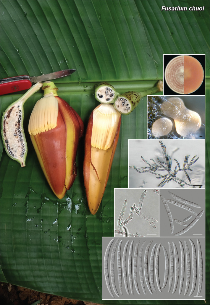
Notes — Fusarium chuoi resides in the Asian clade of the Fusarium fujikuroi species complex (FFSC: O’Donnell et al. 1998, Yilmaz et al. 2021, Crous et al. 2021b). Based on nucleotide searches using the Fusarium Pairwise ID engine on the Fusarioid-ID database (www.fusarium.org, Crous et al. 2021) the closest hit using the ITS sequence was Fusarium siculi (strain CBS 142422; identities = 449/450 (99 %), no gaps). The closest hit using the LSU sequence was F. siculi (strain CBS 142422; identities = 804/805 (99 %), no gaps). Closest hit using the cmdA sequence was Fusarium fractiflexum (strain NRRL 28852; identities = 426/434 (98 %), no gaps). Closest hit using the rpb1 sequence was F. fujikuroi (strain NRRL 13566; identities = 687/702 (98 %), no gaps). Closest hit using the rpb2 sequence was Fusarium globosum (strain CBS 428.97; identities = 856/867 (98 %), no gaps). Closest hit using the tef1 sequence was F. fractiflexum (strain NRRL 28852; identities = 619/643 (96 %), 2 gaps (0.3 %)). The phylogenetic results, however, showed that F. chuoi is not directly related to any of the previously described species of FFSC (see Suppl. material FP1353), clustering as the second basal-most species of that clade after F. sacchari.

Asian Fusarium spp. in the FFSC are characterised by mono- and polyphialides producing oval to ellipsoid, rarely pyriform to globose (i.e., F. annulatum, F. fujikuroi and F. globosum) microconidia organized in chains or false heads; 3–5-septate sporodochial conidia and lacking chlamydospores. The elaborate, profusely branched aerial conidiophores of F. chuoi are comparable to those of F. concentricum, F. lumajangense, F. mangiferae and F. sacchari, all the latter species producing oval, ellipsoidal to allantoid microconidia on false heads. Aerial conidiophores of F. chuoi, however, mostly produce macroconidia, while microconidia grouped on false heads are restricted to short, mostly unbranched and prostrate conidiophores formed on the surface on the culture media.

Several Asian species of the FFSC have been reported from Musa spp. i.e., F. annulatum, F. concentricum, F. fujikuroi, F. lumajangense and F. sacchari (Leslie & Summerell 2006, Maryani et al. 2019, Farr & Rossman 2021). The two strains representing F. chuoi were isolated as endophytes from asymptomatic seeds of wild banana (Musa itinerans), which had been collected predispersal and stored in the Millennium Seed Bank for ~2.5 years at -20 °C prior to isolation.

Colour illustrations. Flowers, fruits, leaves and seeds of Musa itinerans (background photo by D.T. Vu); from top to bottom and left to right: colony on PDA after 14 d at 24 °C in darkness (left = obverse,right = reverse), sporodochia formed on CLA, aerial conidiophore, aerial conidiogenous cells, aerial conidia, sporodochial conidia. Scale bars: black = 20 μm, white = 10 μm.

graphic file with name per-2023-47-6-g065.jpg

Fusarium chuoi

Supplementary material

FP1353

Phylogenetic tree.

per-2023-47-6-SF1353.jpg (640.3KB, jpg)
Persoonia. 2021 Dec 24;47:312–313.

Fungal Planet 1354 – 24 December 2021

Fusarium subflagellisporum T.F. Nóbrega & R.W. Barreto, sp. nov.

Thaisa F Nóbrega 1, Robert W Barreto 2

Etymology. Name refers to the short whip-like projections on apical cells of the sporodochial conidia.

Classification — Nectriaceae, Hypocreales, Sordariomycetes.

Mycelium branched, septate, hyaline, smooth, 2–5 μm diam. Aerial conidiophores branched or simple and reduced to conidiogenous cells. Sporodochia not observed on carnation leaf agar (CLA), occasionally formed on the surface of synthetic nutrient poor agar (SNA) after 14 d in the dark and potato dextrose agar (PDA) after 7 d under a 12 h daily light regime, both at 25 °C. Conidiogenous cells formed on aerial mycelium or in sporodochia, monophialidic, cylindrical to subcylindrical, conical or doliiform, 8–12 × 2–4 μm. Aerial macroconidia formed in SNA, abundant, formed directly on phialides, 1–5-septate, septa distinct, fusiform, mostly straight to slightly curved, 18–39 × 2–6 μm, apical cell curved or conical, basal cell foot-shaped or rounded at base, hyaline, thin-walled, smooth. Sporodochial macroconidia (on SNA), very abundant orange masses, 4–5-septate, elongate, bearing short whip-like projections, curved dorsiventrally, tapering towards both ends, 37–74 × 3–5 μm, apical cell, generally elongate basal cell either typical foot-shaped or, sometimes subconical, hyaline, thin-walled, smooth. Microconidia very rare, formed on phialides of aerial mycelium in SNA, 0–1-septate, ovoid to slightly fusoid, 4–11.5 × 1–4 μm, hyaline, thin-walled, smooth. Chlamydospores abundant, terminal or intercalary, either single or in chains, globose, 8–15 μm, aseptate, firstly subhyaline, becoming brown at maturity, smooth or rugose.

Culture characteristics — Fast-growing (up to 85 mm diam in 7 d on PDA under a 12 h daily light regime at 22 °C); raised, margin entire, aerial mycelium dense, felty, vinaceous centrally with alternating zonate rings white and rose towards the edge, reverse vinaceous; abundant sporulation.

Typus. BRAZIL, Pernambuco, Petrolina, Special Fruit Farm (S9°19’17.4" W40°40’29.9"), isolated from hypertrophied floral and vegetative branches of mango trees, 30 Aug. 2019, T.F. Nóbrega (holotype VIC 47377, ex-type culture COAD 2989, tef1-α and rpb2 sequences GenBank MT774486 and MZ970426, MycoBank MB 841204).

Notes — Fusarium subflagellisporum is a new member from the Brachygibbosum clade (Fusarium brachygibbosum). It belongs to the Fusarium sambucinum species complex as indicated by tef-1α and rpb2 sequence analyses. Isolates MRC 2486 (from peanuts, USA), FRC R-7642 (from sorghum debris, Puerto Rico), FRC R-8881 (from Nigeria), FRC R-9025 (from millet debris on soil, Zimbabwe), and NRRL 66930 (from corn-cultivated soil, USA) which have been reported in another study (O’Donnell et al. 2018, Laraba et al. 2021) all grouped in the same clade with F. subflagellisporum and are regarded here as belonging to the same species. However, no descriptions of morphology were provided for such isolates and no species names were proposed. Instead, only temporary codes were given to those isolates, listed as Fusarium sp. nov. 27 in Laraba et al. (2021) and Fusarium sp. nov. 10 in O’Donnell et al. (2018). Information on those isolates clearly indicates that F. subflagellisporum is widely distributed and occurs on a broad range of substrates. Laraba et al. (2021) also found that all isolates of F. subflagellisporum, listed as Fusarium sp. nov. 27, evaluated in their study produced the mycotoxin neosolaniol. It is not known whether the Brazilian isolate of F. subflagellisporum from mango also produces neosolaniol.

Fusarium subflagellisporum is phylogenetically close to F. brachygibbosum and F. transvaalense. Nevertheless, those two species do not produce sporodochial macroconidia with short whip-like projections on apical cells such as in F. subflagellisporum.

Fusarium subflagellisporum was isolated from a mango sample (stem and inflorescence) bearing typical symptoms of mango malformation disease. Isolation from those samples yielded Fusarium colonies belonging to two morphotypes. One morphotype was identified as F. sterilihyphosum – a well-known etiological agent of mango malformation that belongs to the Fusarium fujikuroi species complex (Lima et al. 2009). The other was described here as F. subflagellisporum. Inoculation on healthy young mango plants with F. sterilihyphosum led to development of mango malformation symptoms whereas inoculations with isolate COAD 2989 did not result in any disease symptoms.

graphic file with name per-2023-47-6-i019.jpg

Bayesian Inference tree constructed from the combined datasets of tef-1α and rpb2 sequences from species belonging to the Fusarium sambucinum species complex, including one isolate of Fusarium subflagellisporum sp. nov. obtained in this study (indicated in bold). Bayesian posterior probabilities (≥ 0.90) and bootstrap support values (≥ 70 %) are given at the nodes. The tree is rooted to Fusarium fujikuroi NRRL 66452.

Colour illustrations. Adult Mangifera indica individual showing hypertrophied inflorescences, typical of mango malformation at plantation in Petrolina, PE, Brazil. Fusarium subflagellisporum colony on PDA plate; sporodochial mass on SNA; globose brown-walled chlamydospores formed on SNA; chlamydospores and conidia formed in aerial mycelium; sporodochia; monophialides in aerial mycelium; conidia formed on aerial mycelium on SNA; conidia formed in sporodochial mass on SNA. Scale bars = 10 μm.

graphic file with name per-2023-47-6-g066.jpg

Fusarium subflagellisporum

Persoonia. 2021 Dec 24;47:314–315.

Fungal Planet 1355 – 24 December 2021

Genea zamorana Cabero, P. Alvarado & B. Martín, sp. nov.

Julio Cabero 1, Pablo Alvarado 2, Berta Martín 3

Etymology. The epithet refers to the province where the holotype was found (Zamora, Spain).

Classification — Pyronemataceae, Pezizales, Ascomycota.

Ascocarps subglobose, broadly lobed, somewhat gibbous; dark brown in colour, darkened with age; surface covered with high warts (0.9–1.1 mm), more or less distant between them, irregular or polygonal (4–6 sides), with a flat top, often cracked, producing an overall rough matt look to the ascocarp; ascocarps measure 1.3–1.6 cm diam; apical orifice conspicuous, irregularly cylindrical; small mycelium tuft tightly attached to the base. Odour not remarkable. Inner chamber not divided; with sinuose foldings of the wall containing whitish hymenium layers; chamber walls about 2 mm wide, covered by a brownish epithecium formed by minute scale-like warts. Peridium formed by two layers: 1) an external pseudoparenchymatic layer about 350 μm thick, composed of subglobose to angular hyaline elements, 25–30 × 35–40 μm mostly arranged perpendicularly to the peridium surface, which is dark brown; the innermost elements (in contact with the next layer) display a conspicuous reddish colour not observed in other species; and 2) an inner prosenchymatic layer about 50 μm thick that fuses with the hymenium. Hymenium arranged as a palisade, composed of asci and cylindrical or filiform septate paraphyses, formed of elements, 35.5–46 × 3.5–4.5 μm, fusing in a epithecium made of subglobose to angular cells. Asci cylindrical, indehiscent, inamyloid, stalked, 215–230 × 20–22 μm, when turgid, collapsing soon; stalk central, straight, lacking a crozier at the base, measuring about 30 × 12 μm (length × width). Ascospores uniseriate, 6–8 per ascus, facing different directions, subglobose to ovoid, measuring (21.5–)23–25.5(–26) × 18.5–20(–20.5) μm (ornamentation excluded); ornamented with tronco-conical or almost cubical warts measuring 2.3–2.8 × 2–3 μm (width × height), developing small secondary warts on them that produce an irregularly crown-like look.

Habitat & Distribution — Ascomata in groups of 2–5 in summer (July–September). Found in central Spain, in a slightly sandy siliceous soil with a pH of 5.5, surrounded by a Quercus pyrenaica forest with Pteridium aquilinum and Erica sp. receiving 650–750 mm rain/year. Altitude about 995 m a.s.l.

Typus. SPAIN, Castilla y León, Zamora, Sanabria, Remesal, 995 m a.s.l., in a slightly sandy siliceous soil with a pH of 5.5, surrounded by a Quercus pyrenaica forest with Pteridium aquilinum and Erica sp. receiving 650–750 mm rain/year, 27 Sept. 2020, J. Cabero (holotype AH 49255, ITS, LSU and tef1 sequences GenBank OK138914, OK138913 and OK143509, isotype JC2020927KY (J. Cabero personal fungarium), MycoBank MB 841174).

Additional materials studied. SPAIN, Castilla y León, Zamora, Sanabria, Ilanes, 995 m a.s.l., in a slightly sandy siliceous soil with a pH of 5.5, surrounded by a Quercus pyrenaica forest with Pteridium aquilinum and Erica sp. receiving 650–750 mm rain/year, 18 July 2017, J. Cabero (JC20170718); Sanabria, Quintana de Sanabria, 995 m a.s.l., in a slightly sandy siliceous soil with a pH of 5.5, surrounded by a Quercus pyrenaica forest with Pteridium aquilinum and Erica sp. receiving 650–750 mm rain/year, 18 July 2017, J. Cabero (JC20140920).

Notes — This species is phylogenetically nested in the clade of G. fragrans, closer related to G. pseudobalsleyi (ITS 90 % similar; LSU 97 % similar; tef1 98 % similar). This clade includes also other species such as G. anthracina, G. amici, G. balsleyi, G. compressa and G. pseudoverrucosa (Alvarado et al. 2016, 2018, 2020). It differs from these taxa because of the remarkable warts in its external surface, which are much smaller in the other species. In addition, G. zamorana presents reddish tones in the innermost rows of the pseudoparenchymatic peridium layer, a feature not observed in any other species of genus Genea.

graphic file with name per-2023-47-6-i020.jpg

Phylogenetic tree. A 50 % majority rule 28S rDNA-tef1 consensus phylogram of the species related to Genea fragrans (with Genea arenaria as outgroup) obtained using MrBayes from 1 200 sampled trees. Nodes were annotated if they were supported by ≥ 0.95 Bayesian posterior probability (left) or ≥ 70 % maximum likelihood bootstrap proportions (right). Nonsignificant support values are exceptionally represented inside parentheses. Samples newly sequenced in this study are in bold.

Colour illustrations. Spain, Castilla y León, Zamora, forest of Quercus pyrenaica. Ascomata; peridium; paraphyses; spores under LM (from the holotype). Scale bars = 20 μm.

graphic file with name per-2023-47-6-g067.jpg

Genea zamorana

Persoonia. 2021 Dec 24;47:316–317.

Fungal Planet 1356 – 24 December 2021

Gymnopus nigrescens Bañares, G. Moreno, P. Alvarado & Antonín, sp. nov.

Angel Bañares 1, Gabriel Moreno 2, Pablo Alvarado 3, Vladimír Antonín 4

Etymology. After the blackish colour of dried lamellae.

Classification — Omphalotaceae, Agaricales, Agaricomycetes.

Basidiocarps annual, with a central stipe. Cap 2–3.5(–4) cm, convex, expanding to convex-subapplanate, sometimes with depressed or subumbilicate centre, light reddish brown, darker at centre and paler towards margin, hygrophanous, translucently striate, rough and sometimes sulcate towards margin, greasy to subviscid when moist. Gills rather distant, L = 24–30, l = 15–25, adnexed to almost free, beige-cream with flesh colour tinge, dark brown to black when dried. Stem 40–60 × 2–3 mm, dark brown to blackish towards base, concolourous to pileus in upper part, cylindrical, sometimes compressed, with longitudinal groove, glabrous, scarcely compressed-tomentose at base, also strigose and subradicant on substrate. Spores (4.4–)4.7–7.2(–7.5) × 2.2–4.5(–5) μm, av. 5.9 × 3.4 μm, Qav = 1.8, n = 50, ellipsoid to lacrimoid. Basidia 4-spored, 23–26 × 7–8 μm. Basidioles very abundant, fusoid to claviform. Cheilocystidia 25–56(–72) × 3.5–8.5(–14) μm, claviform, cylindrical, deformed with wide projections, rarely lobed at apex and sometimes capitate. Pileipellis with lobate terminal cells or poorly developed Dryophila-structure hyphal elements (Antonin & Noordeloos 2010), sometimes with broad lobes, terminal elements up to 21 μm wide, that weakly turn green in KOH. Stipitipellis a cutis of cylindrical elements up to 7 μm wide, that strongly turn green in KOH. Caulocystidia 2.5–5(–8) μm wide, abundant, cylindrical, flexuose, obtuse, septate, thick-walled, sometimes with short to elongated (up to 12 μm) obtuse projections. Clamps present.

Habitat & Distribution — Growing gregarious on bryophytes (Scleropodium touretii) in a dense forest of Erica arborea. Presently known only from the Canary Islands (Spain).

Typus. SPAIN, Canary Islands, Tenerife, Chanajiga (T.M. Los Realejos), N28°20’37.42" W16°34’50.00", 1 170 m a.s.l., 22 Nov. 2019, Á. Bañares & O. Bermúdez (holotype TFC Mic. 25409, isotype in AH 49188; ITS, LSU and rpb2 sequences GenBank MZ542560, MZ542560 and MZ546414, MycoBank MB 840636).

Additional materials examined. Marasmiellus sp. nov. ad int.: FRANCE, Corsica, Valdo Moltifao, roots of Alnus glutinosa, 7 Nov. 2019, G. Moreno, F. Esteve-Raventós, A. Altés, F. Pancorbo & P.A. Moreau (AH 51247, ITS and LSU sequences GenBank MZ542557 and MZ542557). Gymnopus pubipes: SPAIN, Guadalajara, Albalate de Zorita, under Quercus ilex ssp. ballota, 15 Oct. 1999, F. Esteve, M. Villarreal, L. Montoya & V. Bandala (paratypus AH 26931, ITS and LSU sequences GenBank MZ542558 and MZ542558). Gymnopus fuscopurpureus: CZECH REPUBLIC, Moravian Karst, Adamov, Josefovské údolí valley, 13 Sept. 2018, H. Ševčíková (BRNM 809119, ITS, LSU and rpb2 sequences GenBank MZ542559, MZ542559 and MZ546413).

Notes — Gymnopus nigrescens is characterised by its light reddish brown pileus, flesh coloured lamellae that turn blackish when dried, poorly-developed Dryophila-structure in the pileipellis, and walls of all hyphae turning green in alkali.

Based on these characters, G. nigrescens belongs to sect. Levipedes, subsect. Alkalivirentes (Antonin & Noordeloos 2010). Other related species with fleshy coloured lamellae are Gymnopus fuscopurpureus, which differs by its darker pileus, lamellae not blackening, densely tomentose stipe and differently shaped caulocystidia, and G. obscuroides with a long stipe (to 9 cm) and a light brown reduced pileus of 0.9–1.8 cm wide. The sequences of G. fuscopurpureus obtained in the present work from specimen BRNM 809119 were significantly similar to those of G. nigrescens, but displayed a considerable distance in most markers (ITS: 93 % similar mainly due to a single 30 bp insertion, LSU: 99.5 % similar, rpb2: 95 % similar). In turn, the ITS sequences of G. obscuroides available in public databases (GenBank KX958398 and KX958399) were identical to others identified as G. alkalivirens (GenBank GU234141 and GU234022), which nests inside the genus Marasmiellus. Other similar genetic sequences in public databases were obtained from specimens identified as Collybia brunneola (GenBank MH874036, 99.32 % similar to LSU of G. nigrescens) and Gymnopus spongiosus (GenBank KY026706, also 99.32 % similar to LSU of G. nigrescens). Collybia brunneola is considered an independent species related to G. dryophilus (Vilgalys & Miller 1983), so the exact identity of the sample identified with this name needs to be confirmed. Gymnopus spongiosus is an American species with white to cream-white or creamy-buff lamellae, a stipe surface subglabrous at apex, and tomentose or strigose towards the base, slightly larger basidiospores (6.2–8.4 × 3.5–4.2 μm), inconspicuous, often collapsed, 24.5–50 μm long, cylindric to strangulated or irregularly lobed and knobbed cheilocystidia, a pileipellis composed of branched hyphae, often bifurcate, not diverticulate or coralloid, coarsely encrusted with a yellow brown pigment, caulocystidia cylindric to contorted, branched, septate (Halling 1996). According to Mata et al. (2006) it is similar to G. erythropus.

Colour illustrations. Spain, Canary Islands, Tenerife, Chanajiga, on bryophytes in a dense forest of Erica arborea, where the holotype was collected. Basidiocarps in situ; basidiocarps in herbarium; pileipellis with Dryophila-structure and clamps; cheilocystidia; caulocystidia; spores (from the holotype). Scale bars = 1 cm (basidiomata), 0.5 cm (basidiomata in herbarium), 10 μm (pileipellis, cheilocystidia and spores under LM), 10 μm (caulocystidia, left 20 μm), 10 μm (spores).

graphic file with name per-2023-47-6-g068.jpg

Gymnopus nigrescens

Supplementary material

FP1356

Phylogenetic tree.

per-2023-47-6-SF1356.jpg (773.7KB, jpg)
Persoonia. 2021 Dec 24;47:318–319.

Fungal Planet 1357 – 24 December 2021

Hesperomyces parexochomi Mironova & Haelew., sp. nov.

Danny Haelewaters 1, Polina Mironova 2, Pieter Asselman 2, Jerzy Romanowski 3, Piotr Ceryngier 3

Etymology. Referring to the host genus, Parexochomus.

Classification — Laboulbeniaceae, Laboulbeniales, Laboulbeniomycetes.

Associated with ladybird hosts in the genus Parexochomus (Coccinellidae, Coleoptera). Thallus (251–)299–348–396(–441) μm long from foot to perithecial apex; coloured yellowish. Cell I irregularly quadrilateral, broadening distally, 2.3 × longer than broad, with very oblique septum I–II and short septum I–III. Cell II 2.3 × longer than broad, (34–)38–43–48(–53) × (15–)17–19–21(–24) μm, subtrapezoidal in section. Cell III always smaller than cell II, (8.5–)9.3–12.0–14.4(–18.9) × 10.3–14.3–18.9 μm, with rounded dorsal cell wall. Primary appendage consisting of 4–6 superposed cells, (49–)53–59–66(–73) μm long; in the same axis as cell I and III, separated from the latter by the constricted primary septum; its basal cell as long as broad, cup-shaped, larger than each of the remaining cells; second to terminal cells each carrying a single antheridium externally, the terminal cell also carrying a second upwardly directed antheridium. Antheridia flask-shaped, with slightly curved efferent necks; the original ascospore apex still present as a short, pointed process between the fourth-to-last and second-to-last antheridium. Cell VI 2 × longer than broad, (34–)42–58–74(–108) × (21–)25–29–32(–36) μm, with subparallel margins to broadening distally, septum VI–VII oblique. Perithecium (165–)215–253–292(–314) × (47–)56–63–70(–79) μm, asymmetrical, with the anterior margin convex and the posterior one almost straight; broadest at the second and third tiers, then gradually tapering towards the apex; remainder of the trichogyne apex visible at basis of fifth tier cell; apex a complex with two short lower lobes, two upper (terminal) lobes, and two prominent lips surrounding the ostiole; lower lobes fingertip-shaped; terminal lobes unicellular, elongated, 34–40–44 μm in length, curved upwards and outwardly; ostiole with two lips, one lip triangular, the other slightly shorter, and blunt or rounded. Ascospores (59–)63–66–70(–74) μm long, with conspicuous slime sheath only surrounding the larger cell.

Typus. SPAIN, Canary Islands, Las Palmas, Fuerteventura Island, desert near Costa Calma, N28°09’56.2" W14°13’27.4", on Parexochomus quadriplagiatus (Coccinelidae, Coleoptera), 27 Mar. 2017, J. Romanowski, slide D. Haelew. 1465c (holotype at GENT, one subadult and five adult thalli from sternites; isotype DH1465b, three adult thalli from sternites, SSU, ITS and LSU sequences GenBank MZ994889, MZ994868 and MZ994879, MycoBank MB 840989).

Additional material examined. SPAIN, Canary Islands, Las Palmas, Fuerteventura Island, Caleta de Fuste, N28°23’11.4" W13°51’50.9", on Parexochomus nigripennis (Coccinelidae, Coleoptera), 13 Feb. 2017, J. Romanowski, slides D. Haelew. 1377a (GENT, one juvenile and seven adult thalli from left metatrochanter) and 1377b (GENT, nine thalli from sternites); ibid., desert near Costa Calma, N28°09’56.2" W14°13’27.4", on P. quadriplagiatus, 27 Mar. 2017, J. Romanowski, slide D. Haelew. 1466a (GENT, one subadult thallus from right epipleuron and one adult thallus from sternite); ibid., Lajares, N28°40’44.1" W13°56’13.5", on P. nigripennis, 20 Oct. 2018, J. Romanowski, slide D. Haelew. 1690e (GENT, one adult thallus from right elytron); ibid., slide D. Haelew. 1691e (GENT, two adult thalli from elytral tips); ibid., slides D. Haelew. 1693c (GENT, one juvenile and 11 adult thalli from left elytral tip) and 1693d (GENT, 14 adult thalli from tip of right elytron); ibid., slide D. Haelew. 1694b (GENT, two adult thalli from left antenna).

Additional materials sequenced. See Supplementary material page.

Notes — This species is a part of the near-cryptic Hesperomyces virescens species complex (Haelewaters et al. 2018). Hesperomyces parexochomi is phylogenetically distinct from its closest relatives. Morphologically, it is very similar to other taxa within H. virescens s.lat., except for the position of the original spore apex. It is situated between the fourth-to-last and second-to-last antheridium in H. parexochomi whereas it occurs on the last or penultimate antheridium in other members of the complex. Thalli removed from Pareochomus nigripennis and P. quadriplagitus are morphologically similar and form a single phylogenetic clade. This is in line with the hypothesis that the host specificity within the H. virescens complex exists at the generic level (Haelewaters et al. 2018, Haelewaters & De Kesel 2020).

Based on a megablast search of NCBIs GenBank nucleotide database, the closest hits using the ITS sequence had highest similarity to H. virescens ex Harmonia axyridis (isolate D. Haelew. 1268d, GenBank MG757830.1; Identities = 789/853 (92 %), 13 gaps (1 %)), H. virescens ex Harmonia axyridis (isolate D. Haelew. 1268b, GenBank MG757829.1; Identities = 789/853 (92 %), 13 gaps (1 %)), and H. virescens ex Harmonia axyridis (isolate D. Haelew. 943b, GenBank MGM757810.1; Identities = 789/853 (92 %), 13 gaps (1 %)). Closest hits using the LSU sequence are H. virescens ex Adalia bipunctata (isolate D. Haelew. 1232a, GenBank MG745351.1; Identities = 884/899 (98 %), one gap (0 %)), H. virescens ex Adalia bipunctata (isolate D. Haelew. 1199h, GenBank MG745347.1; Identities = 884/899 (98 %), one gap (0 %)), H. virescens ex Adalia bipunctata (isolate D. Haelew. 1231a, GenBank MG745350.1; Identities = 884/900 (98 %), two gaps (0 %)) and H. virescens ex Psyllobora vigintimaculata (isolate D. Haelew. 1251b, GenBank MG745356.1: Identities = 884/901 (98 %), no gaps).

Colour illustrations. Xerophilous plants, habitat for Parexochomus nigripennis. Parexochomus nigripennis with thalli of Hesperomyces parexochomi on elytral tips; P. quadriplagiatus with thalli of H. parexochomi on left elytron; decorative plants at Caleta de Fuste, habitat for P. quadriplagiatus; primary appendage, with arrowhead pointing at original spore apex; adult thallus. Scale bars = 200 μm (hosts), 50 μm (thallus), 10 μm (appendage).

graphic file with name per-2023-47-6-g069.jpg

Hesperomyces parexochomi

Supplementary material

FP1357-1

Phylogenetic tree.

per-2023-47-6-SF1357-1.jpg (1,009.7KB, jpg)
FP1357-2

Table. Species of Hesperomyces included in this study.

Persoonia. 2021 Dec 24;47:320–321.

Fungal Planet 1358 – 24 December 2021

Heterophoma rehmanniae A. Shibata, Tonegawa, Sei. Kato, Toy. Sato & Hirooka, sp. nov.

Aoi Shibata 1, Chie Tonegawa 1, Seiji Kato 1, Yuuri Hirooka 1, Toyozo Sato 2

Etymology. Named after the host, Rehmannia glutinosa, from which our fungus was isolated.

Classification — Didymellaceae, Pleosporales, Dothideomycetes.

Conidiomata on oatmeal agar (OA) pycnidial, solitary or aggregated, sub-globose, glabrous, superficial and immersed into the agar, 71–441 × 55–253 μm in size. Ostiole single, slightly papillate or non-papillate. Mycelium hyaline, thin-walled, smooth, becoming dark-brown and hypertrophic with age. Conidiogenous cells phialidic, hyaline, smooth, globose to lageniform, 5.2–9.3 × 3.7–7.7 μm in size. Conidia ellipsoidal to cylindrical, hyaline, thin-walled, smooth, aseptate, (2.9–)3.4–4.2(–4.6) × (1.2–)1.4–1.7(–1.8) μm with small polar guttles. Chlamydospores absent.

Culture characteristics — Colonies on malt extract agar (MEA) reaching 51–61 mm at 20 °C and 61–67 mm diam at 25 °C after 7 d, margin regular, floccose, (pale) olivaceous grey to dull green; reverse dark olivaceous green near centre. Colonies on OA reaching 46–50 mm at 20 °C and 58–60 mm at 25 °C after 7 d, margin regular, woolly, pale olivaceous grey, sometimes black pycnidia formed; reverse concolourous. NaOH test negative.

Typus. JAPAN, Tokyo, Kodaira city, on leaves of Rehmannia glutinosa f. hueichingensis (Scrophulariaceae), Aug. 2015, A. Shibata (holotype TNS-F-93697, culture ex-type HM15-014C = MAFF247505; ITS, LSU, rpb2 and tub2 sequences GenBank LC656204, LC656208, LC656201 and LC656212, MycoBank MB 841842).
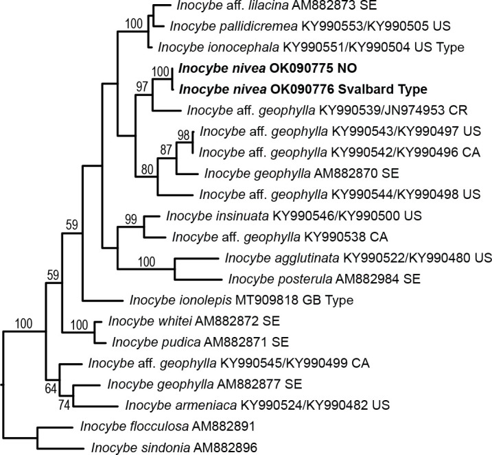
Additional materials examined. JAPAN, Tokyo, Kodaira city, on leaves of R. glutinosa var. purpurea, Aug. 2015, A. Shibata (HM15-015 = 15-M0092 = TNS-F-93698, culture HM15-015C = MAFF247506; ITS, LSU, rpb2 and tub2 sequences GenBank LC656205, LC656209, LC656202 and LC656213); Toyama, Toyama city, on leaves of R. glutinosa f. hueichingensis, Sept. 2015, T. Sato (HM15-362 = TKP2 = TNS-F-93699, culture HM15-362C = MAFF 245313; ITS, LSU, rpb2 and tub2 sequences GenBank LC656206, LC656210, LC656203 and LC656214); Toyama, Toyama city, on leaves of R. glutinosa var. purpurea, Sept. 2015, T. Sato (HM15-364 = TAP1 = TNS-F-93700, cultures HM15-364C = MAFF 245316; ITS, LSU and tub2 sequences GenBank LC656207, LC656211 and LC656215).
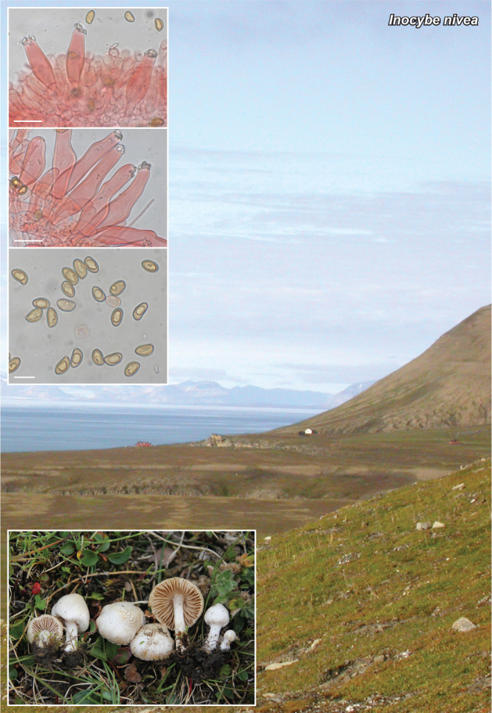
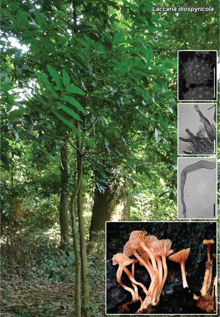
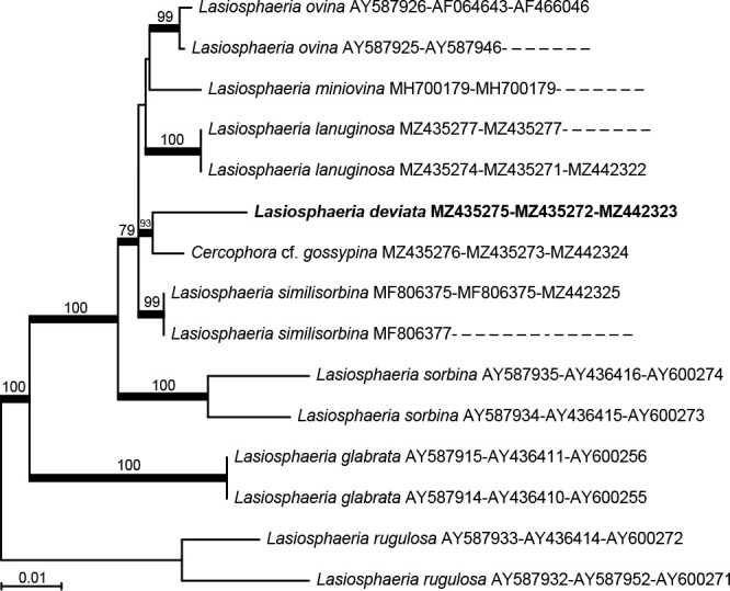
Notes — In our Maximum Likelihood tree using LSU, ITS, rpb2 and tub2, our four isolates represented an independent clade in the genus Heterophoma. The isolates were different from seven existing species of Heterophoma, i.e., H. nobilis, H. poolensis, H. adonidis, H. sylvatica, H. verbascicola, H. verbasci-densiflori and H. novae-verbascicola, based on shape and size of conidia, and NaOH reaction (see Table in Supplementary material; De Gruyter & Noordeloos 1992, De Gruyter et al. 1993, Chen et al. 2015, 2017, Hou et al. 2020a).

graphic file with name per-2023-47-6-i021.jpg

Phylogenetic tree of the combined LSU, ITS, rpb2 and tub2 MAFFT-aligned datasets obtained using Maximum Likelihood. A heuristic search was performed in RAxML-NG (Kozlov et al. 2019) with support at the nodes calculated using bootstrap analyses with 100 replicates. The ML bootstrap support values ≥ 70 % are indicated at the nodes. Stagonosporopsis hortensis (CBS 104.42 and CBS 572.85) was used as outgroup. T: ex-type strain; R: representative strain.

Colour illustrations. Flowers of Rehmannia glutinosa. Upper row: colony on OA after 7 d at 20 °C. Middle row: colony on MEA after 7 d at 20 °C; conidioma on leaf spot. Bottom row: conidiomata; conidiogenous cell; conidia on OA. Scale bars = 200 μm (conidiomata bottom row), 50 μm (conidioma middle row), 5 μm (conidiogenous cell and conidia).

graphic file with name per-2023-47-6-g070.jpg

Heterophoma rehmanniae

Supplementary material

FP1358

Table. Morphological characteristics of Heterophoma rehmanniae and published descriptions of Heterophoma spp.

per-2023-47-6-SF1358.jpg (205.6KB, jpg)
Persoonia. 2021 Dec 24;47:322–323.

Fungal Planet 1359 – 24 December 2021

Hyphodermella pallidostraminea Bukharova & Volobuev, sp. nov.

Sergey V Volobuev 1, Nadezhda V Bukharova 2

Etymology. Name refers to the pale straw-yellow colour of hymenial surface.

Classification — Phanerochaetaceae, Polyporales, Agaricomycetes.

Basidiomata annual, resupinate, adnate, effuse, ceraceous to crustaceous when dry, at first orbicular, then confluent, up to 15 mm diam. Margin white, determinate, narrow, fibrillose, with age thinning out. Hymenial surface yellowish, tiger yellow (090 90 50), fresh yellow (095 90 40) or pea green (095 80 40) (all colour codes are given following the RAL Design colour chart), smooth to slightly tuberculate, cracked. Subiculum thin, indistinct, white. Hyphal system monomitic; generative hyphae simple-septate. Subicular hyphae 3.5–5 μm diam, hyaline, thick-walled or with slightly thickened walls, encrusted with numerous prismatic crystals, loosely interwoven, more or less parallel arranged, occasionally branched. Subhymenial hyphae 2.5–3.5 μm diam, hyaline, thin-walled, smooth, compactly interwoven, frequently ramified. Cystidia absent, but cylindrical to fusoid, cystidioid hyphal ends occasionally present, thin-walled, not encrusted, projecting above the hymenium up to 35 μm. Basidia 17–25 × 5.2–6 μm, clavate, with a simple septum at the base, 4-sterigmate. Basidiospores (5.0–)5.4–6.6(–6.7) × (2.9–)3.0–3.5(–3.6) μm, n = 30, L = 5.96, W = 3.23, Q = 1.82–1.87, smooth, hyaline, thin-walled, ellipsoid, inamyloid, non-dextrinoid, with tiny oil drops.

Habitat & Distribution — On dry, dead Actinidia sp. branch, in a herbaceous mixed coniferous-broadleaf forest. Hitherto only known from the type locality in the south of the Russian Far East.

Typus. RUSSIA, Jewish Autonomous Oblast, Obluchensky District, Bastak State Nature Reserve, 15 km north-northeastward from the Kirga settlement, at the foot of Mount Skalistaya, N49°00’47.3" E132°53’53.6", on dry dead branch of Actinidia sp. (Actinidiaceae) in mixed coniferous-broadleaf forest, 24 Aug. 2009, N. Bukharova (Vasilyeva) (holotype LE 286968, isotype VLA M-22719, ITS and LSU sequences GenBank OK138912 and OK138911, MycoBank MB 841840).

Notes — Hyphodermella pallidostraminea is a minute-sized corticioid fungus characterised macroscopically by pale-yellowish smooth to tuberculate hymenophore and white pruinose margin of basidiocarps. The main distinguishing microscopic feature among other representatives of the genus Hyphodermella is the spore size (up to 3.5 μm in width) that is overlapping only with the recently described H. zixishanensis, but the latter species differs by the reddish to brown hymenial surface and the absence of cystidioid hyphal ends projecting above the hymenium (Wang et al. 2021). The previous attempt to find a taxonomic interpretation for the studied specimen of H. pallidostraminea without a molecular analysis led to the fact that this material was erroneously identified as Sistotremastrum niveocremeum (Bukharova & Zmitrovich 2014), also being a deciduous-dwelling fungus having a monomitic hyphal system, similar spore sizes and shape, but differing by clamps on hyphae.

Based on a megablast search of NCBIs GenBank nucleotide database, the closest hits using the ITS sequence had highest similarity to H. rosae (strain FP-150552, GenBank KP134978; Iden-tities = 625/657 (95 %), 13 gaps (1 %)), H. corrugata (voucher K(M) 237661, GenBank MK496919; Identities = 633/666 (95 %), 13 gaps (1 %)) and H. rosae (voucher CFMR DLL2011-177, Gen-Bank KJ140672; Identities = 613/646 (95 %), 13 gaps (2 %)). Closest hits using the LSU sequence are Geliporus exilisporus (voucher TNM GC 1702-15, GenBank LC379153; Identities = 847/859 (99 %), no gaps), H. rosae (strain GC 1604-113, GenBank MZ637147; Identities = 849/862 (98 %), no gaps) and H. corrugata (voucher MA-Fungi 5527, GenBank JN939597; Identities = 849/862 (98 %), no gaps).

graphic file with name per-2023-47-6-i022.jpg

Phylogenetic tree derived from Maximum Likelihood analysis based on nrITS1-5.8S-ITS2 data. The analysis was performed in the IQ-TREE Web Server (Trifinopoulos et al. 2016) with 1 000 ultrafast bootstrap replicates. Maximum Likelihood bootstrap support values shown above branches (BS > 80 %). The new species described in this study is in bold face. The scale bar represents the expected number of nucleotide changes per site.

Colour illustrations. Coniferous-broadleaf mixed forest in the Bastak State Nature Reserve (Jewish Autonomous Oblast, Russia). Basidiocarp; basidia and spores (all from holotype). Scale bars = 2 mm (basidiocarp), 10 μm (cystidioid hyphal ends), 5 μm (spores and basidia).

graphic file with name per-2023-47-6-g071.jpg

Hyphodermella pallidostraminea

Persoonia. 2021 Dec 24;47:324–325.

Fungal Planet 1360 – 24 December 2021

Inocybe corsica Esteve-Rav., Pancorbo & G. Moreno, sp. nov.

Fernando Esteve-Raventós 1, Gabriel Moreno 1, Alberto Altés 1, Fermín Pancorbo 2, Ibai Olariaga 3

Etymology. Name refers to the island of Corsica (Corse), where the samples were collected.

Classification — Inocybaceae, Agaricales, Agaricomycetes.

Basidiomata agaricoid and stipitate. Pileus 10–25 mm, at first conical-campanulate, then convex to plano-convex, broadly umbonate to subumbonate, slightly hygrophanous; margin straight, regular to hardly wavy with age, fissurate at times, velipellis not present; colour yellow ochraceous (Mu 10YR 6/8, 7/6) when young or moistened, from uniform to light ochraceous in the centre (Mu 10YR 8/2) in some basidiomata; surface radially fibrillose, smooth, not rimose, slightly striated towards the margin in adult basidiomata, with greasy aspect, not sticky when moist. Lamellae moderately crowded (L = 35–45; l = 1–2), free to adnate, ventricose, initially whitish, becoming pale grey to beige, then pale brown, edge concolourous, entire to finely crenulate. Stipe 22–40 × 3–4 mm, straight to curved towards base, bulbous to bulbous-napiform, not marginately bulbous; colour whitish, becoming beige to ochraceous with age (Mu 10YR 8/2; 7/3); surface densely pruinose along the entire surface. Cortina absent. Context fibrose, whitish, concolourous to the stipe surface. Smell herbaceous, taste not recorded. Colour of exsiccatum ochre-brownish (pileus and stipe), not blackening. Spores (8.0–)8.7–9.7–10.9(–12.2) × (5.4–)6.7–7.7–8.7(–9.1) μm, Qm: (1.0–)1.09–1.25–1.42(–1.7) (n = 193 / N = 2), mostly subisodiametric to sometimes subheterodiametric, distinctly nodulose (though exceptionally sub-entolomatoid) under the optical microscope, provided with 7–10 distinct knobs variable in height (1.3–2.3 μm high), yellowish, apicula distinct. Basidia (24–)27–34.8–41(–49) × (10.9–)11.3–13.2–15.8(–17.1) μm; Qm: (1.81–)2.0–2.6–3.2(–3.4), 4-spored, rarely 2-spored, clavate, sterigmata 2.5–7 μm long. Lamella edge practically sterile, composed by numerous protruding hyaline to brownish cheilocystidia mixed with abundant mostly hyaline to brownish clavate paracystidia. Pleurocystidia abundant, (38–)45–58.1–72(–80) × (11.3–)12.7–16.5–21.2(–22.4) μm, Qm: (1.78–)2.5–3.6–4.8(–5.6), (n = 110 / N = 2), broadly lageniform to rarely fusiform, hyaline or with brownish content, base often attenuate but not pedicellate, rather crystalliferous at the apex, walls (1.0–)1.2–1.6–2.2(–2.4) μm thick, reaching –3 μm at the apex, hyaline to hardly yellowish in 10 % NH4OH. Cheilocystidia numerous, (43–)47–58.2–73(–79) × (11.0–)13.3–16.5–20.3(–22.7) μm, Qm: (2.21–)2.7–3.5–4.8(–7.1), similar in size and shape to pleurocystidia. Stipitipellis a cutis of parallel hyphae (3.5–)4.3–6.0–7.9(–10.3) μm, bearing numerous caulocystidia along the entire length of the stipe, (45–)49–63.2–83(–90) × (8.3–)8.8–12.2–15.8(–16.8) μm, Qm: (3.8–)4.0–5.2–7.6(–8.4) (n = 73 / N = 2), more slender but similar in shape to hymenial cystidia, mixed with numerous clavate to subcylindrical hyaline paracystidia. Pileipellis a cutis formed by parallel cylindrical cells, 4.5–13 μm wide, somewhat constricted at septa, showing minute pale yellow ochraceous pigment, minutely encrusting and also diffuse intracellular. Lamellar trama of parallel hyphae, 6–16 μm broad, showing diffuse yellowish intracellular pigment. Clamp connections abundant in all tissues.

Habitat & Distribution — Gregarious in acidic soil; in humid and herbaceous areas, next to willows (Salix atrocinerea), in somewhat muddy and nitrified soils; the collecting area is located in a Mediterranean environment of holm oak (Quercus ilex subsp. ilex) and cork oak (Quercus suber) mixed forest. No sequence similar to the ones generated from our material are available in GenBank.

Typus. FRANCE, Corsica, Corti, Saint-Jean, Maison San Giovanni, N42°17’38.04" W9°10’04.61", 427 m a.s.l., along a ditch on mossy and wet ground, near Salix atrocinerea, with some Juncaceae and Rosa sp. scrub, close to Quercus suber-Q. ilex subsp. ilex forest, in acidic sandy soil, 8 Nov. 2019, F. Pancorbo, A. Altés, G. Moreno & F. Esteve-Raventós (holotype AH 51900, ITS and LSU sequences GenBank MZ308644 and MZ308646, MycoBank MB 839085).

Additional material examined. Same location, date, habitat and collectors, AH 55201 (paratypus), ITS and LSU sequences GenBank MZ308645 and MZ308647).

Notes — Colour codes are taken from Munsell (1994), terminology follows Vellinga (1988) and Kuyper (1986). Inocybe corsica is probably a species of Mediterranean distribution linked to Salicaceae (found under Salix atrocinerea), of humid and waterlogged areas in acidic soils; macroscopically it resembles several species of the Xanthomelas clade (Esteve-Raventós et al. 2015, Vauras & Larsson 2016, Bandini et al. 2019, 2020) because of its medium-small size, and is characterised by the presence of a non-marginate napiform bulb and flesh that does not blacken markedly with age or desiccation. Our analyses of the ITS region (Supplementary Fig. 1) show that I. corsica nests in the well supported I. sect. Marginatae clade (ML-BS 98 %, BPP 1), within which it forms a clade together with a sequence of I. diabolica (ML-BS 95 %, BPP 0.98). The two sequences of I. corsica obtained are identical and form a fully-supported clade (ML-BS 100 %, BPP 1). From the group of species linked to the Salicaceae, both I. salicis (= I. straminipes) and I. lacunarum (Vauras & Larsson 2016), show different spores, provided with numerous (17–22) prominent knobs (Kühner 1955); another species bound to Salix and Populus and discovered in Japan is I. populea, very close to I. salicis in morphological features but showing an indistinctly marginate to claviform stipe base and smaller cystidia (Kobayashi & Courtecuisse 2000).

(text continues on Supplementary material page FP1360)

Colour illustrations. France, Corsica, Corti, Saint-Jean, Maison San Giovanni, Salix atrocinerea with Quercus suber in muddy and mossy soil, near a dirt road, locality where the samples were collected. Basidiomata; pleurocystidia; cheilocystidia; caulocystidia at stipe base; spores under optical microscope; spores under SEM (bottom right). Scale bars = 50 μm (cystidia), 10 μm (spores), 2 μm (spores under SEM).

graphic file with name per-2023-47-6-g072.jpg

Inocybe corsica

Supplementary material

FP1360

Phylogenetic tree.

per-2023-47-6-SF1360.jpg (652.5KB, jpg)
Persoonia. 2021 Dec 24;47:326–327.

Fungal Planet 1361 – 24 December 2021

Inocybe nivea E. Larss., sp. nov.

Ellen Larsson 1

Etymology. Refers to the white colour of the mushroom.

Classification — Inocybaceae, Agaricales, Agaricomycetes.

Pileus 5–20 mm diam, as young campanulate, conical to convex mostly with an obtuse to broad umbo, sometimes more prominent, margin incurved, later becoming convex to plano-convex with broad or a more prominent umbo, initially pure white, sericeous smooth, silky, with age becoming pale buff to isabelline, when affected by harsh weather cracking and becoming scaly, velipellis indistinct. Cortina white visible and rather abundant in young basidiomata. Lamellae moderately crowded, interspersed with lamellulae, L = 25–40, 2–3 mm broad, ventricose, sinuate to emarginate, first pale beige with a greyish tone, later yellowish brown, edge fimbriate white. Stipe 15–35 × 1–4 mm, equal to slightly bulbous but without bulb, solid, whitish, at apex pruinose descending to about 1/3, downwards smooth, in lower part fibrillose. Context whitish to pale buff. Smell spermatic, taste indistinct. Basidiospores (8.5–)9.6–9.7–9.8(–11.1) × (4.6–)5.7–5.8–5.9(–6.5) μm, n = 80, Q = (1.5–)1.62–1.65–1.66(–1.9), smooth ellipsoid, amygdaliform to subamygdaliform with obtuse apex and a small distinct apiculus, yellowish brown. Basidia 30–35 × 9–11 μm, n = 20, subclavate to clavate, 4-spored, hyaline, sterigmata 4.5–5.8 μm. Pleurocystidia 45–65 × 11–18 μm, n = 50, fusiform, lageniform to utriform with a slender pedicel, thick walled, 1–3 μm thick, hyaline, crystalliferous at apex. Cheilocystidia similar to pleurocystidia but shorter, 38–52 × 13–18 μm, n = 20, lageniform to broadly utriform, densely arranged, thick walled, 2.5–4.5 μm thick. Paracystidia hyaline, pyriform to clavate 15–28 × 10–13 μm, n = 10. Caulocystidia at apex similar to pleurocystidia, abundant, with crystals, less so further down 45–65 × 11–18 μm, n = 10, fusiform to more cylindrical, caulocystidioid hairs thin-walled, sometimes septate, 30–60 × 5–12 μm, cauloparacystidia few. Pileipellis an interwoven cutis of smooth hyaline hyphae, 6–12 μm wide. Clamp connections present.

Ecology & Distribution — Associated with Salix polaris and Salix spp. on nutrient rich and somewhat calcareous soils. Basidiomata so far only known from an arctic habitat on Svalbard where it grew in a rather moist area among mosses and herbs associated with S. polaris, and from a subalpine Betula pubescens forest in Norway, where it grew in a moist area associated with Salix spp. Blast search of NCBIs GenBank nucleotide database and the UNITE database gave no further information on distribution and occurrence as no matches of additional ITS sequence data were available.

Typus. SVALBARD AND JAN MAYEN, Spitsbergen, Revneset, arctic habitat with mosses and herbs, associated with Salix polaris, 12 Aug. 2015, E. Larsson, EL76-15 (holotype GB-0207613, isotype O, ITS-LSU sequence GenBank OK090776, MycoBank MB 841189).

Additional materials examined. SVALBARD AND JAN MAYEN, Spitsbergen, Revneset, arctic habitat with mosses and herbs, associated with Salix polaris, 12 Aug. 2015, E. Larsson, EL81-15, GB-0207614. – NORWAY, Nordland, Saltdal, Jukerdalsura NR, subalpine area on gravelly ground with Betula pubescens, Alnus incana, and Salix spp., 10 Aug. 2012, E. Larsson, EL43-12, GB-0207612 (ITS-LSU sequence GenBank OK090775).

Notes — Inocybe nivea belongs in the I. geophylla group, a group that is identified to host a high phylogenetic diversity and the name I. geophylla has been widely applied to many taxa in North America and Europe (Ryberg et al. 2008, Matheny & Swenie 2018), and in addition I. geophylla is reported associated with a large number of frondose and coniferous trees (Kuyper 1986). In macro- and micro-morphology they all are rather similar, but the geographic distribution and ecological preferences may explain part of the observed phylogenetic diversity. Further work on the group is needed.

Inocybe nivea is a rather small species within the group, characterised by a conical to plano-convex white pileus, about 15–20 mm diam in mature basidiomata, and seems to be associated with Salix spp. The pileus is silky and smooth but can in the arctic zone often be affected by cold and windy weather conditions that makes the cutis break up and form large scales. This was not observed in the subalpine basidiomata. In I. geophylla the pileus is usually much larger and they have different host preferences and ecology.

graphic file with name per-2023-47-6-i023.jpg

Phylogram obtained using PAUP v. 4.0a (Swofford 2003) based on ITS and LSU data showing the position of I. nivea in the I. geophylla group. Heuristic searches with 1 000 random-addition sequence replicates and tree bisection-reconnection (TBR) branch swapping were performed. Relative robustness of clades was assessed by the bootstrap method using 1 000 heuristic search replicates with 100 random taxon addition sequence replicates and TBR branch swapping. Bootstrap support values are indicated on branches. Inocybe nivea is marked in bold and the holotype is indicated.

Colour illustrations. Inocybe nivea habitat in the arctic zone growing associated with Salix polaris, on Svalbard, Spitsbergen, Revneset. In situ basidiomata of the holotype (GB-0207613); hymenial pleurocystidia; caulocystidia from stipe apex; basidiospores. Scale bars = 20 μm (pleuro- and caulo-cystidia), 10 μm (spores).

graphic file with name per-2023-47-6-g073.jpg

Inocybe nivea

Persoonia. 2021 Dec 24;47:328–329.

Fungal Planet 1362 – 24 December 2021

Laccaria diospyricola Salna Nanu & T.K.A. Kumar, sp. nov.

Salna Nanu 1, TK Arun Kumar 1

Etymology. Basidiomata of the species were always found among roots of Diospyros paniculata.

Classification — Hydnangiaceae, Agaricales, Agaricomycetes.

Pileus 5–15 mm diam, slightly depressed when young, becoming concave to infundibuliform at maturity; surface flesh coloured (Methuen 6B5–6B6, 7A2–7A4), cream (6A2–6A3) towards the centre, with occasional scattered violaceous spots, hygrophanous, minutely pubescent throughout, and covered with small superficial patches of whitish mycelium; margin uplifted at maturity. Lamellae adnate to slightly decurrent, concolourous with the pileus (6B5–6B6), thick, distant, up to 1.5 mm wide, with lamellulae in 2–3 tiers; edge serrate, concolourous with the sides. Stipe 10–30 × 1–3 mm, central, terete, almost equal, slightly tapering towards the base, flesh coloured (6B5–6B6) without violaceous spots, pubescent; base with mycelial pad. Basidiospores (two observations two collections, five sporocarps, 20 basidiospores) 7–9 × 6–8 μm (excluding the spines), Q = 1.0–1.28, Qm = 1.15, subglobose to globose, echinulate, thin-walled, hyaline, inamyloid; echinulae up to 1 μm long. Basidia 23–66 × 9–15 μm, clavate, thin-walled, 4-spored. Sterigmata up to 11 μm long. Cheilocystidia 40–90 × 4–10 μm, flexuose, cylindrical to narrowly clavate, hyaline, thin-walled, inamyloid. Lamellar trama subregular; hyphae 3–6 μm wide, hyaline, inamyloid, slightly thick-walled (up to 1 μm thick). Pileipellis an agglutinated cutis, occasionally disrupted by trichodermal patches of ascending or erect hyphae; hyphae 2–7 μm wide, thin-walled. Stipitipellis a cutis frequently disrupted by clustered or scattered caulocystidia; hyphae 2–6 μm wide, inamyloid, slightly thick-walled (up to 1 μm thick) with pale yellow content. Caulocystidia 38–185 × 8–16 μm, flexuose, inamyloid, hyaline, slightly thick-walled (up to 1 μm thick). Stipe context composed of 3–10 μm wide hyphae which are hyaline, inamyloid, slightly thick-walled (up to 1 μm thick), with yellowish brown plasmatic pigment. Clamp connections observed on all hyphae.

Habitat & Distribution — Scattered or in small groups, on sandy soil, in the root bed of Diospyros paniculata. Basidiocarps collected from sacred groves of two different regions of Kerala State, India.

Typus. INDIA, Kerala State, Kozhikode District, Thurayilkavu, on the ground under Diospyros paniculata (Ebenaceae), 10 Aug. 2018, S. Nanu (holotype CAL1771, nrITS sequence GenBank MK776767, MycoBank MB 833411).

Additional material examined. INDIA, Kerala State, Kannur District, Poongottukavu, 12 Sept. 2018, S. Nanu (CAL1770).

Notes — Small basidiomata, infunduliformis to applanate pileus, absence of two-spored basidia, cutis type of pileipellis with trichodermal patches and presence of caulocystidia are the diagnostic features of L. diospyricola. This species is closely related to L. violaceotincta and L. vinaceoavellanea. Laccaria diospyricola is found near or on the root bed of D. paniculata, whereas, L. violaceotincta was reported from a freshwater swamp forest dominated with Myristica species (Latha et al. 2019). Laccaria diospyricola differs from L. violaceotincta by having small basidiomata, absence of 2-spored basidia, and a cutis type of pileipellis disrupted by trichodermal patches. Hongo (1971) originally described L. vinaceoavellanea from Japan, having brownish to vinaceaous basidiomata, greyish red to white pileus, distant and decurrent gills, tough, solid and striate stipe, globose and inamyloid spores with ornamentation, and presence of clamp connections in all hyphae. Laccaria diospyricola basidiomata show some resemblance with L. vinaceoavellanea in having globose to subglobose basidiospores with spines, and in having clamp connections in all hyphae. Different colour of basidiomata, absence of tough, solid and equal stipe, and molecular data are the characters that distinguish L. diospyricola from L. vinaceoavellanea. Laccaria yunnanensis also shows similarities with L. diospyricola. However, the presence of adnate to slightly decurrent lamellae, densely pubescent and terete stipe, larger cheilocystidia, and caulocystidia separates L. diospyricola from L. yunnanensis.

A BLAST search in GenBank nucleotide database using the newly generated ITS sequence of L. diospyricola, resulted in a close hit with Laccaria violaceotincta (GenBank MK141034), showing a sequence similarity of 94.26 %. The second closest hit (93.74 %) in the BLAST search was with L. vinaceoavellanea (GenBank LC098741). Phylogenetic analyses using maximum parsimony (MP) and maximum likelihood (ML) methods generated trees that were congruent with regards to the topology and the relative positions of the taxa. The bootstrap support values from the MP analysis were similar to ML support values. In both the MP and ML trees, Laccaria diospyricola settled in a separate well-supported clade along with other tropical species, L. violaceotincta, L. yunnanensis, and L. vinaceoavellanea. Laccaria diospyricola resolved in a branch distinct from all other species, and the clade received a bootstrap support value of 99 % in the ML analysis and MP analysis.

Colour illustrations. India, Kerala State, Kozhikode district, Thurayilkavu, a sacred grove with different plants including Diospyros paniculata. SEM photograph of basidiospores; cheilocystidia; caulocystidia; basidiomata. Scale bars = 10 mm (basidiomes), 10 μm (all others).

graphic file with name per-2023-47-6-g074.jpg

Laccaria diospyricola

Supplementary material

FP1362

Phylogenetic tree.

per-2023-47-6-SF1362.jpg (368.2KB, jpg)
Persoonia. 2021 Dec 24;47:330–331.

Fungal Planet 1363 – 24 December 2021

Lasiosphaeria deviata A.N. Mill. & Læssøe, sp. nov.

Andrew N Miller 1, Thomas Læssøe 2

Etymology. The specific epithet refers to the aberrant ascospores that deviate from the typical ascospores found in Lasiosphaeria.

Classification — Lasiosphaeriaceae, Sordariales, Sordariomycetes.

Ascomata ampulliform to ovoid, papillate, 300–500 μm diam, 410–560 μm high, scattered to gregarious, superficial, tomentose, pale sulphur-yellow; tomentum becoming appressed, waxy and darker yellow with age; neck conical, glabrous, black. Ascomatal wall of textura angularis in surface view, in longitudinal section 3-layered, 50–75 μm thick, inner layer pseudoparenchymatous, 6–10 μm thick, composed of 2–3 layers of elongate, flattened, hyaline to pale brown cells, middle layer pseudoparenchymatous, 30–37 μm thick, composed of 5–8 layers of polygonal to angular, brown cells, outer layer prosenchymatous, 14–28 μm thick, composed of few to several layers of hyphae depending on age of ascomata; hyphae 2–3 μm wide, hyaline to pale yellow, septate, thin-walled. Ascomatal apex with periphyses. Centrum without yellow pigments. Paraphyses filiform, 2–5 μm wide, longer than asci, hyaline, numerous, septate, unbranched, persistent. Asci cylindrical, 210–270 × 17–22 μm, stipitate; stipe 45–90 μm long; ring and subapical globule absent, with 8 (occasionally 7), uniseriate ascospores. Ascospores ellipsoid, basal end rounded, apical end slightly pointed, with a single germ pore at apical end, 20–27 × 13–18 μm (av. 23.5 ± 2.0 × 15.0 ± 1.5), aseptate, pale brown becoming dark brown, multiguttulate or occasionally containing a single globose, refractive oil droplet when young, lash-like appendages at both ends, 21–34 × 2–3.5 μm, of similar size, form and structure, appendage at pointed apical end covering the germ pore.

Culture characteristics — Colonies (of holotype) moderately slow-growing at 3 wk on water agar (WA), malt extract agar (MEA), potato carrot agar (PCA) and oatmeal agar (OA), 24–27 mm diam on WA, 21–28 mm diam on MEA, 30 mm diam on PCA, 32 mm diam on OA, subfelty on WA and PCA, downy on MEA, felty on OA, hyaline on WA, light orange to brownish yellow (5A4–5C7; Kornerup & Wanscher 1978) on MEA, light orange (5A4) on PCA, white (1A1) on OA; margin even on WA, MEA, PCA, wavy and exuding a brownish orange (5C6) pigment on OA, appressed; reverse same as the mat. Asexual morph: Hyphae largely undifferentiated, 2–4 μm wide, thin-walled, hyaline; thin-walled, hyaline globose cells produced terminally or intercalarily on MEA, PCA and OA. Conidiogenous cells phialides, occasionally produced from hyphae as single terminal phialides on WA and OA, not seen on MEA or PCA, delimited by a basal septum, monophialidic, cylindrical to lageniform, 5.5–21 × 2–4.5 μm at widest part, hyaline; collarette absent; conidia occasionally produced from hyphal branches. Conidia pyriform to obclavate, truncate at base, 3.3–5 × 2–4 μm, hyaline. Perithecial initials observed on WA after 3 wk.

Habitat & Distribution — Decorticated wood and herbaceous debris in cattle grazed old meadow partly overgrown with Crataegus. Known only from the type locality in Denmark.

Typus. DENMARK, Sjælland, Amager Fælled, on pieces of wood and herbaceous debris, N55.6464 W12.5838, 22 Nov. 2020, T. Læssøe, DMS-10152277 (holotype C-F-141768, isotype ILLS00121498, culture ex-type CBS 148319, ITS, LSU and Btub sequences GenBank MZ435275, MZ435272 and MZ442323, MycoBank MB 841761).

Notes — Lasiosphaeria deviata possesses the typical tomentose ascomata with a 3-layered ascomal wall known for the genus but lacks yellow centrum pigments (Miller & Huhndorf 2004a, b). It can be distinguished from all other species of Lasiosphaeria by its aberrant arnium-like ascospores, which are dark brown, ellipsoid and possess hyaline, lash-like appendages at each end of the ascospore. Arnium has been shown to be highly polyphyletic (Kruys et al. 2015). Other tomentose species of Arnium include A. olerum, A. sulphureum and A. tomentosum (Lundquist 1972). Arnium olerum possesses a whitish tomentum, whereas A. tomentosum possesses a brown tomentum. Arnium sulphureum possesses a yellow tomentum, but only around the ascomal neck, is semi-immersed, has larger ascospores and grows on horse dung (Muroi et al. 1987). Neither the holotype at NHL nor the isotype at TNS of A. sulphureum could be found, so this name should be considered a nomen dubium. Lasiosphaeria deviata occurs as a sister taxon to Cercophora gossypina, which has a white tomentum and typical cercophora-like ascospores.

graphic file with name per-2023-47-6-i024.jpg

Maximum likelihood tree generated from a PhyML analysis of a combined ITS-LSU-Btub sequence alignment in Seaview v. 4.5.4 (Gouy et al. 2010). Missing loci are shown as dashes. Lasiosphaeria deviata is in bold. Bootstrap branch support ≥ 75 % are shown above or below nodes and thickened branches indicate Bayesian posterior probabilities ≥ 95 %. GenBank accession numbers for the combined ITS-LSU-Btub alignment are given after taxon names.

Colour illustrations. Cattle-grazed old meadow partly overgrown with Crataegus. Ascomata; ascomal neck; longitudinal section through ascomal wall; asci; ascospores and phialides. Photo credits: Nikolaj Gervig Correll (habitat), Andrew N. Miller & Thomas Læssøe. Scale bars = 500 μm (ascomata), 10 μm (all others).

graphic file with name per-2023-47-6-g075.jpg

Lasiosphaeria deviata

Persoonia. 2021 Dec 24;47:332–333.

Fungal Planet 1364 – 24 December 2021

Lepiota sayanensis E.F. Malysheva & Malysheva, sp. nov.

Ekaterina F Malysheva 1, Vera F Malysheva 1

Etymology. The specific epithet refers to the geographic name of type locality, the Sayan Mountains in southern Siberia.

Classification — Verrucosporaceae, Agaricales, Agaricomycetes.

Basidiocarps small. Pileus 10–15 mm diam, convex, plano-convex with low prominent umbo, beige (RAL 1001) (the RAL colour standard – K7 colour fan, edition 2019, https://www.ral-farben.de/en/ral-k7-colour-fan, was used for basidiocarps description) or sand yellow (RAL 1002), around centre beige red (RAL 3012) or orange-brown (RAL 8023), smooth and glabrous, with contrasting cream (RAL 9001) or whitish, felted to fringed margin. Lamellae crowded, free, ventricose, whitish at first and soon cream coloured (RAL 9001), with concolourous even edge. Stipe 30–40 × 1–3 mm, cylindrical or slightly wider at base, cream (RAL 9001) or sand yellow (RAL 1002), ochre brown (RAL 8001) or beige brown (RAL 8024) at utmost base, smooth, hollow, with white tomentum at base. Annulus whitish and distinct, funnel-shaped, persistent, with flaky rim. Context thin, cream or white. Odour indistinct. Basidiospores 4.1–5.2 × (2.0–)2.7–3.4 μm, (n = 60, Q = 1.3–1.7, Qav = 1.6), ellipsoid to oblong, slightly thick-walled, smooth, without germ pore, non-dextrinoid. Basidia 16–20 × 4–6 μm, 2- and 4-spored, with clamp connection. Lamella edge fertile. Cheilocystidia absent. Pleurocystidia absent. Pileipellis hymeniderm, made up of cells, 24–45 × 10–22 μm, varying in shape: narrowly to broadly clavate, broadly utriform, or sphaeropedunculate, with short to long pedicel, thick-walled, with pale yellowish intracellular pigment Clamp connections present, numerous.

Habitat & Distribution — Growing solitary on litter and soil. So far known only from type locality.

Typus. RUSSIA, Krasnoyarsk Territory, Sayano-Shushensky Biosphere Reserve, vicinity of Kerema field station, the mouth of the Bolshaya Kerema River, N52°07’07.6" E92°13’35.8", mixed forest (Betula pendula with Pinus sylvestris and Larix sibirica), on litter, 23 Aug. 2015, E. Malysheva (holotype LE 312933, ITS and LSU sequences GenBank MZ997391 and MZ997392, MycoBank MB 841097).

Notes — Lepiota sayanensis is characterised by its small basidiocarps, pale coloured pileus with white, fringed margin, persistent and funnel-shaped annulus, ellipsoid to oblong, non-dextrinoid basidiospores, fertile lamellae edge without cheilo- and pleurocystidia, and hymeniderm pileipellis with tightly packed elements.

This new species is very similar and morphologically almost indistinguishable from Lepiota neophana (including var. europaea and f. papillata). The only differences of our species from the latter are a smooth, non-wrinkled pileus, persistent and funnel-shaped annulus on the stem, which is flimsy and evanescent in L. neophana, and the presence of 2-spored basidia. According to E.C. Vellinga (Vellinga 2010), L. neophana most likely represents a species complex and may combine several species that differ phylogenetically and in their geographic distribution, and our species is very close to this complex (see phylogenetic tree below). The ITS sequences from L. neophana show a clear genetic distance from L. sayanensis that reaches 4 %.

Morphologically close species, Lepiota psalion, with similar small and pale-coloured basidiocarps, differs in shorter basidiospores (3.0–4.5 × 2.5–3.5 μm; Vellinga 2001, Vizzini et al. 2019) and the presence of cheilocystidia. The new species differ similarly from L. coloratipes (Lepiota rufipes f. phaeophylla), which has cheilocystidia, shorter basidiospores (3.0–3.6(–4) × 2.0–2.8 μm) and evanescent annulus on stipe (Vizzini et al. 2014). Another morphologically close species, Lepiota recondita, can be distinguished by an incomplete, often disappearing with age, annulus, and the presence of cheilocystidia (Vizzini et al. 2019).

graphic file with name per-2023-47-6-i025.jpg

Maximum likelihood phylogeny from a nrITS alignment performed on RAxML server v. 1.0.0 (https://raxml-ng.vital-it.ch/#/) (Kozlov et al. 2019) with 100 rapid bootstrap (BS) replicates, under the GTR+G model, showing relationships of some Lepiota species with a hymeniform pileipellis, with Chamaemyces fracidus as outgroup. A Bayesian analysis was performed with MrBayes v. 3.2.5 (Ronquist et al. 2012) with 7 M generations under the same model. Support values (BS/PP) are given above the branches. All tips are labelled with taxon name and GenBank accession number. The newly generated sequence is in bold.

Colour illustrations. Russia, bank of Yenisei River in Sayano-Shushenskiy Reserve. Mature basidiocarp; pileipellis elements; basidiospores (all from holotype). Scale bar = 0.5 cm (basidiocarp), 10 μm (all others).

graphic file with name per-2023-47-6-g076.jpg

Lepiota sayanensis

Persoonia. 2021 Dec 24;47:334–335.

Fungal Planet 1365 – 24 December 2021

Micropsalliota albofelina D. D. Ivanova & O.V. Morozova, sp. nov.

Daria D Ivanova 1, Olga V Morozova 2

Etymology. From Latin ‘albus’ (white) and ‘felis’ (cat), referring to the surface of the basidiomata, covered with white hairs that resemble white feline fur.

Classification — Agaricaceae, Agaricales, Agaricomycetes.

Basidiomata delicate and tiny, pristine white when fresh, discolouring to brown, brown in exsiccata, reddish brown (6C5, 6D5–6; Kornerup & Wanscher 1978) when bruised or cut. Pileus 4–5 mm diam, convex, surface hirsute due to presence of thin hairs, pure white. Context less than 1 mm. Lamellae free, with 1–2 series of lamellulae, 0.5–0.7 mm broad, white as the whole basidiomata. Annulus absent. Stipe 17–29 × 1.0–1.5 mm, cylindrical, similarly thick across the whole length, concolourous with the pileus, surface hirsute. Odour faint, taste not reported. Basidiospores 5.5–7.5 × 3.5–4 μm, Q = 1.4–2, Qav = 1.8 (n = 21) ellipsoid, with apical thickening. Basidia 12.5–17.5 × 6.5–8 μm, 4-spored, clavate, hyaline, smooth (in the light microscope). Cheilocystidia 33–34.5 × 10⁻¹³ μm, irregularly lecythiform, capitate, with a long, narrow and fragile neck (3–4 μm diam) and thickened base (10–13 μm diam), capitulum 4–6 μm diam, pigment absent. Pleurocystidia none. Pileipellis a cutis with transition to a trichoderm, composed of cylindrical hyphae 8–15 μm wide, a bit constricted in septa, slightly incrusted. Stipitipellis of cylindrical hairs 5–15 μm wide, some with capitate apices.

Habit, Habitat & Distribution — On naked soil in tropical evergreen mixed forests. Known from Vietnam.

Typus. VIETNAM, Binh Phuoc Province, Bu Gia Map District, Bu Gia Map National Park N12.19155° E107.20422°, 520 m a.s.l., on naked soil of bank of stream in tropical evergreen mixed forests with the participation of palms, 20 Aug. 2011, O.V. Morozova (holotype LE 312536, ITS and LSU sequences GenBank OK257212 and OK257209, MycoBank MB 841774).

Notes — Micropsalliota albofelina is characterised by the following features: delicate, tiny pristine white discolouring to brown basidiocarps, convex pileus, cylindrical stipe without annulus, and cheilocystidia with long neck and well-distinguished capitulum. The most distinctive feature of this species is the presence of thin white hairs which cover the entire basidioma.

There are some species of Micropsalliota with tiny white basidiocarps: M. alba, M. albella, M. albocericea, M. dentatomarginata, M. pudica, and M. pusillissima. However, none of them is marked by so peculiarly a fuzzy surface and most of them (with the exception of M. pudica) possess the stipe with an annulus. Besides, Micropsalliota alba (described from India) differs by having wider conical pilei (4–8 mm diam), although has similar cheilocystidia with a capitulum and long neck to that of M. albofelina (Heinemann & Little Flower 1983). Micropsalliota albella (from Thailand) is distinguished by non-capitate cheilocystidia (He et al. 2020). Micropsalliota albosericea (known from India and Thailand) is characterised by distant ventricose lamellae, cymbiform spores and ventricose-capitate cheilocystidia (Zhao et al. 2010). Micropsalliota dentatomarginata (from China) has convex pilei and cheilocystidia with thickened base, long neck and capitate apex, but differs by wider pilei (12–15 mm diam) with appendiculate dentate margin (Li et al. 2021). Micropsalliota pusillissima (from Thailand) differs by smaller size of basidiomata (pileus 1–3 mm, stipe 5–12 × 0.2 mm with annulus), and ventricose-capitate cheilocystidia (Zhao et al. 2010). Micropsalliota pudica (from India) lacks an annulus and possesses capitate cheilocystidia, but is distinguished from M. albofelina by a convex-campanulate silky smooth pileus with appendiculate margin and yellow-orange discolouring of the basidiomata (Heinemann & Leelavathy 1991).

graphic file with name per-2023-47-6-i026.jpg

Phylogenetic tree derived from a Bayesian analysis, based on nrITS1-5.8S-ITS2 data. Analysis was performed under a GTR model of evolution, for 3 M generations, using MrBayes v. 3.2.1 (Ronquist et al. 2012). Posterior probability (PP > 0.95) values from the Bayesian analysis are shown at the nodes. The scale bar represents the expected number of nucleotide changes per site. The novel species is shown in bold.

Colour illustrations. Vietnam, Binh Phuoc Province, Bu Gia Map District, Bu Gia Map National Park, evergreen mixed forest, type locality. Spores; basidium; stipitipellis; cheilocystidia; pileipellis; basidiomata in situ; discolouring basidiomata (all from holotype). Scale bars = 1 cm (basidiomata), 10 μm (all others).

graphic file with name per-2023-47-6-g077.jpg

Micropsalliota albofelina

Persoonia. 2021 Dec 24;47:182–183.

Fungal Planet 1366 – 24 December 2021

Mollisia inferiseptata Matočec, I. Kušan, Pošta, Tkalčec & Mešić, sp. nov.

Neven Matočec 1, Ivana Kušan 1, Ana Pošta 1, Zdenko Tkalčec 1, Armin Mešić 1

Etymology. Named after the septate spores occupying lower positions of pars sporifera while still inside living mature asci.

Classification — Mollisiaceae, Helotiales, Leotiomycetes.

Ascomata apothecial, plate shaped when young, becoming richly lobed when fully mature, superficial, sessile, irregular from the top view, *0.5–1.8 mm diam, solitary or few apothecia merged, thin-fleshed in relation to size, notably leathery in consistence. Hymenium ashy grey, undulate; margin sharp, brownish grey, very finely mealy, entire, lobed, fully stretched out in maturity; excipular surface ochre-brown from apothecial base to the margin, mealy. Basal hyphae not visible. Hymenium *85–97 μm thick. Asci cylindrical with conical-subtruncate to subpapillate apex, *69.8–80.5 × 6.6–7.2 μm with pars sporifera *25.8–36.4 μm, 58.3–68.7 × 5–6 μm, 8-spored, base pleurorhynchous, arising from repetitive croziers, apical apparatus refractive and readily visible already in water, in Lugol’s solution (IKI) apical ring medium to strongly amyloid (2–3bb) of Calycina-type. Ascospores4–6(–8) in lower positions of pars sporifera inside *mature asci 2-celled and *10.4–16.4 × 2.5–3.2 μm, *Q = 3.6–5.3, while the remaining (0–)2–4 upper ones are 1-celled and *8.2–11.7 × 2.6–3 μm, *Q = 2.8–4.1, (Σn = 40), subscutuliform or ellipsoid-bacilliform when septate, piscioid when 1-celled, ± heteropolar, slightly bent, hyaline, smooth, binucleate in 2-celled spores and uninucleate in 1-celled spores, freshly ejected with temporary fine sheath, biseriate inside *asci, containing very low amount of minute lipid bodies (LBs) and containing two to several small to medium sized slightly refractive globose bodies per cell, clearly visible in dark field technique; in IKI and brilliant cresyl blue (CRB) sporoplasm unstained. Paraphyses subcylindrical, rounded at apex, apical cell *(18.2–)22–43.3(–60.6) × (3.0–)3.3–4.1 μm, 2.5–3.1 μm wide, straight, simple, *containing single or several cylindrical vacuolar bodies (VBs), wall thin and hyaline; in KOH without yellow reaction; in IKI VBs not stained, in CRB turquoise blue to deep blue, immediately collapsing after adding KOH. Subhymenium *11–15 μm thick, hyaline, richly beset with highly repetitive croziers, composed of hyaline densely packed epidermoid and shortly cylindrical cells *3–5.3 μm wide. Medullary excipulum *24–36 μm thick at the middle flank, composed of rather compact hyaline textura porrecta-intricata, cells *2.1–3.4 μm wide, notably gelatinous, intercellular spaces lilac in CRB, devoid of crystals and KOH-soluble cytoplasmic bodies. Ectal excipulum *31–43 μm thick at the middle flank, composed of textura globulosa-angularis, cells *9.7–16.8 × 6.7–12.6 μm, may contain both VBs and LBs, in the inner part walls brownish, in the outer part thickened, hazel brown, *0.6–0.8 μm thick. Outermost cells with very thick blackish-brown plaque on the surface exposed to the outer environment, firmly cementing together all surface cells; excipulum devoid of intra- or intercellular crystals. Marginal tissue *26–29 μm thick, hairless, composed of brownish clavate cells, running at the low angle towards the surface, terminal cells cylindrical-clavate, thick-walled, cemented together with dark brown matter on upper portions, *8.2–13.3 × 4.5–5.8 μm, may contain VBs. Subicular hyphae always present but of limited extent, not reaching upper flank, hyphae sparsely branched, anastomosed, and septate, *3.1–5.5 μm diam, thick-walled; walls brown and rough, *(0.8–)1.1–1.7 μm thick.

Colonies after 30 d in the dark at 20 °C on wort agar (2 % sucrose) 19.1–20 mm diam, flat discoid, slightly elevated in centre, with sparse very loosely woven whitish grey covering; margin densely radially diffuse, milky cream, forming lobed outline; underlying layer more compact, brown at the centre and near the margin, greyish-black in middle portion with slight concentric pattern; reverse margin as above, middle portion dull grey, creamy ochre at the centre, without diffuse pigmentation around colonies. Exudate drops and crystalline matter absent, but colonies produce large quantities of irregular, dark rusty brown to dark purplish brown to blackish matter. Conidiogenesis absent even within 9 mo. Aerial, covering loosely woven hyphae hyaline to subhyaline; underlying hyphae ± compact, porrectoid, pale ochre, but becoming substromatal, with thickened walls in old cultures; all hyphae smooth-walled, septate, sparsely branched, nearly without refractive content, *2.3–4 μm wide, wall *0.4–0.6 μm thick, often producing loops without any ectochroic pigment. Conidiophores and conidia absent. Asterisk (*) denotes living and cross () dead state. Ascus amyloidity is termed after Baral (1987), spore shape after Kušan et al. (2014), and wort agar is prepared after Fassatiová (1986).

Distribution & Habitat — Known so far only from the type locality in the Konavle area, Croatia. Type collection was found on rotten trunk of Laurus nobilis on the forest floor, exposed to a continual water spraying under a dense canopy in the evergreen thermo-Mediterranean forest.

Typus. CROATIA, Dubrovnik-Neretva County, Konavle area, 20 m S from the Ljuta river spring, N42°32’22.5" E18°22’49.1", 70 m a.s.l., on rotten decorticated lying Laurus nobilis trunk, in a forest of Laurus nobilis, with Ligustrum vulgare, Smilax aspera, Asparagus sp. and Hedera helix, 20 Dec. 2020, N. Matočec, (holotype CNF 2/11123, ex-type culture CBS 147848, ITS and LSU sequences GenBank OK135173 and OK135174,MycoBank MB 841175).

Notes — Based on our phylogenetic analysis resulting in the concatenated ITS-LSU phylogram (see Supplementary material FP1366), our new species Mollisia inferiseptata belongs to the Mollisia s.str. species group. This clade is characterised by lignicolous apothecial species, or caulicolous as in Neomollisia gelatinosa (Ekanayaka et al. 2019), having a grey coloured hymenium, or greyish blue in M. novobrunsvicensis and M. rava (Tanney & Seifert 2020), or some shade of olivaceous grey in M. cinereo-olivascens (Saccardo 1889, Le Gal & Mangenot 1961), and ectal excipulum brown or greyish brown at least to the marginal rim. All species of this clade have an euamyloid apical ring.

(Notes continues on Supplementary material page FP1366)

Colour illustrations. Croatia, Konavle area, forest of Laurus nobilis near the Ljuta river spring (type locality). Apothecia; mature living asci, crozier and paraphyses in *H2O; paraphyses in *CRB; ascus tips in IKI; freshly ejected mature ascospores in *H2O (dark and bright field, all from the same asci); subicular hyphae in *H2O; middle excipular flank in *H2O; marginal texture in *H2O; 30-d-old colonies on wort agar (two pictures). Scale bars = 10 mm (colonies), 5 mm (single colony), 1 mm (apothecia), 10 μm (microscopic elements).

graphic file with name per-2023-47-6-g078.jpg

Mollisia inferiseptata

Supplementary material

FP1366

Phylogenetic tree.

Persoonia. 2021 Dec 24;47:338–339.

Fungal Planet 1367 – 24 December 2021

Murispora kazachstanica Akhmetova, Kovács & D.G. Knapp, sp. nov.

Galiya Akhmetova 1, Gábor M Kovács 1, Dániel G Knapp 1

Etymology. Referring to the origin country of the species, Kazakhstan.

Classification — Amniculicolaceae, Pleosporales, Dothideomycetes.

Murispora kazachstanicadiffers from its closest phylogenetic neighbour, Murispora fagicola (MFLUCC 13-0600, ITS: NR_155379.1; LSU: NG_060797.1; SSU: NG_065639.1; tef1: KT709188.1), by unique fixed alleles in the ITS, LSU, SSU and tef1 loci based on alignments of the separate loci deposited in TreeBASE as study 28727: ITS positions: 1 (A), 2 (A), 7 (T), 25 (A), 27 (C), 47 (C), 53 (T), 56 (G), 63 (T), 64 (T), 65 (A), 66 (T), 67 (C), 69 (A), 70 (T), 77 (A), 87 (A), 88 (A), 92 (T), 103 (C), 114 (A), 284 (C), 318 (T), 320 (A), 321 (G), 324 (T), 325 (C), 349 (T), 362 (C), 363 (C), 365 (T), 366 (T), 390 (A), 399 (A), 401 (T), 402 (T), 403 (T), 404 (T), 405 (T), 411 (del.), 415 (G), 417 (T), 419 (A); LSU positions: 44 (A), 135 (C), 147 (T), 150 (T), 152 (A), 234 (C), 365 (T), 376 (T), 432 (T), 433 (C), 477 (T), 478 (C), 647 (T), 835 (T); SSU positions: 51 (A), 978 (G); tef1 positions: 39 (C), 63 (T), 96 (C), 165 (C), 192 (C), 199 (C), 200 (A), 201 (C), 222 (C), 225 (C), 252 (T), 312 (C), 513 (T), 549 (C), 615 (T), 678 (T), 687 (T), 702 (T), 729 (C), 774 (T), 789 (A), 795 (T), 798 (C), 801 (A).

Culture characteristics — Cultures covered the Petri dish on potato dextrose agar (PDA), malt extract agar (MEA) and oatmeal agar (OA) media within 3 wk at 25 °C in the dark. Diameter of the colonies on synthetic nutrient-poor agar (SNA) and Modified Melin-Norkrans (MMN) media are less than 20 mm. The margin is usually undulate on PDA, MEA, and MMN, and entire on PDA, MEA. Cultures sterile, hyphae are pigmented and usually form chlamydospores. Isolates stain the agar from orange-brown to deep red on PDA.

Typus. KAZAKHSTAN, Shortandy, from healthy roots of Triticum aestivum, 10 Sept. 2018, G. Akhmetova (holotype BP111491), barcode HNHM-MYC-009908, culture ex-type KG103 = CBS 148424, ITS, LSU, SSU and tef1 sequences GenBank MZ996880, MZ997323, MZ996882 and OK019410, MycoBank MB 841081.

Additional material examined. KAZAKHSTAN, Shortandy, from roots of Hordeum vulgare, 10 Sept. 2018, G. Akhmetova paratype BP111490, barcode HNHM-MYC-009907, culture KG93 = CBS 148424, ITS, LSU, SSU and tef1 sequences GenBank MZ996879, MZ997322, MZ996881 and OK019409.

Notes — Based on a megablast search of NCBIs GenBank nucleotide database, the closest hits of Murispora kazachstanica (KG103 = CBS 148425) using the ITS sequence are Murispora aquatica (GenBank MN325085.1; Identities = 423/450 (94 %), six gaps (1 %)), Murispora fagicola (GenBank NR_155379.1; Identities = 349/376 (93 %), five gaps (1 %)), and Murispora medicaginicola (GenBank NR_155380.1; Identities = 401/436 (92 %), four gaps (0 %)). The closest hits using the LSU sequence are Murispora galii (GenBank KT709175.1; Identities = 887/898 (99 %), one gap (0 %)), Murispora cardui (GenBank NG_059607.1; Identities = 885/898 (99 %), no gaps), and Murispora hawksworthii (GenBank KT709180.1; Identities = 885/898 (99 %), one gap (0 %). The closest hits using the SSU sequence are Murispora hawksworthii (GenBank NG_063592.1; Identities = 1051/1052 (99 %), one gap (0 %)), Murispora cicognanii (GenBank NG_063591.1; Identities = 1050/1052 (99 %), one gap (0 %)), and Murispora medicaginicola (GenBank NG_063590.1; Identities = 1048/1052 (99 %), three gaps (0 %)). The closest hits using the tef1 sequence are Murispora medicaginicola (GenBank KT709191.1; Identities = 761/779 (98 %), no gaps), Murispora aquatica (GenBank MN337969.1; Identities = 708/725 (98 %), no gaps), and Murispora cicognanii (GenBank MK109804.1; Identities = 783/802 (98 %), no gaps).

No sporulation of both Murispora kazachstanica isolates was observed in any of the media PDA, MEA, SNA, OM, MMN and WA supplemented with various autoclaved plant tissues (methods described in Knapp et al. 2015).

graphic file with name per-2023-47-6-i027.jpg

Maximum Likelihood (RAxML) tree of concatenated ITS, LSU, SSU, and tef1 sequence alignments of Murispora species and representative taxa of related families sensu Wanasinghe et al. (2015) and Bao et al. (2019). All the GenBank accession numbers of the sequences used for the analysis can be found in Bao et al. (2019). Murispora kazachstanica isolates are highlighted in bold. The RAxML analysis was performed by raxmlGUI v. 1.3 (Silvestro & Michalak 2012), bootstrap support values (≥ 70 %) are shown above branches and before slashes. The Bayesian analysis was performed with MrBayes v. 3.2.1 (Ronquist et al. 2012) and Bayesian posterior probabilities (≥ 0.90) are shown below branches and after slashes. Quadricrura septentrionalis (CBS 125428), Tetraploa sasicola (KT 563) and Triplosphaeria maxima (KT 870) served as multiple outgroups. The scale bar indicates the expected number of changes per site.

Colour illustrations. The barley (Hordeum vulgare) field in Shortandy, Northern Kazakhstan, where Murispora kazachstanica strain KG93 was isolated in 2018 from healthy roots of the plant. Colony of KG103 (upper) and KG93 (middle) on PDA media; pigmented septate hyphae and chlamydospores of strain KG93 (lower) on PDA media. Scale bar = 20 μm.

graphic file with name per-2023-47-6-g079.jpg

Murispora kazachstanica

Persoonia. 2021 Dec 24;47:340–341.

Fungal Planet 1368 – 24 December 2021

Neodevriesia kalakoutskii Kochkina, Kachalkin, Ivanushkina, Trubitsyn & Ozerskaya, sp. nov.

Galina A Kochkina 1, Nataliya E Ivanushkina 1, Vladimir E Trubitsyn 1, Svetlana M Ozerskaya 1, Aleksey V Kachalkin 2

Etymology. Named in honour of Lev Kalakoutskii, a famous Russian microbiologist, for his contributions to the study of microbial diversity and development of the All-Russian Collection of Microorganisms.

Classification — Neodevriesiaceae, Mycosphaerellales, Dothideomycetes.

Mycelium consisting of branched, septate, pale brown, smooth, 2–3 μm wide hyphae. Conidiophores rarely micronematous, predominantly macronematous, solitary, erect, arising from superficial mycelium, subcylindrical, predominantly unbranched, medium brown, smooth, straight, thick-walled, 10–50 × 2–3 μm, 0–2-septate. Conidiogenous cells terminal, medium to dark brown, subcylindrical, smooth, 5–15 × 2–3 μm; rarely proliferating sympodially; conidiogenous loci, thickened and darkened, 1–1.5 μm wide. Ramoconidia medium brown, smooth, subcylindrical, (9–)11–15(–17) × 2.5–3.5 μm, 0–1-septate, predominantly with two conidiogenous loci, scars darkened and thickened, 1 μm diam. Conidia occurring in long branched chains, pale to medium brown, smooth, subcylindrical to narrowly fusoid-ellipsoidal or obclavate, 0(–1)-septate, hila darkened, 1–1.5 μm wide, sometimes situated on denticles up to 2.5 μm long, intercalary conidia with flattened ends, (6–)7–11(–15) × 2.5–3.5 μm; apical conidia with rounded apex, (4–)5–7(–9) × 2–2.5 μm. Fragmenting conidial segments from aerial hyphae abundant.

Culture characteristics — On potato dextrose agar (PDA), 20 °C after 1 mo colonies grey (3E1; Kornerup & Wanscher 1978), dense, aerial mycelium abundant, felty to woolly, erumpent in the centre, flat and entire at the margin, reverse olive grey (3F2); colonies reaching 15–16 mm diam (at 20 °C), 12–14 mm diam (at 15 °C), 3–4 mm diam (at 5 °C), no growth (at 25 °C).

Typus. NORWAY, West Spitsbergen, near Barentsburg on the southern bank of Isfjorden, isolated from permafrost, 30 Oct. 2020, V.E. Trubitsyn, WS-F1 (holotype VKM H-0002 preserved in a metabolically inactive state, culture ex-type VKM F-4872 = CBS 148320, SSU, ITS-D1/D3 domains of LSU nrDNA, tef1, rpb1 and rpb2 sequences GenBank MZ317563, MZ025963, OD937129, OU070195 and OU070196, MycoBank MB 840057).

Notes — Based on a blastn search of NCBIs GenBank nucleotide database, the closest hits to Neodevriesia kalakoutskii using the ITS sequence are uncultured fungus clones RS-Apr-ITS18 and RS-Oct-ITS03 from saline efflorescence on rock shelter (GenBank KY096661 and KY096647; Identities = 516/517 (99.81 %), one gap), N. metrosideri (strain CBS 145084, GenBank NR_161141; Identities = 497/518 (95.95 %), eight gaps) and N. lagerstroemiae (strain CPC 14403, GenBank GU214634; Identities = 494/518 (95.37 %), six gaps); using the LSU sequence it is N. modesta (strain CCFEE 5672, GenBank KF310026; Identities = 756/758 (99.74 %), two gaps); using the SSU sequence these are Neodevriesia sp. (strain MUT 4891, GenBank KT587311; Identities = 1599/1601 (99.88 %), no gaps) and Neodevriesia sp. (strain CPC 11876, GenBank GU214564; Identities = 1595/1601 (99.63 %), no gaps); using tef1 it is N. strelitziae (strain CBS 122379, GenBank GU349049; Identities = 841/900 (93.44 %), two gaps); using rpb2 it is N. strelitziae (strain CBS 122379, GenBank GU371738; Identities = 849/1071 (79.27 %), 14 gaps); using rpb1 it is ‘Capnodiales’ sp. (strain S2, GenBank GU250946; Identities = 669/770 (86.88 %), 15 gaps).

Neodevriesia kalakoutskii micromorphologically differs from the phylogenetically closely related species N. modesta (Egidi et al. 2014), also growing at lower temperatures, in having distinctly differentiated conidiophores bearing long branched chains and having ramoconidia. Conidia are predominantly aseptate and not so uniform in size and shape.

graphic file with name per-2023-47-6-i028.jpg

Maximum likelihood (ML) tree obtained from the combined analysis of ITS and LSU sequence data. Bootstrap support values above 55 % are shown at the nodes. The alignment included 1 386 bp and was performed with MAFFT v. 7 (Katoh et al. 2019). The General Time Reversible model (GTR) with Gamma distribution and invariant sites (G+I) was used as the best nucleotide substitution model. Phylogenetic analysis was conducted in MEGA v. 6 (Tamura et al. 2013). Parastagonospora nodorum (KF251177/EU754175) was used as outgroup (hidden). The novel species is highlighted with bold font.

Colour illustrations. Dry-drilling and sampling site in West Spitsbergen. Neodevriesia kalakoutskii VKM F-4872: colony after 30 d at 20 °C on PDA; conidiophores; conidia and fragmenting conidial segments from aerial hyphae. Scale bars = 10 μm.

graphic file with name per-2023-47-6-g080.jpg

Neodevriesia kalakoutskii

Persoonia. 2021 Dec 24;47:342–343.

Fungal Planet 1369 – 24 December 2021

Papiliotrema horticola Kachalkin, A.M. Glushakova & M.A. Tomashevskaya, sp. nov.

Aleksey V Kachalkin 1, Anna M Glushakova 2, Maria A Tomashevskaya 3

Etymology. Named after ‘hortus’, as many known strains were isolated from the horticultural plants.

Classification — Rhynchogastremataceae, Tremellales, Tre-mellomycetes.

On glucose peptone yeast extract agar (GPYA) and 5 % malt extract agar (MEA), after 7 d at 21 °C, streak is cream to ivory coloured, smooth and semi-glossy, with an entire margin. Cells are subglobose and ellipsoidal (1.8–4.5 × 3.2–8.0 μm), occurring singly or in pairs, dividing by polar budding. Sexual structures, pseudohyphae, true hyphae and ballistoconidia have not been observed during 6 wk at 21 °C in culture (pure cultures and in mating test) grown on GPYA, MEA, potato dextrose agar (PDA), cornmeal agar (CMA), McClary acetate agar. Glucose is not fermented. Glucose, galactose, sucrose, maltose, trehalose, lactose, melibiose, cellobiose, raffinose, melezitose, soluble starch (delayed and weak), D-xylose, L-arabinose, D-arabinose, D-ribose, L-rhamnose, D-glucosamine, ethanol, glycerol, ribitol, galactitol, D-mannitol, D-glucitol, myo-inositol, methyl alpha-D-glucoside, salicin, DL-lactic acid (weak), succinic acid, citric acid (weak), 2-keto-D-gluconate, 5-keto-D-gluconate, arbutin, L-malic acid and saccharic acid are assimilated; no growth occurs on L-sorbose, inulin, erythritol, methanol, m-hydroxybenzoic acid, p-hydroxybenzoic acid, gallic acid, ferulic acid, vanillic acid, protocatechuic acid and veratric acid. Assimilation of nitrogen compounds: positive for ammonium sulphate, L-lysine and cadaverine, and negative for potassium nitrate. Growth on vitamin-free medium, on 50 % w/w glucose / yeast extract (0.5 %) agar, on MEA with 10 % NaCl is positive. Growth with 0.01 % and 0.1 % cycloheximide is negative. Starch-like compounds are produced. Diazonium blue B colour and urease reactions are positive. Maximum growth temperature is 25–26 °C.

Typus. RUSSIA, Vladimir Oblast, isolated as endophyte from the hypanthium of apples (Malus communis), Oct. 2019, A.M. Glushakova, ok6-19 (holotype KBP Y-6685, preserved in a metabolically inactive state, culture ex-type VKM Y-3061 = DSM 111206 = CBS 16715, ITS-D1/D2 domains of LSU nrDNA, SSU, tef1, rpb1 and rpb2 sequences GenBank MW579431, MT636882, LR814014, LR814015 and HG993247, MycoBank MB 840888).

Additional materials examined. RUSSIA, Moscow, isolated as endophyte from leaves of Lactuca sativa, Apr. 2020, A.M. Glushakova, KBP YE-0975, ITS and D1 domain of LSU nrDNA sequence GenBank MZ749747; isolated as endophyte from leaves of Brassica rapa subsp. pekinensis, Apr. 2020, A.M. Glushakova, KBP YE-0998, ITS and D1 domain of LSU nrDNA sequence GenBank MZ749748.

Notes — Based on a blastn search of NCBIs GenBank nu-cleotide database, the closest hits using the ITS sequence are ‘Cryptococcus’ sp. from Vitis vinifera collected in USA (P40A001, GenBank JX188145; Identities = 527/527 (100 %)), strain under patent deposit Papiliotrema cf. aureus (NRRL Y-30215, GenBank EF056299; Identities = 384/385 (99 %), no gaps) and endophytic strains Papiliotrema aurea from wheat collected in Italy (AD12 and AD17, GenBank MN913559 and MN913560; Identities = 484/485 (99 %), no gaps), but this result differs from the ex-type strain of P. aurea (CBS 318T, GenBank AB035045; Identities = 424/440 (97 %), one gap (0 %)). The closest hits using the LSU sequence are ‘Cryptococcus’ sp. (P40A001, GenBank JX188145; Identities = 844/844 (100 %)) and P. aurea (CBS 12077, GenBank KY108729; Identities = 842/844 (99 %), no gaps), but this result also differs from the ex-type strain of P. aurea (CBS 318T, GenBank AB035041; Identities = 562/570 (99 %), no gaps); using SSU it is P. aurea (CBS 318T, GenBank NG_062615; Identities = 1658/1664 (99 %), no gaps), using tef1 it is P. aurea (CBS 318T, GenBank KF037035; Identities = 480/531 (90 %), 12 gaps (2 %)), using rpb1 it is P. aurea (CBS 318T, GenBank LK024017; Identities = 621/680 (91 %), two gaps (0 %)), using rpb2 it is P. aurea (CBS 318T, GenBank KF036763; Identities = 744/825 (90 %), seven gaps (0 %)). Based on the thresholds for nrDNA as DNA barcodes (Vu et al. 2016), strains P. aurea CBS 318T and KBP Y-6685 belong to different species. Additionally, the study of the neighbouring clade including P. flavescens and P. terrestris strains (Yurkov et al. 2015) was able to reveal that the pairwise sequence alignments using blast have intraspecific differences in Papiliotrema much less than 10 % for the tef1 and rpb1 sequences. The phylogenetic position of the new species is demonstrated using the combined ITS and LSU rDNA phylogeny and multi-locus phylogeny based on the ribosomal DNA cystron, rpb1 and tef1 sequences. Papiliotrema horticola sp. nov. can be phenotypically distinguished from the closest species P. aurea by assimilation ethanol, DL-lactic acid, saccharic acid (data for ex-type strain VKM Y-328), growth on vitamin-free medium, on media with 50 % glucose and 10 % NaCl, maximum temperature of growth, and size of cells.

Colour illustrations. Russia, Vladimir Oblast, apples on local market. Papiliotrema horticola KBP Y-6685: growth of yeast colonies and yeast cells on MEA (after 7 d at 21 °C). Scale bar = 5 μm.

graphic file with name per-2023-47-6-g081.jpg

Papiliotrema horticola

Supplementary material

FP1369

Phylogenetic tree.

per-2023-47-6-SF1369.jpg (564.7KB, jpg)
Persoonia. 2021 Dec 24;47:344–345.

Fungal Planet 1370 – 24 December 2021

Paraphoma variabilis Magaña-Dueñas, Cano-Lira & Stchigel, sp. nov.

Viridiana Magaña-Dueñas 1, Alberto M Stchigel 1, José F Cano-Lira 1

Etymology. From Latin variabilis, due to the variable shape of the conidia.

Classification — Didymellaceae, Pleosporales, Dothideomycetes.

Hyphae hyaline, septate, smooth- and thin-walled, 1.5–3 μm wide. Conidiomata pycnidial, immersed to semi-immersed, solitary, scattered, pale brown at the base, with an ostiolate neck, subglobose to pyriform, 150–180 × 175–200 μm, covered with very short setae. Conidiomatal wall 3–5-layered, 10–20 μm wide, translucent, of textura angularis, composed by an outer layer of very pale brown to dark brown, flattened polygonal cells of 4–6 μm diam; setae subhyaline, rounded at the tip, finger-like, 7–25 × 2.5–3.0 μm, ostiolar neck brown to dark brown, conical-truncate, 55–60 × 25–30 μm. Conidiophores absent. Conidiogenous cells phialidic, ampulliform to cylindrical, determinate, hyaline, smooth-walled, 5–8 × 1.5–3 μm. Conidia aseptate, hyaline, smooth- and thin-walled, variable in shape, cylindrical, ellipsoidal, ovate, reniform, sigmoid, 4–8 × 2–3 μm, truncate at the base.

Culture characteristics — (after 7 d at 25 °C). Colonies on PDA reaching 37–41 mm diam, radiated, velvety, margin undulate, with abundant aerial mycelium, grey to yellowish grey (29F1/29C1; Methuen Handbook of Colour); reverse bluish grey to yellowish grey (23F3/4B2), diffusible pigment not produced. Colonies on OA reaching 40 mm diam, convex, velvety, margin regular, with abundant aerial mycelium, surface and reverse grey (20A1), diffusible pigment not produced. Colonies on MEA 2 % reaching 25 mm diam, flattened, velvety, margin lobate, with abundant aerial mycelium, grey to orange grey (30E1/5B1); reverse grey to dark grey (30D1/30F1), diffusible pigment not produced. Cardinal growing temperatures: optimum 25 °C, maximum 30 °C, minimum 5 °C

Typus. SPAIN, Tarragona Province, Els Ports de Tortosa-Beseit, from dung, Oct. 2017, coll. I.A. Iturrieta-González, isol. V. Magaña-Dueñas (holotype CBS H-24765, cultures ex-type CBS 147695 = FMR 17160; ITS, LSU, rpb2 and tub2 sequences GenBank LR993310, LR993311, LR993313 and LR993314, MycoBank MB 839143).
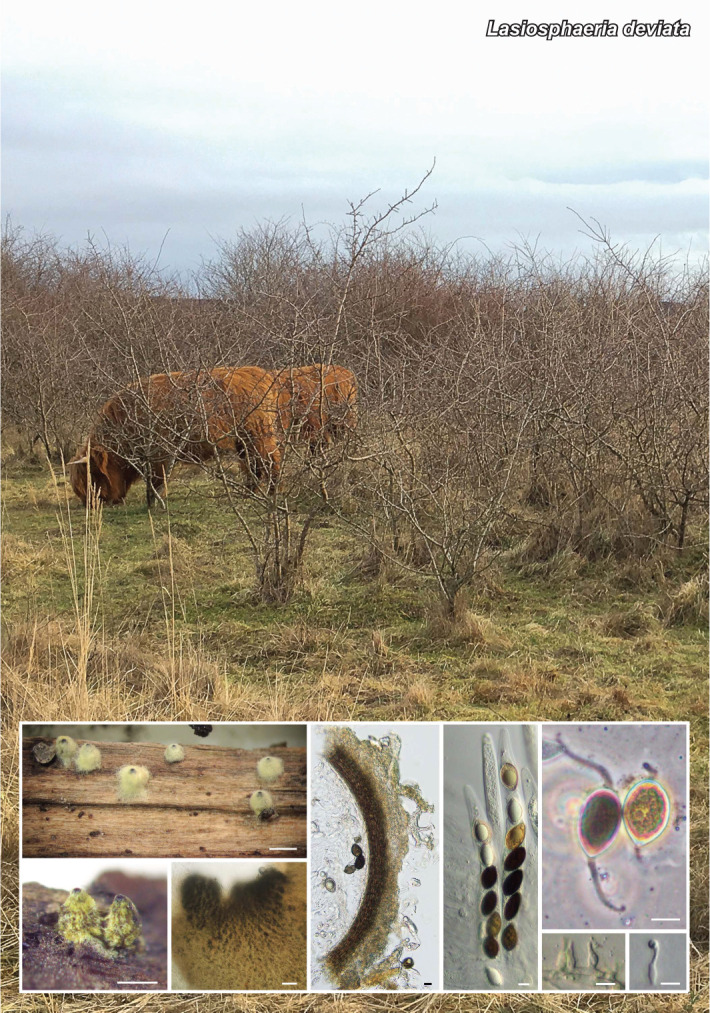
Notes — Based on a megablast search of NCBIs GenBank nucleotide database, the closest hit using the LSU sequence was Phaeosphaeriopsis nolinae (strain CBS 102205, GenBank KY090667; Identities = 552/552 (100 %), no gaps). The closest hit using the ITS sequence was Paraphoma fimeti (strain UTHSC DI16-296, GenBank LT796872; Identities = 528/585 (90 %), eight gaps (1 %)). The closest hit using the rpb2 sequence was Paraphoma fimeti (strain UTHSC DI16-296, GenBank LT797032; Identities = 842/905 (93 %), no gaps). The closest hit using the tub2 sequence was Paraphoma fimeti (strain UTHSC DI16-296, GenBank LT796952; Identities = 294/320 (92 %), five gaps (1 %)). Based on the combined analysis of ITS, LSU, rpb2 and tub2 sequences, the closest relative of Paraphoma variabilis is Paraphoma fimeti. The latter differs from our novel species in that it produces smaller (3–5 × 2–3 μm vs 4–8 × 2–3 μm) ellipsoidal conidia (Boerema et al. 2004), which are cylindrical, ellipsoidal, ovate, reniform and sigmoid, and truncated at the base, in P. variabilis. Also, P. fimeti produce a yellowish diffusible pigment, which is absent in P. variabilis.

graphic file with name per-2023-47-6-i029.jpg

Maximum likelihood tree obtained from the combined DNA sequences dataset from four loci (ITS, LSU, rpb2 and tub2) of our isolate and sequences retrieved from the GenBank database. Ex-type strains of the different species are indicated with superscript T. The new species proposed in this study is indicated in bold. The RAxML v. 8.2.10 (Stamatakis 2014) bootstrap support values (> 70 %) are provided at the nodes. Neophaeosphaeria agaves CPC 21264 and Neophaeosphaeria filamentosa CBS 102202 were used as outgroup.

Colour illustrations. Ports de Totosa-Beseit natural park, Tarragona, Spain. Colony on PDA and OA after 14 d at 25 + 1 °C; pyriform conidioma; conidiogenous cells; conidia. Scale bars = 25 μm (conidioma) and 10 μm (all others).

graphic file with name per-2023-47-6-g082.jpg

Paraphoma variabilis

Persoonia. 2021 Dec 24;47:346–347.

Fungal Planet 1371 – 24 December 2021

Phaeococcomyces kinklidomatophilus Sastoque, Stchigel & Cano-Lira, sp. nov.

Angie Sastoque 1, Alberto M Stchigel 1, José F Cano-Lira 1

Etymology. From Greek κιΓκλInline graphicδωμα-, railing, and - ϕInline graphicλΟζ, friend, because the environmental origin of the fungus.

Classification — Phaeococcomycetaceae, Lichenostigmatales, Arthoniomycetes.

Colonies lacking mycelium but consisting of a mass of yeast-like cells embedded in a mucous matrix, which are aseptate, hyaline, smooth- and thin-walled, ellipsoid to globose, 3–6 × 2–5 μm when young, producing blastoconidia via multilateral budding, becoming brown, verrucose and globose with the age, 5–8 μm diam, remaining attached to one another to form up to 5-celled chains.

Culture characteristics — (after 14 d at 25 °C in the dark). Colonies reaching 3.5 mm diam on malt extract agar (MEA), 2.0 mm on oatmeal agar (OA) and potato carrot agar (PCA), and 2.5 mm on potato dextrose agar (PDA), circular, margins entire, flat to slightly convex, smooth and glistening on OA and PCA, but crater-like, dry and slightly roughened and tough with lobate margins when older on MEA and PDA. Surface and reverse black on all culture media tested. Minimum, optimum and maximum temperature of growth: 5 °C, 15–25 °C, and 25 °C, respectively.

Typus. SPAIN, Tarragona Province, Els Pallaresos, isolated from a blackened metal railing of an industrial warehouse, 20 Nov. 2020, coll. J. F. Cano-Lira & A. M. Stchigel, isol. A.P. Sastoque (holotype CBS H-24766, culture ex-type FMR 18615 = CBS 147696; ITS and LSU sequences GenBank HG995460 and HG995431, MycoBank MB 839137).

Notes — Based on a megablast search of NCBIs GenBank nucleotide database, the closest hit using the ITS sequence was Phaeococcomyces eucalypti (strain CPC 17606, GenBank NR_120226; Identities = 494/535 (92.34 %), eight gaps (1 %)), and using the LSU sequence it was Phaeococcomyces eucalypti (strain CPC 17606, GenBank NG_042748; Identities = 463/473 (97.89 %), two gaps (0 %)). Our ITS phylogenetic tree corroborated the placement of our isolate as a new species of the genus, being phylogenetically close to P. eucalypti. Phaeococcomyces kinklidomatophilus was isolated by swabbing from the surface of a metal railing in Els Pallaresos village, Tarragona province, Catalonia, Spain. Phaeococcomyces is a genus of black yeasts with slimy, folded, slow-growing colonies lacking of aerial mycelium. The genus currently contains four species: P. eucalypti, P. mexicanus, P. nigricans (the type species) and P. rothmanniae. Phaeococcomyces kinklidomatophilus is morphologically very similar to other species of the genus, but differs from P. eucalypti, its phylogenetically closest species, in size (3.5 mm vs 25 mm diam on MEA), shape (crater-shaped vs folded on MEA and PDA) and colour (nearly black vs iron-grey on MEA and PDA) of the colonies. Moreover, P. kinklidomatophilus is molecularly easily distinguishable from the other species of the genus.

graphic file with name per-2023-47-6-i030.jpg

Maximum likelihood tree obtained from the ITS alignment of our isolate and sequences retrieved from GenBank. The tree was built by using RAxML CIPRES (http://www.phylo.org/sub_sections/portal/) (Miller et al. 2010). Bootstrap support values ≥ 70 % are presented at the nodes. Anteaglonium gordoniae SGSF172 and Flammeascoma lignicola MFLUCC 10-0128a were used as outgroups. The new species proposed in this study is indicated in bold. TRepresents the ex-type strain of the species.

Colour illustrations. Railing with chromatic alteration in Els Pallaresos village, Tarragona Province, Spain. Colonies growing on PDA culture media (at 25 °C); blastoconidia (DIC and PH respectively). Scale bars = 10 μm.

graphic file with name per-2023-47-6-g083.jpg

Phaeococcomyces kinklidomatophilus

Persoonia. 2021 Dec 24;47:348–349.

Fungal Planet 1372 – 24 December 2021

Penicillium xyleborini Visagie & W.J. Nel, sp. nov.

Cobus M Visagie 1, Wilma Nel 1

Etymology. Latin, xyleborini, refers to the genus of the beetle host (Xyleborinus saxesenii) from which this species was isolated.

Classification — Aspergillaceae, Eurotiales, Eurotiomycetes.

Conidiophores biverticillate to terverticillate; stipes smooth-walled, 100–200(–500) × 2.5–4(–4.5) μm; branches 10–30 μm; metulae 4–6 per stipe/branch, (8–)10–16 × 3–4 μm; phialides 6–10 per metula, 8–11 × 3–4 μm (av. = 9.5 ± 0.23 × 3.3 ± 0.2), av. length metula/phialide 1.3; conidia smooth-walled, globose to subglobose, 2.5–3.5 × 2.5–3.5 μm (av. = 3.1 ± 0.2 × 2.94 ± 0.2), av. width/length = 0.95, n = 57.

Culture characteristics — (25 °C, 7 d): On Czapek yeast autolysate agar (CYA): colonies moderately deep, concentrically and radially sulcate, crateriform; margins low, narrow (1 mm), entire; mycelia white; texture velutinous; sporulation moderately dense, conidia en masse greyish to dull green (26D4–F4–5; Kornerup & Wanscher 1967); soluble pigments absent; exudates clear to inconspicuously yellow; reverse greyish yellow, olive brown (4B3, 4E4–5). On malt extract agar (MEA): colonies moderately deep, concentrically sulcate, crateriform; margins low, narrow (1 mm), somewhat irregular; mycelia white; texture velutinous; sporulation moderately dense, conidia en masse dull to greyish green (25D4–5); soluble pigments absent; exudates absent; reverse brown (6E6), dark brown (6F6). On yeast extract sucrose agar (YES): colonies deep, radially and concentrically sulcate, crateriform; margins low, narrow (1 mm), somewhat irregular; mycelia white; texture velutinous; sporulation moderately dense, conidia en masse greyish green (25C4); soluble pigments absent; exudates absent; reverse yellowish white, greyish yellow, olive brown (4A2, 4B3, 4E4–5). On dichloran 18 % glycerol agar (DG18): colonies moderately deep, radially and concentrically sulcate; margins low, narrow (1 mm), entire; mycelia white; texture velutinous; sporulation moderately dense, conidia en masse greyish to dull green (26D4–F4–5); soluble pigments inconspicuously yellow; exudates absent; reverse greyish yellow, greenish white (4B4–C4, 29A2–30A2). On oatmeal agar (OA): colonies low, plane; margins low, narrow (1 mm), entire; mycelia white, inconspicuously yellow; texture velutinous; sporulation moderately dense, conidia en masse dull green (27D4–F5); soluble pigments absent; exudates clear; reverse yellowish white, (2A2), olive yellow (2C6). On creatine sucrose agar (CREA): colonies weak growth, no acid production. Colony diam (in mm, after 7 d, at 25 °C): CYA 15–16; CYA 15 °C 18–20; CYA 20 °C 20–24; CYA 30 °C no growth; CYA 37 °C no growth; CYAS 18–21; MEA 12–14; DG18 15–18; YES 18–20; OA 15–18; CREA 13–18.

Typus. SOUTH AFRICA, Limpopo Province, Tzaneen, (-23.708192, 30.099344), from an ambrosia beetle (Xyleborinus saxesenii), 30 Sept. 2019, W. Nel (holotype PREM 63078 (dried specimen), culture ex-type CMW 56800 = CN 001D4 = WN 2019.10.32; LSU, ITS, BenA, CaM and rpb2 sequences GenBank MW504290, MW504356, MW480817, MW480823 and MW480824, MycoBank MB 840990).
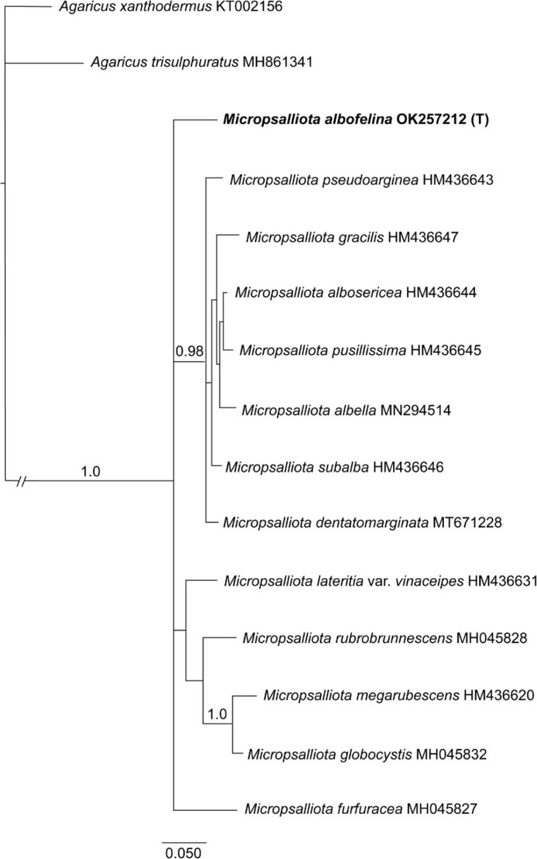
Notes — A multigene phylogeny resolves Penicillium xyleborini as a close relative of P. lusitanum in sect Ramosum series Soppiorum (Houbraken et al. 2020). The new species produces greyish to dull green conidia, compared to the dark green conidia of P. lusitanum. Also, P. xyleborini conidiophores are more commonly terverticillate, with longer phialides and larger conidia when compared with P. lusitanum (Gonçalves et al. 2019). Other Penicillium species isolated from ambrosia beetles or their galleries during this project included P. brevicompactum, P. cairnsense, P. citreonigrum, P. coprophilum, P. glabrum, P. ibericum, P. pancosmium, P. pasqualense, P. soliforme, P. ubiquetum, P. viticola and a new section Sclerotiorum species that will be described elsewhere. Sequences of these species were submitted to GenBank and accessioned with numbers MW480792–MW480820.

graphic file with name per-2023-47-6-i031.jpg

Combined phylogeny of Penicillium section Ramosum based on ITS, BenA, CaM and rpb2. Aligned data sets (MAFFT v. 7.454; Katoh et al. 2013) were analysed using maximum likelihood (IQ-TREE v. 1.6.12; Minh et al. 2020) and Bayesian Inference (MrBayes v. 3.2.7a; Ronquist et al. 2012). Maximum likelihood bootstrap support values (≥ 80 %) and Bayesian posterior probability values (≥ 0.95) are given above branches. The new species is indicated by bold text, T = ex-type strain and GenBank accession numbers are shown in a smaller font next to the culture accession number (ITS = green, BenA = blue, CaM = red, rpb2 = purple).

Colour illustrations. The ambrosia beetle (Xyleborinus saxesenii) from which the new species was isolated. Penicillium xyleborini colonies on CYA15C, CYA20C, CYA, CYAS, MEA, YES, DG18, OA, CREA (from top to bottom); conidiophores; conidia. Scale bars = 10 μm.

graphic file with name per-2023-47-6-g084.jpg

Penicillium xyleborini

Persoonia. 2021 Dec 24;47:350–351.

Fungal Planet 1373 – 24 December 2021

Phylloporia tamilnadensis Sridhar, Palani & Decock, sp. nov.

Pichai Sridhar 1, Perumal Palani 1, Cony Decock 2

Etymology. Name refers to the southwestern State of Tamil Nadu, where this fungus was collected.

Classification — Hymenochaetaceae, Hymenochaetales, Basidiomycota.

Basidiomata annual, mostly solitary, pileate, sessile; pileus spongy to leathery when fresh, turning horny to brittle on drying, initially arises as small dot gradually widening in all direction to form distinct basidiomata, infrequently laterally fused with adjacent basidiomata, easily separable from host, 0.8–20 × 0.6–10 mm, 0.8–20 mm thick near point of attachment, applanate to circular, hoof-shaped, broadly to narrowly attached, more or less bent at margin, pale orange (5A3, Kornerup & Wanscher 1978); pilear surface tomentose to velvety, irregularly zonate, pitted, melon (5A6) to pale orange (5A3); margin 0.6–1 mm wide, sterile, entire, more or less lobed at maturity, reddish blond (5C4) to pale orange; hymenophore poroid; pores round to angular mostly, 3–4 per mm; dissepiments thin, entire to lacerate; context up to 3 mm thick, distinctly duplex, the lower park homogenous, dense, brownish orange (5C4), very thin into margin up to 0.4–1.0 mm at base, with a distinct to indistinct thin black line subtending an upper tomentum, porous spongy; tubes 0.6–10 mm long, orange when fresh (6A8), the tubes starting 0.3 mm from the margin. Hyphal system monomitic in all parts; generative hyphae simple septate, thin- to slightly thick-walled, hyaline, yellowish to light golden brown, darker, brownish in KOH, scarcely ramified, the branches constricted at their emergence point, soon growing parallel to mother hyphae; in the tomentum, hyphae parallel, adpressed first, soon erected, loosely packed, straight to sinuous, mostly unbranched, slightly thick-walled, sub-hyaline to pale golden brown, from 3.5 μm diam near the base, gradually enlarging up to 4.5–8.0 μm; in the context hyphae adpressed to oblique but mostly erected, with a near parallel orientation, moderately thick-walled with the lumen widely open, septate, but with long aseptate segments, 3.5–4.0 μm diam; in the hymenophoral trama hyphae with a subparallel disposition, straight, occasionally geniculated, the lumen widely open, septate, with aseptate segments or with occasional secondary septa, 2.5–3.5 μm diam. Hymenium: basidia ~11 × 5.5 μm, clavate, septate at base, with four sterigmata up to 5 μm long; basidioles ~10.5 × 5 μm; basidiospores 3.5–5.0 × 2.3–3.6 μm (av. 4.3 × 2.9 μm), Q = 1.3, ellipsoid, moderately thick-walled, smooth, hyaline to pale yellow, inamyloid, acyanophilic.

Typus. INDIA, Tamil Nadu State, Ranipet District, Sholinghur Taluk, Mettu kunnathur Village, N13°02’17" E79°31’10", growing on thin branches of living plant of Catunaregam spinosa (Rubiaceae), 10 Dec. 2017, P. Sridhar, MKRF 05 (holotype CAL #1840, LSU sequence GenBank MN525278, MycoBank MB 838470).

Additional material examined. INDIA, Tamil Nadu State, Ranipet District, Sholinghur Taluk, Mettu kunnathur Village, N13°02’17" E79°31’10", growing on thin branches of living plant of C. spinosa, 10 Nov. 2018, P. Sridhar, MKRF 21 (LSU sequence GenBank MN525279).

Notes — The olive yellow cap and ash grey dots on the stipe surface, as well as larger spores, distinguish S. praetermissus from the closest species of the Suillus acidus group. According to phylogenetic analysis, the nearest species is S. subalutaceus, p-distance is 1.8 %, 12 nucleotide substitutions per 666 alignment positions of ITS1-5.8S-ITS2. According to a microscopic examination of the type specimen of S. subalutaceus (MICH 50221) carried out by E. Zvyagina, its spores are much shorter with the same width. The cap surface of S. subalutaceus is ‘pinkish-buff’ (Smith & Thiers 1964). Suillus subalutaceus forms mycorrhiza with two-needle pines (Nguyen et al. 2016), while the new taxon S. praetermissus is associated with five-needle pines. In our study all examined specimens were collected under Pinus sibirica. However, for specimens and mycorrhizal samples from Japan, of which sequences were used in the phylogenetic tree, an association with P. corayensis and P. pumila was also noted. Suillus subalutaceus is an American species, while S. praetermissus has an Asian distribution.

Colour illustrations. India, Tamil Nadu State, Ranipet District, Sholinghur Taluk, Catunaregam spinosa growing in Mettu kunnathur village. Habitat; basidiomata pore view; transverse section of basidiomata; pilear surface details; basidium and basidiospores. Scale bars = 1 cm (basidiomata, pore view and T.S. of basidiomata); 50 μm (pilear surface details); 10 μm (basidium and basidiospores).

graphic file with name per-2023-47-6-g085.jpg

Phylloporia tamilnadensis

Supplementary material

FP1373

Phylogenetic tree.

per-2023-47-6-SF1373.jpg (653.2KB, jpg)
Persoonia. 2021 Dec 24;47:352–353.

Fungal Planet 1374 – 24 December 2021

Phytophthora docyniae Q.N. Dang, T.H. Nguyen & T.I. Burgess, sp. nov.

Quynh N Dang 1, Thu H Nguyen 1, Hung X Tran 1, Thu Q Pham 1, Treena I Burgess 2

Etymology. Refers to the genus name of the only known host, Docynia indica.

Classification — Peronosporaceae, Peronosporidae, Oomycota.

Sporangia produced abundantly in non-sterile soil extract after 2 d, terminally borne on unbranched sporangiophores, non-papillate, ellipsoid to ovoid, 34.5 ± 2.7 × 23.8 ± 2.7 μm (overall range 30–39.5 × 19.8–27.9 μm), length/breadth ratio 1.45 ± 0.9, with internal proliferation sporangia in both a nested and extended way. Direct germination of sporangia with unusual shapes was often observed. Hyphal swellings are globose, subglobose, irregular, mostly catenulate and sometimes clustered. Chlamydosporesproduced abundantly on all media (potato dextrose agar (PDA), carrot agar (CA), V8 agar) and in soil extract, mostly intercalary, globose, subglobose and irregular, 43.4–96.3 μm diam, very thick-walled on PDA, 4.2 ± 1.1 (2.0–6.9 μm). Chlamydospores often germinated and produced sporangia in soil extract. Gametangianot produced in single culture or when paired with A1 and A2 tester strains of P. cinnamomi (MP75, DCE60). Radial growth rates on V8 agar at optimum temperature (20–32.5 °C) and near the maximum temperature (35 °C), 4.2 ± 0.8 mm/d and 2.2 ± 0.13 mm/d, respectively. Minimum temperature for growth is 15 °C.

Culture characteristics — Colonies are chrysanthemum on PDA and V8 agar, while growth on CA showed no distinct pattern. Growth on CA and V8A was faster than on PDA.

Typus. VIETNAM, Yen Bai Province, Mu Cang Chai, isolated from the soil and root of Docynia indica (Rosaceae), Oct. 2019, V.L. Binh & X.T. Hung (holotype CBS H-24780, culture ex-type CBS 148031 = FP77, ITS, coxI, ß-tubulin and LSU sequences GenBank MZ420209, MZ435257, MZ435255 and MZ435254, MycoBank MB 840241).

Additional material examined. VIETNAM, Yen Bai Province, Mu Cang Chai, isolated from the soil and roots of D. indica, Oct. 2019, V.L. Binh & X.T. Hung, culture FP78.

Notes — Phylogenetically, Phytophthora docyniae resides in clade 9c of the Phytophthora phylogeny (Abad et al. 2019) most closely related to P. macrochlamydospora, P. quininea and P. richardiae. These species have a few similar features, such as hypha swelling and unbranched sporangiophores; however, there are many differences. Phytophthora docynia, P. quininea, and P. richardiae produce non-papillate sporangia, while P. macrochlamydospora has semi-papillate sporangia (Buisman 1927, Crandall 1947, Irwin 1991). The size of sporangia varies; the smallest belong to P. docynia, followed by P. macrochlamydospora, P. quininea and then P. richardiae. Chlamydospores are absent in P. richardiae and very large for P. macrochlamydospora. Furthermore, a distinguishing feature of P. docyniae is that the chlamydospores frequently germinate and immediately produce sporangia and direct germination of sporangia with unusual shapes was often observed.

graphic file with name per-2023-47-6-i032.jpg

Maximum likelihood phylogeny produced using RAxML v. 8.2.11 (Stamatakis 2014) from concatenated ITS, BT and cox1 using the GTR + I model showing the phylogenetic position of Phytophthora docyniae in clade 9c. Sequences are all available from IDphy (Abad et al, 2019) and Bose et al. (2021). Bayesian analysis was conducted on the same dataset with MrBayes v. 3.2.6 (Ronquist & Huelsenbeck 2003) and resulted in the same topology. Numbers above the branches reflect support obtained from the analysis of the same dataset (bootstrap values estimated by RAxML/Bayesian posterior probabilities only values above 60 % or > 0.6 are shown). Representative species from other clades were used as outgroup taxa (not shown). The scale bar corresponds to expected number of substitutions per nucleotide site.

Colour illustrations. A flowering shoot of Docynia indica, the host of Phytophthora docyniae. Top to bottom: chlamydospores germinating to produce sporangia; chlamydospores and hyphal swelling with projections; chrysanthemum colony morphology on PDA; ovoid sporangia with direct germination; internal nested and extended proliferation; intercalary chlamydospore; thick-walled chlamydospore on PDA. Scale bars = 40 μm.

graphic file with name per-2023-47-6-g086.jpg

Phytophthora docyniae

Persoonia. 2021 Dec 24;47:354–355.

Fungal Planet 1375 – 24 December 2021

Pluteus brunneovenosus Ferisin, Fellin, Justo & Dovana, sp. nov.

Francesco Dovana 1, Giuliano Ferisin 2, Alfredo Justo 3, Alessandro Fellin 4

Etymology. The epithet ‘brunneovenosus’ refers to the colour of the cap with brown veins at the centre.

Classification — Pluteaceae, Agaricales, Agaricomycetes.

Basidiomata medium-sized, agaricoid. Pileus up to 35 mm diam, initially campanulate, then expanded to applanate, with low umbo at centre; surface glabrous, brown (Mu 10YR6/2) to dark brown (Mu 10YR3/2, 2/2) in centre pallescent towards the margin, hygrophanous, weakly to strongly venose at centre, strongly striate-sulcate at the margin up to the half of the radius. Lamellae fairly distant, free, slightly ventricose, up to 4 mm broad, first whitish later pink with whitish edge. Stipe 30–40 × 3–4 mm, cylindrical usually broadened towards base, striate lengthwise and flocculose, whitish in the upper or totally greyish, dark grey at the base. Context white, greyish in the stipe base. Smell and taste not distinctive. Basidia 26–30 × 8–10 μm, clavate, 4-spored. Basidiospores (6.5–)7.0–7.4–7.9(–8.5) × (5.0–)5.5–5.9–6.3(–7.0) μm, Q = (1.06–)1.21–1.26–1.35(–1.64); mainly broadly ellipsoid, more rarely ellipsoid, thick-walled, non-amyloid, cyanophilous. Pleurocystidia scattered and rare, similar to cheilocystidia. Cheilocystidia 30–140 × 15–31 μm, so numerous as to make the lamellar edge sterile; shape variable from clavate to utriform, fusiform, fusiform with rostrate apex, (rostrum length from 10 up to 35 μm), hyaline or with oily contents, slightly thick-walled. Pileipellis a trichohymeniderm made up of appressed clavate to fusiform elements 40–120 × 20–32 μm, sometimes with rostrate apex, (rostrum length up to 25 μm), pigment intracellular pale brown to brown. Stipitipellis a cutis of pale brown-grey hyphae with oily contents, 4–10 μm wide. Caulocystidia 40–60 × 18–22 μm, clavate to fusiform generally with rostrate apex (rostrum length up to 25 μm), generally in small clusters in the apical part of the stipe, rare towards the base, but present in all stipe length, hyaline or with oily contents. Clamp connections absent in all tissues.

Habitat & Distribution — Generally solitary or in small groups, on twigs of broadleaved trees (willow, oak, and poplar). Found, so far as we know, only in North Italy in Trentino Alto Adige and Friuli Venezia Giulia Region.

Typus. ITALY, Farra d’Isonzo, Bosco di Sotto, on twigs of broadleaved trees on the ground, 23 June 2020, G. Ferisin (holotype GDOR5090, ITS sequence GenBank OK090867, MycoBank MB 841180).

Additional materials examined. ITALY, Friuli-Venezia Giulia, Farra d’Isonzo, Bosco di Sotto, on twigs of broadleaved trees on the ground, 14 Oct. 2019, G. Ferisin, GDOR5091, ITS sequence GenBank OK090865; Trentino-Alto Adige, Ton, Masi di Vigo, on twigs on the ground, 11 Aug. 2019, A. Fellin, GDOR5089, ITS sequence GenBank OK090866.

Notes — Terminology for descriptive terms is according to Vellinga (1988) and colour codes are taken from Munsell (1994). Spore dimensions are expressed as (a–)b–c–d(–e), where (a) = minimum value, b = (average - standard deviation), c = average, d = (average + standard deviation) and (e) = maximum value. Maximum-likelihood analysis of the ITS region was performed with RAxML v. 8.2.11 (Stamatakis 2014) using the GTR+G model as implemented in Geneious v. 11.1.5. Pluteus brunneovenosusis macroscopically characterised by its relatively small-sized basidiomes with pale brown to brown pileus, strongly veined in the centre; stipe cylindrical, pubescent, with grey to dark grey base; broadly ellipsoid spores; pileipellis a trichohymeniderm made up of clavate to fusiform elements sometime with rostrate apex, cheilocystidia utriform to broadly fusiform also with rostrated apex, pleurocystidia absent; caulocystidia present in all the stipe length. In the ITS phylogenetic tree, the three collections of P. brunneovenosus clustered in a fully supported clade (maximum likelihood bootstrap support value (ML-BS) = 100 %) which is sister (ML-BS = 95 %) to another clade that includes two sequences (GenBank FJ816665 of P. dominicanus var. hyalinus and GenBank KM983712, coll. AJ491, which is probably an undescribed species from Virgin Islands in the USA). Pluteus dominicanus var. hyalinus differs mainly for the very rare cheilocystidia, the absence of cystidia with rostrate apex and, for having an hymeniderm pileipellis made of sphaeropedunculate elements (Menolli et al. 2010). Phylogenetically, P. brunneovenosus is also close to sequences named P. thomsonii (GenBank HM562053 and HM562197) and P. diverticulatus (GenBank FJ375247). Pluteus thomsonii is very similar to P. brunneovenosus and can be distinguished on account of the more developed veins on the pileus and for the absence of rostrate terminal elements of the pileipellis (Vellinga 1990). Pluteus diverticulatus, originally described from France in 2003 and subsequently considered Pluteus thomsonii var. diverticulatus by Ludwig (2007) differs mainly by the presence of diverticulate caulocystidia, cheilocystidia and terminal elements of the pileipellis (Corriol 2003). Morphologically, P. brunneovenosus is also similar to P. insidiosus, that differs mainly for having a hymenidermal pileipellis made of sphaeropedunculate or narrowly clavate elements and the absence of caulocystidia (Vellinga 1990).

Colour illustrations. Italy, Farra d’Isonzo, deciduous natural forest where the holotype collection was collected. Fresh basidiomes in field (holotype GDOR5090); pileipellis; stipitipellis with caulocystidia; cheilocystidia and basidiospores. Scale bars = 20 μm.

graphic file with name per-2023-47-6-g087.jpg

Pluteus brunneovenosus

Supplementary material

FP1375

Phylogenetic tree.

per-2023-47-6-SF1375.jpg (473.3KB, jpg)
Persoonia. 2021 Dec 24;47:356–357.

Fungal Planet 1376 – 24 December 2021

Pythium serotinoosporum Abrinbana, Abdollahz. & Badali, sp. nov.

Masoud Abrinbana 1, Farzaneh Badali 1, Jafar Abdollahzadeh 2

Etymology. Name refers to the late production of oospores in culture.

Classification — Pythiaceae, Peronosporales, Oomycota, Straminipila.

Main hyphae up to 8 μm diam. Sporangia rarely produced zoospores through short discharge tubes, globose to subglobose, internally proliferating, terminal, occasionally intercalary, 15–33 μm diam (av. 25.21 μm diam). Hyphal swellings mostly germinating with 1–4 germ tubes, variously shaped, typically globose, subglobose, ellipsoidal, limoniform, ovoid, pyriform or irregular in shape, intercalary, occasionally terminal, occasionally in chains of 2–3, globose hyphal swellings 14–42 μm diam (av. 29.23 μm diam), elongated hyphal swellings 20–50 × 10–34 μm (av. 32.82 × 22.19 μm). Sexual organs produced infrequently after at least 3 wk in water culture. Oogonia smooth-walled, globose, subglobose, rarely elongated, terminal or intercalary, globose oogonia 17–29 μm diam (av. 22.65 μm diam), elongated oogonia up to 50 μm long. Antheridia 1–2(–7) per oogonium, diclinous. Antheridial cells globose, clavate, swollen, crooked or elongated. Oospores mainly globose, rarely subglobose or ovoid, plerotic or nearly plerotic, occasionally aplerotic, mostly one, but sometimes two per oogonium, 17–28 μm diam (av. 22.52 μm diam), the oospore wall up to 1 μm thick.

Culture characteristics — Colonies on potato carrot agar (PCA) submerged with a narrow chrysanthemum pattern, on cornmeal agar (CMA) submerged with a vague chrysanthemum pattern, on potato dextrose agar (PDA) profuse and dense aerial mycelium with no special pattern. Daily growth on PCA was 3 mm at 5 °C, 7 mm at 10 °C, 10 mm at 15 °C, 11 mm at 20 °C, 17 mm at 25 °C, 15 mm at 30 °C, 14 mm at 35 °C and 0 mm at 40 °C. The isolates were not viable at 40 °C.

Typus. IRAN, West Azarbaijan Province, Mahabad, from soil under Prunus dulcis (Rosaceae), 15 Aug. 2012, M. Abrinbana (holotypeIRAN 16694 F, ex-type IRAN 2393 C, ITS, COXI, COXII and LSU sequences GenBank MG182707, MG182700, OK482595, OK428852, MycoBank MB 823188).

Additional material examined. IRAN, West Azarbaijan Province, Mahabad, from soil under Prunus dulcis, 15 Aug. 2012, M. Abrinbana (IRAN 2394 C = CBS 143583, ITS, COXI, COXII and LSU sequences GenBank MG182708, MG182701, OK482596, OK428853).

Notes — Phylogenetic analyses of DNA sequence data supported the phylogenetic position of P. serotinoosporum in clade E2 sensu Lévesque & de Cock (2004). This species is indistinguishable from P. middletonii by ITS but, in the combined COXI and COXII phylogeny, P. serotinoosporum isolates form a fully-supported distinct clade sister to P. middletonii with 8 and 22 bp differences in COXI and COXII sequences, respectively. Morphologically, it differs from P. middletonii by having hyphal swellings, mostly plerotic oospores and a higher number of antheridia per oogonium (1–7 vs 1–2 per oogonium) and the absence of hypogynous as well as sessile antheridia.

graphic file with name per-2023-47-6-i033.jpg

The 50 % majority rule phylograms inferred from the Bayesian analysis of the ITS (a) and concatenated COXI and COXII (b) sequences alignment of Pythium clade E2, indicating the position of P. serotinoosporum. Bayesian posterior probability/maximum parsimony bootstrap support values are given at the nodes. Bayesian and maximum parsimony analyses were performed using MrBayes v. 3.2.7 (Ronquist et al. 2012) and PAUP v. 4.0b10 (Swofford 2003), respectively. The new species is indicated in bold face and a coloured block. Pythium acanthophoron (CBS 33729) from clade J was used as outgroup. T indicates ex-type strains. The scale bars represent 0.1 (a) and 0.01 (b) expected changes per site.

Colour illustrations. Almond orchard in West Azarbaijan Province. Various shapes of hyphal swellings; empty sporangium and zoospores; empty sporangium with internal proliferation; diclinous antheridium with crook-necked antheridial cell and oogonium; two diclinous antheridia applied to intercalary oogonium; six diclinous antheridia attached to oogonium; plerotic oospore; aplerotic oospores; double oospore; ovoid oospore. Scale bars = 10 μm.

graphic file with name per-2023-47-6-g088.jpg

Pythium serotinoosporum

Persoonia. 2021 Dec 24;47:358–359.

Fungal Planet 1377 – 24 December 2021

Thecaphora stajsicii J. Kruse, R.G. Shivas & McTaggart, sp. nov.

Julia Kruse 1, Roger G Shivas 2, Alistair R McTaggart 3

Etymology. In acknowledgement of Val Stajsic, an Australian botanist based at the National Herbarium of Victoria (MEL), who first recognised and collected smut fungi on Oxalis in Australia.

Classification — Glomosporiaceae, Ustilaginales, Ustilaginomycetes.

Sori in all or most capsules on an infected plant, replace the seeds with a powdery, cinnamon-brown spore mass causing the capsules to split longitudinally. Spores solitary when mature, globose to subglobose, 12–16 μm diam, pale to medium yellowish brown; wall even, 1–2 μm thick including moderately dense and coarse verruculose, warts c. 1 μm high. Asexual morph not seen.

Typus. AUSTRALIA, Australian Capital Territory, Canberra, Pialligo Redwood Forest, S35°19’12" E149°12’00", 595 m a.s.l., in capsules of Oxalis radicosa (Oxalidaceae), 13 Nov. 2018, J. Kruse 945 (holotype BRIP 68991, isotype MEL 2496300A; ITS sequence GenBank MW892416, MycoBank MB 839329).

Notes — Thecaphora stajsicii is the second species described on Oxalis in Australia. The first, T. australiensis, was described from Oxalis exilis in Victoria (Crous et al. 2018b). Capsules of O. radicosa infected by T. stajsicii are about half the length of healthy capsules. Oxalis exilis and O. radicosa are both native species in Australia (Stajsic 2020a), where 30 species of Oxalis have been recorded, of which 23 are introduced (Stajsic 2020b). Worldwide, two other species, T. oxalidis and T. capensis, are known to infect Oxalis (Vánky et al. 2008). Thecaphora stajsicii has smaller spores than T. australiensis (14–26 × 14–19 μm), and has spores similar in size to those of T. oxalidis (12–17 × 13.5–21(–24) μm) (Vánky 2011). The ITS sequence of T. australiensis (GenBank MW892417) was obtained from the ex-type specimen for the phylogenetic analysis. The four Thecaphora species on Oxalis form a monophyletic clade. Thecaphora australiensis and T. stajsicii are closely related, which may indicate further biodiversity of host specific species on Oxalis.

graphic file with name per-2023-47-6-i034.jpg

Phylogram based on an alignment of the ITS region of rDNA. Tree searched in IQTree v. 2.1.3 (Minh et al. 2020) with a model test (command -m TEST), and support from 10 000 replicates of an approximate likelihood ratio test (≥ 0.9) and ultra-fast bootstraps (≥ 95 %) above nodes. Phylogenetic network made in SplitsTree v. 4.14.8 (Huson & Kloepper 2005) showing genetic distance and putative recombination events among species of Thecaphora.

Colour illustrations. Oxalis radicosa infected with Thecaphora stajsicii, Canberra. Fungarium specimen; spores emerging from capsules; spores (equatorial and upper surface view). Scale bars = 1 cm (plant material), 10 μm (spores).

graphic file with name per-2023-47-6-g089.jpg

Thecaphora stajsicii

Persoonia. 2021 Dec 24;47:360–361.

Fungal Planet 1378 – 24 December 2021

Trechispora patawaensis V. Papp, Dima & L. Nagy, sp. nov.

Viktor Papp 1, Bálint Dima 2, László G Nagy 3

Etymology. Named after the place (Patawa camp) where the type specimen was collected.

Classification — Hydnodontaceae, Trechisporales, Agaricomycetes.

Basidiocarp annual, resupinate, soft and fragile, easily separable from the substrate; margin arachnoid, consisting of radiating hyphal mats with cord-like structures; context soft; hymenophore poroid, pore surface when fresh pure white, becoming pale yellowish when dried; the pores round to slightly irregular with thin dissepiments, small sized (6–7 per mm); hyphal system monomitic, thin-walled subicular hyphae hyaline, branched, with clamp connections and ampullate septate, up to 5 μm wide; Subhymenial hyphae thin-walled, branched, short-celled, isodiametric, up to 4 μm wide, with clamps in all septa. Single, rhomboid crystals abundantly present in subiculum; cystidia or other sterile hymenial elements not observed. Basidia short, cylindrical with four sterigmata and a basal clamp connection, 8–10 × 4–5 μm; basidiospores ovoid to subglobose (Qav = 1.36), hyaline, echinulate, (3.01–)3.07–3.35(–3.50) × (2.11–)2.21–2.44(–2.60) μm (excluding the ornamentation), L = 3.18 μm, W = 2.3 μm (n = 30), not cyanophilous.

(Line-drawn micromorphological structure of T. patawaensis basidiomata in FP1378 suppl. mat.)

Habitat & Distribution — The type was found in the Kaw Mts of French Guiana, on decayed dead branch of unknown angiosperm wood lying on the ground. A similar unpublished ITS sequence of a specimen collected in Ecuador (deposited at the UNITE database, no. UDB014091; Identities = 604/617 (98 %), seven gaps (1 %)), suggests that T. patawaensis presumably occurs throughout the Amazon basin.

Typus. FRENCH GUIANA, Kaw Mts, Camp Patawa, N4.54437° W52.15277°, on decayed branch of unknown angiosperm tree, 19 Dec. 2019, V. Papp & B. Dima (holotype WU-Mykol. 44791,WU); ITS and LSU sequences GenBank OL314550 and OL314546, MycoBank MB841876).

Notes — In our phylogenetic analysis based on ITS and LSU sequences, T. patawaensis is located in a unique fully-supported clade (phylogenetic tree shown on FP1378 & 1379 suppl. mat.). In our phylogram, T. patawaensis clusters together with T. farinacea (MA-Fungi 79474, KHL 8454), T. araneosa (KHL 8570), T. thelephora (1984a AMV, MVL109) and T. hymenocystis (KHL 8795) in a well-supported clade. Trechispora hymenocystis has similar rhomboid crystals in the subiculum and cords but differs from T. patawaensis by its fairly large sphaerocysts on the subicular hyphae, smaller pores and larger basidiospores (Larsson 1994). The poroid Trechispora species formerly described from tropical regions, are easily distinguished from T. patawaensis, because T. brasiliensis has a dimitic hyphal system, T. polygonospora is characterised by angular and mainly hexagonal basidiospores, while T. regularis is well delimited by its cystidia-like incrusted hyphal ends (Larsson 1994). According to Hjortstam & Ryvarden (2007), three further poroid species originally described from Europe were reported from tropical areas: T. candidissima, T. clancularis and T. mollusca. The new species T. patawaensis morphologically differs from these species by the rhomboid crystal structures, smaller pores and smaller basidiospores. Trechispora suberosa was described from Guangxi (Southern China) and also has a poroid hymenophore, but differs from T. patawaensis by corky basidiocarps, thin- to slightly thick-walled generative hyphae without ampullate septa, and by lacking crystals (Yuan & Dai 2012).

Colour illustrations. Camp Patawa, Kaw Mts, French Guiana (type locality). Basidiomata in situ (holotype). Scale bar = 1 mm.

graphic file with name per-2023-47-6-g090.jpg

Trechispora patawaensis

Supplementary material

FP1378

Line-drawn micromorphological structure of Trechispora patawaense basidiomata (WU-Mykol. 44791, type). a. Hyphae from subiculum; b. hyphae from cords; c. hyphae from dissepiment edge; d. subhymenium and hymenium; e. basidiospores. Scale bars = 10 μm (a–d), 5 μm (e).

per-2023-47-6-SF1378.jpg (382.6KB, jpg)
FP1378 & 1379

Phylogenetic tree.

per-2023-47-6-SF1378.jpg (382.6KB, jpg)
Persoonia. 2021 Dec 24;47:362–363.

Fungal Planet 1379 – 24 December 2021

Trechispora subregularis V. Papp, Dima & L. Nagy, sp. nov.

Viktor Papp 1, Bálint Dima 2, László G Nagy 3

Etymology. The specific epithet ‘subregularis’ refers to the morphological similarity to Trechispora regularis.

Classification — Hydnodontaceae, Trechisporales, Agaricomycetes.

Basidiocarp annual, effused, resupinate, soft and fragile, easily separable from the substrate, margin abrupt, slightly byssoid; hymenophore poroid, pores up to 0.5 mm long; pore surface whitish when young, turns pale yellowish in spots when dried; the pores angular, small sized, 6–7 per mm; hyphal system monomitic, thin-walled subicular hyphae hyaline, branched, with clamp connections and ampullate septate, up to 6 μm wide; crystals formed as jagged ridges along the hyphae (in subiculum and cords) and thick-walled cystidia-like hyphal segments. Basidia short, cylindrical with four sterigmata and a basal clamp connection, 9–11 × 4–5 μm; basidiospores ovoid to subglobose (Qav = 1.34), hyaline, echinulate, (3.10–)3.22–3.41(–3.45) × (2.32–)2.41–2.55(–2.62) μm (excluding the ornamentation), L = 3.26 μm, W = 2.5 μm (n = 30), not cyanophilous.

(Line-drawn micromorphological structure of T. subregularis basidiomata in FP1379 suppl. mat.)

Habitat & Distribution — Only known from the type locality in the Kaw Mts of French Guiana, tropical South America, on dead log of unknown angiosperm wood.

Typus. FRENCH GUIANA, Kaw Mts, near Camp Patawa, N4.55515° W52.14227°, on decayed log of unknown angiosperm tree, 21 Dec. 2019, V. Papp & B. Dima (holotype WU-Mykol. 44792, WU); ITS and LSU sequences GenBank OL331097 and OL314548, MycoBank MB 841878).

Notes — BLASTn searches based on ITS rDNA did not closely match with any known Trechispora species in the UNITE and GenBank databases. Our phylogenetic analysis based on ITS and LSU sequences (phylogenetic tree shown on FP1378 & 1379 suppl. mat.), the holotype of T. subregularis forms a unique lineage, while the collection of T. regularis (GenBank AF347087) from Jamaica is closely related. Amongst the eight poroid species currently accepted in Trechispora, only T. regularis has cystidia-like incrusted hyphal ends like T. subregularis (Larsson 1994, Yuan & Dai 2012). Trechispora subregularis is rather similar to T. regularis, but the latter has larger pores as 3–4 per mm (Larsson 1994), and is phylogenetically well delimited. Based on thorough morphological examinations, Larsson (1994) concluded, that the specimens of T. regularis are not homogenous, and the studied collections may even contain several species. Our recent findings supported this observation, and suggest that further studies are needed to clarify the taxonomic position of those species previously synonymised with T. regularis (Lowe 1966, Liberta 1973, Rajchenberg 1987, Meiras-Ottoni et al. 2021).

Colour illustrations. Fragment of tropical forest in Kaw Mts, French Guiana (type locality). Basidiomata in situ (holotype). Scale bar = 1 mm.

graphic file with name per-2023-47-6-g091.jpg

Trechispora subregularis

Supplementary material

FP1379

Line-drawn micromorphological structure of Trechispora subregularis basidiomata (WU-Mykol. 44792, type). a. Hyphae from subiculum; b. hyphae from cords; c. hyphae from dissepiment edge; d.cystidia-like hyphal segment; e. subhymenium and hymenium; f. basidiospores. Scale bars = 10 μm (a–e), 5 μm (f).

FP1378 & 1379

Phylogenetic tree.

per-2023-47-6-SF1378.jpg (382.6KB, jpg)
Persoonia. 2021 Dec 24;47:364–365.

Fungal Planet 1380 – 24 December 2021

Tuber suaveolens Ant. Rodr. & Morte, sp. nov.

Antonio Rodríguez 1, Alfonso Navarro-Ródenas 1, Francisco Arenas 1, Angel Luigi Guarnizo 1, Asunción Morte 1

Etymology. Referring to its mild odour.

Classification — Tuberaceae, Pezizales, Pezizomycetes.

Ascomata hypogeous, 1–3 cm in size, subglobose, sometimes with a rudimentary basal cavity, covered with brown-black pyramidal warts, 4–6-sided, 2–4(–5) mm across, 1–3 mm high, often depressed at the apex. Peridium 150–300 μm thick, pseudoparenchymatous, composed of subglobose, angular cells, 10–20 μm diam, dark red-brown and thickened walls in the outermost layers, pale yellow and thin-walled in the innermost layers, merging with interwoven hyphae of glebal tissue. Gleba firm, solid, white when immature, becoming dark brown at maturity, marbled with numerous, thin, white, meandering veins. Mild phenolic odour. Bitter taste. Asci inamyloid, 50–90 × 50–70 μm, walls thickened, 1–2 μm, ellipsoid to subglobose, with a short stalk, 10–30 × 5–7 μm, 1–5(–6)-spored. Ascospores 25–55 × 25–37 μm, Q = 1.1–1.4, excluding ornamentation, yellowish, ellipsoid to subglobose, ornamented with a coarse irregular reticulum, 3–5 μm high, sometimes bending at the top. Meshes variable, usually 3–5 across width of spore and often with incomplete secondary crests inside.

Ecology & Distribution — Tuber suaveolens grows in calcareous soil of limestone mountains of the southeast of the Iberian Peninsula, associated to Quercus spp. in winter.

Typus. SPAIN, Albacete, Nerpio, in calcareous soil, under Quercus faginea (Fagaceae), 23 Jan. 2008, A. Rodríguez (holotype MUB Fung-931; ITS and LSU sequences GenBank MZ616376 and MZ618623, MycoBank MB 840650).

Additional material examined. SPAIN, Albacete, Nerpio, under Quercus ilex subsp. ballota, 11 Feb. 2009, A. Rodríguez, MUB Fung-1013; ITS sequence GenBank MZ616377.

Notes — Tuber suaveolens is a black truffle of the aestivum clade characterised by its brown-black warty peridium, brown gleba marbled with thin white veins, mild odour and reticulate-alveolate spores. It resembles T. mesentericum, but in addition to genetic differences, it differs from T. mesentericum (Vittadini 1831) by having a mild phenolic odour, rudimentary or absent basal cavity and different vein arrangement. It also resembles T. alcaracense, but T. alcaracense has a pleasant odour and lacking a basal cavity (Crous et al. 2020b).

graphic file with name per-2023-47-6-i035.jpg

Maximum likelihood (ML) phylogenetic tree inferred from ITS sequences, using RAxML-HPC v. 8 (Stamatakis 2014) on XSEDE in the CIPRES science gateway (Miller et al. 2010). GTR + G was selected as model of evolution for analysis. The sequences obtained in the present study are highlighted in bold and the novel species with a coloured box. Bootstrap support values (≥ 70 %) are indicated at the nodes. Tuber macrosporum AF106885 was used as outgroup. The scale bar indicates the expected number of changes per site.

Colour illustrations. Spain, Nerpio (Albacete), wild truffière of Quercus faginea. Ascocarp; mature ascospores. Scale bar = 20 μm.

graphic file with name per-2023-47-6-g092.jpg

Tuber suaveolens

Persoonia. 2021 Dec 24;47:366–367.

Fungal Planet 1381 – 24 December 2021

Variabilispora viridis V.A. Iliushin, I.Y. Kirtsideli & E.G. Lukina, sp. nov.

Vadim A Iliushin 1, Irina Y Kirtsideli 1, Elizaveta G Lukina 2

Etymology. Named after its green (Lat.: viridis) colonies.

Classification — Tympanidaceae, Helotiales, Leotiomycetes.

Vegetative hyphae hyaline, smooth-walled, septate, branched, 1.5–3.8 μm wide. Sporulation abundant, conidia formed on hyphal cells and by microcyclic conidiation. Conidiophores on hyphae reduced to conidiogenous cells. Conidiogenous cells enteroblastic, hyaline, smooth-walled, terminally and intercalary, reduced to short necks or collarettes formed directly on hyphal cells; discrete phialides often observed, cylindrical to ampulliform, 5–10 × 1.5–3.5 μm; collarettes cylindrical, thin-walled, inconspicuous, 0.5–1 μm long, opening 0.5–1.2 μm wide; necks cylindrical, 0.5–2.5 μm long, 0.5–1.5 μm wide. Conidia aggregated in masses around the hyphae, hyaline, smooth-walled, aseptate, subglobose, ellipsoidal, obovate, 7–10.5 × 2.5–5 μm. Conidiomata and endoconidia not observed. Microcyclic conidiation occurs from collarettes at one or sometimes both ends of conidia that have developed into mother cells, often thick-walled, sometimes septate, > 8 μm long, 3.5–5.5 μm wide.

Culture characteristics — Colonies on Czapek agar (CZ) after 4 wk at 23 °C reaching 20–29 mm diam, flat to low convex with dentate to fimbriate margin, moist, lacking aerial mycelium; zonate, successively deep olive green (#232f00; ISCC-NBS Centroid Color Charts) and moderate olive green (#4a5d23) from the centre towards the edge; reverse deep olive green (#232f00). Colonies on malt extract agar (MEA) after 4 wk at 23 °C reaching 10–20 mm diam, moist, lacking aerial mycelium; non-zonate, deep olive green (#232f00); reverse same colour. Colonies on oatmeal agar (OA) after 4 wk at 23 °C reaching 20–32 mm diam, flat, moist, lacking aerial mycelium; successively moderate olive green (#4a5d23) and light greenish yellow (#eae679) from the centre towards the edge; reverse same colour. Minimum temperature for growth 8 °C, optimum 23 °C, maximum 30 °C.

Typus. NORWAY, Svalbard Archipelago, coastal area of the Grunfjord of the Greenland Sea, from driftwood of Picea abies (Pinaceae), 2020, E.G. Lukina (holotype LE F-341002, culture ex-type CBS 148030, ITS and LSU sequences GenBank MW201500.1 and MZ798432.1, MycoBank MB 840872).

Notes — Bien et al. (2020) introduced a new genus Variabilispora for collophorina-like fungi. Phylogenetic analyses show V. viridis as the closest species to V. flava, but several morphological differences were found. Morphologically, V. viridis is different from V. flava in size of the phialides (2–9 × 1.5–2.5 μm vs 5–10 × 1.5–3.5 μm in V. viridis), conidia (2.5–6.5 × 1.5–2 μm vs 7–10.5 × 2.5–5 μm in V. viridis), and mother cells of microcyclic conidiation (> 5 × 2.5–3.5 μm vs > 8 × 3.5–5.5 μm in V. viridis). Furthermore, V. viridis also differs from V. flava by its moderate olive green to light greenish yellow colour on OA (V. flava is sulphur yellow to pure yellow). Based on a megablast search of NCBIs GenBank nucleotide database, the closest hit using the ITS sequence is with the ex-type strain of V. flava (culture CBS 144845, GenBank NR_165906.1; Identities = 460/477 (96 %), two gaps (0 %)); and using the LSU sequence it is with the same strain of V. flava (GenBank NG_068608.1; Identities = 526/542 (97 %), no gaps).

graphic file with name per-2023-47-6-i036.jpg

Maximum likelihood tree obtained from the combined DNA sequences dataset from two loci (LSU and ITS) of our isolate and sequences retrieved from the GenBank nucleotide database. The tree was built using MEGA X (Kumar et al. 2018). Bootstrap support values ≥ 50 % are presented at the nodes. Cadophora luteo-olivacea CBS 141.41 was used as outgroup. The new species proposed in this study is indicated in bold.

Colour illustrations. Svalbard Archipelago, coastal area of the Grunfjord of the Greenland Sea with driftwoods. Colony on CZ and OA after 4 wk at 23 °C; conidiogenous cells formed on hyphal cells; conidia; mother cell. Scale bars = 20 μm.

graphic file with name per-2023-47-6-g093.jpg

Variabilispora viridis

Persoonia. 2021 Dec 24;47:368–369.

Fungal Planet 1382 – 24 December 2021

Microdochium maculosum A.P. de Souza, B.W. Ferreira, R.W. Barreto & B.S. Vieira, sp. nov.

Adriany P de Souza 1, Bruno S Vieira 1, Bruno W Ferreira 2, Robert W Barreto 2

Etymology. Referring to leaf spots (maculas) caused by the fungus.

Classification — Microdochiaceae, Xylariales, Sordariomycetes.

Lesion on living leaves, starting as small necrotic dots, becoming irregular, 5–50 mm diam, pale brown, with dark brown margins, surrounded by a yellow halo, coalescing and leading to extensive leaf blight. Mycelium superficial and immersed; hyaline to pale brown, branched, septate, hyphae 1–4 μm diam. Conidiophores cylindrical to filiform, 7–55 × 1–3 μm, sometimes reduced to conidiogenous cells borne directly from the hyphae. Conidiogenous cells terminal or intercalary, holoblastic, mono- or polyblastic, cylindrical to filiform, denticulate, sympodial, 7–39 × 1–3 μm, hyaline, smooth. Conidia fusoid, straight or curved, 6–15 × 2–4 μm, 1–3-septate, hyaline, occasionally curved at the tip, base tapering to a distinct hilum, 1–2 × 1–2 μm. Sexual morph not observed. Chlamydospores not observed.

Culture characteristics — Reaching up to 55 mm diam on oatmeal agar (OA) and 50 mm diam on malt extract agar (MEA), after 7 d at 25 °C. Colony flat, edge entire, aerial mycelium dense and felt-like; on OA, centre olivaceous to grey olivaceous and white toward the periphery; on MEA, centre flesh to rosy buff, margins, reverse rosy buff. Sporulation abundant on OA.

Typus. BRAZIL, Minas Gerais, Viçosa, Floricultura, campus of the Universidade Federal de Viçosa, on living leaves of Digitaria insularis (Poaceae), 21 May 2020, R.W. Barreto (holotype VIC 47532, culture ex-type COAD 3358, ITS, LSU and rpb2 sequences GenBank OK966953, OK966954 and OL310501, MycoBank MB 841887).
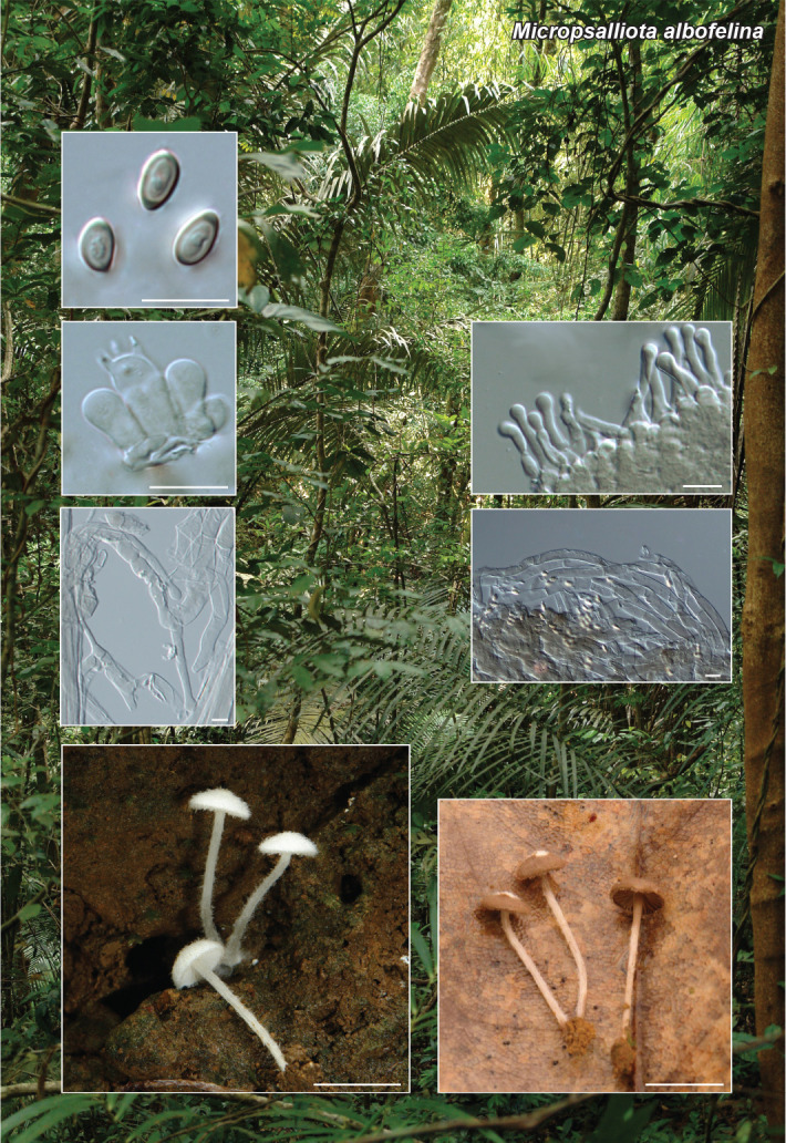
Notes — Based on a megablast search of NCBIs GenBank nucleotide database, the LSU and rpb2 sequences of Microdochium maculosum showed a similarity of 99.77 % and 91.31 % respectively, with the sequences of Microdochium indocalami (SAUCC1016, GenBank MT199878 and MT510550); whereas the ITS sequence was 98.61 % similar to that of Microdochium sp. (JP103, GenBank AB255278). Microdochium maculosum had a similarity of 97.62 % with ITS sequence of Microdochium fisheri strain CBS 242.91 (GenBank NR_155374), 98.70 % similarity with LSU and 89.29 % with rpb2 of M. fisheri strain CBS 242.90, (GenBank KP858951 and KP859124). The phylogenetic reconstruction using ITS, LSU and rpb2 of accepted Microdochium species, showed that the new species was located in an highly-supported clade together with M. indocalami, M. fisheri, M. lycopodinum, M. phragmitis, and M. rhopalostylidis, being closely related with the former two species. However, M. maculosum has clear morphological differences distinguishing it from M. fisheri and M. indocalami. Microdochium fisheri has fusoid, obovoid, subpyriform to clavate (7–12 × 3–4 μm, 0–1-septate) conidia (Hernández-Restrepo et al. 2016a), and M. indocalami has conidia which are cylindrical, clavate to obovoid (13–15.5 × 3.5–5.5 μm, 1–3-septate) and have a flattened base and no distinct hila (Huang et al. 2020). On the other hand, the new species M. maculosum has fusoid, straight or curved conidia (6–15 × 2–4 μm, 1–3-septate). Microdochium fisheri is known from Oryza sativa in India and the UK, and M. indocalami is known from Indocalamus longiauritus in China (Farr & Rossman 2021). No species of Microdochium have ever been reported on members of Digitaria prior to the present work.

graphic file with name per-2023-47-6-i037.jpg

Bayesian tree inferred from the combined datasets of ITS, LSU and rpb2 sequences from species belonging to the genus Microdochium, including the specimen of Microdochium maculosum obtained in this study (indicated in bold). The analysis was performed with 10 M generations in MrBayes v. 3.2.6 (Ronquist et al. 2012). Bayesian probabilities > 0.50 are given at each node. The tree is rooted to Idriella lunata CBS 204.56. GenBank accession numbers of the sequences used in this study are from Huang et al. (2020).

Colour illustrations. Microdochium maculosum causing leaf spots in Digitaria insularis. Conidiophores, with denticulate conidiogenous cells; fusiform to allantoid conidia; colonies in OA and MEA media. Scale bars = 10 μm.

graphic file with name per-2023-47-6-g094.jpg

Microdochium maculosum

Associated Data

    This section collects any data citations, data availability statements, or supplementary materials included in this article.

    Supplementary Materials

    FP1284

    Phylogenetic tree.

    See the phylogenetic tree provided with the supplementary material FP1284.

    FP1287

    Phylogenetic tree.

    per-2023-47-6-SF1287.jpg (643.7KB, jpg)
    FP1290

    Phylogenetic tree.

    per-2023-47-6-SF1290.jpg (974.6KB, jpg)
    FP1292

    Phylogenetic tree.

    per-2023-47-6-SF1292.jpg (729.3KB, jpg)
    FP1293

    Phylogenetic tree.

    per-2023-47-6-SF1293.jpg (589.8KB, jpg)
    FP1296

    Phylogenetic tree.

    per-2023-47-6-SF1296.jpg (704.1KB, jpg)
    FP1298

    Phylogenetic tree.

    FP1299

    Phylogenetic tree.

    per-2023-47-6-SF1299.jpg (588.8KB, jpg)
    FP1300

    Phylogenetic tree.

    per-2023-47-6-SF1300.jpg (1,022.3KB, jpg)

    See the phylogenetic tree provided with the supplementary material FP1300.

    per-2023-47-6-SF1300.jpg (1,022.3KB, jpg)
    FP1302

    Phylogenetic tree.

    per-2023-47-6-SF1302.jpg (909.1KB, jpg)
    FP1303

    Phylogenetic tree.

    per-2023-47-6-SF1303.jpg (518.6KB, jpg)
    FP1304

    Phylogenetic tree.

    per-2023-47-6-SF1304.jpg (419.8KB, jpg)
    FP1305

    Phylogenetic tree.

    per-2023-47-6-SF1306.jpg (903.4KB, jpg)

    See the phylogenetic tree provided with the supplementary material FP1290.

    per-2023-47-6-SF1290.jpg (974.6KB, jpg)

    See the phylogenetic tree provided with the supplementary material FP1290.

    per-2023-47-6-SF1290.jpg (974.6KB, jpg)

    See the phylogenetic tree provided with the supplementary material FP1290.

    per-2023-47-6-SF1290.jpg (974.6KB, jpg)

    See the phylogenetic tree provided with the supplementary material FP1293.

    per-2023-47-6-SF1293.jpg (589.8KB, jpg)

    See the phylogenetic tree provided with the supplementary material FP1298.

    FP1311

    Phylogenetic tree.

    per-2023-47-6-SF1311.jpg (474.4KB, jpg)

    See the phylogenetic tree provided with the supplementary material FP1293.

    per-2023-47-6-SF1293.jpg (589.8KB, jpg)

    See the phylogenetic tree provided with the supplementary material FP1290.

    per-2023-47-6-SF1290.jpg (974.6KB, jpg)

    See the phylogenetic tree provided with the supplementary material FP1293.

    per-2023-47-6-SF1293.jpg (589.8KB, jpg)

    See the phylogenetic tree provided with the supplementary material FP1293.

    per-2023-47-6-SF1293.jpg (589.8KB, jpg)

    Also see the phylogenetic tree provided with the supplementary material FP1284.

    FP1318

    Phylogenetic tree.

    per-2023-47-6-SF1318.jpg (974.5KB, jpg)

    See the phylogenetic tree provided with the supplementary material FP1284.

    See the phylogenetic tree provided with the supplementary material FP1284.

    See the phylogenetic tree provided with the supplementary material FP1284.

    See the phylogenetic tree provided with the supplementary material FP1303.

    per-2023-47-6-SF1303.jpg (518.6KB, jpg)

    See the phylogenetic tree provided with the supplementary material FP1284.

    FP1325

    Phylogenetic tree.

    per-2023-47-6-SF1325.jpg (580.9KB, jpg)

    See the phylogenetic tree provided with the supplementary material FP1284.

    See the phylogenetic tree provided with the supplementary material FP1304.

    per-2023-47-6-SF1304.jpg (419.8KB, jpg)
    FP1328

    Phylogenetic tree.

    See the phylogenetic tree provided with the supplementary material FP1318.

    per-2023-47-6-SF1318.jpg (974.5KB, jpg)
    FP1331

    Phylogenetic tree.

    per-2023-47-6-SF1331.jpg (732.5KB, jpg)
    FP1332

    Phylogenetic tree.

    per-2023-47-6-SF1332.jpg (999.3KB, jpg)
    FP1333

    Phylogenetic tree.

    per-2023-47-6-SF1333.jpg (589.6KB, jpg)
    FP1337

    Phylogenetic tree.

    per-2023-47-6-SF1337.jpg (502.7KB, jpg)
    FP1338

    Phylogenetic tree.

    per-2023-47-6-SF1338.jpg (415.3KB, jpg)
    FP1339

    Phylogenetic tree.

    per-2023-47-6-SF1339.jpg (364.8KB, jpg)
    FP1340

    Phylogenetic tree.

    per-2023-47-6-SF1340.jpg (373.5KB, jpg)
    FP1344

    Phylogenetic tree.

    per-2023-47-6-SF1344.jpg (321.9KB, jpg)
    FP1347

    Phylogenetic tree.

    per-2023-47-6-SF1347.jpg (650.4KB, jpg)
    FP1352-1

    Phylogenetic tree.

    FP1352-2

    Phylogenetic tree.

    FP1353

    Phylogenetic tree.

    per-2023-47-6-SF1353.jpg (640.3KB, jpg)
    FP1356

    Phylogenetic tree.

    per-2023-47-6-SF1356.jpg (773.7KB, jpg)
    FP1357-1

    Phylogenetic tree.

    per-2023-47-6-SF1357-1.jpg (1,009.7KB, jpg)
    FP1357-2

    Table. Species of Hesperomyces included in this study.

    FP1358

    Table. Morphological characteristics of Heterophoma rehmanniae and published descriptions of Heterophoma spp.

    per-2023-47-6-SF1358.jpg (205.6KB, jpg)
    FP1360

    Phylogenetic tree.

    per-2023-47-6-SF1360.jpg (652.5KB, jpg)
    FP1362

    Phylogenetic tree.

    per-2023-47-6-SF1362.jpg (368.2KB, jpg)
    FP1366

    Phylogenetic tree.

    FP1369

    Phylogenetic tree.

    per-2023-47-6-SF1369.jpg (564.7KB, jpg)
    FP1373

    Phylogenetic tree.

    per-2023-47-6-SF1373.jpg (653.2KB, jpg)
    FP1375

    Phylogenetic tree.

    per-2023-47-6-SF1375.jpg (473.3KB, jpg)
    FP1378

    Line-drawn micromorphological structure of Trechispora patawaense basidiomata (WU-Mykol. 44791, type). a. Hyphae from subiculum; b. hyphae from cords; c. hyphae from dissepiment edge; d. subhymenium and hymenium; e. basidiospores. Scale bars = 10 μm (a–d), 5 μm (e).

    per-2023-47-6-SF1378.jpg (382.6KB, jpg)
    FP1378 & 1379

    Phylogenetic tree.

    per-2023-47-6-SF1378.jpg (382.6KB, jpg)
    FP1379

    Line-drawn micromorphological structure of Trechispora subregularis basidiomata (WU-Mykol. 44792, type). a. Hyphae from subiculum; b. hyphae from cords; c. hyphae from dissepiment edge; d.cystidia-like hyphal segment; e. subhymenium and hymenium; f. basidiospores. Scale bars = 10 μm (a–e), 5 μm (f).

    FP1378 & 1379

    Phylogenetic tree.

    per-2023-47-6-SF1378.jpg (382.6KB, jpg)

    Articles from Persoonia : Molecular Phylogeny and Evolution of Fungi are provided here courtesy of Naturalis Biodiversity Center & Centraalbureau voor Schimmelcultures

    RESOURCES