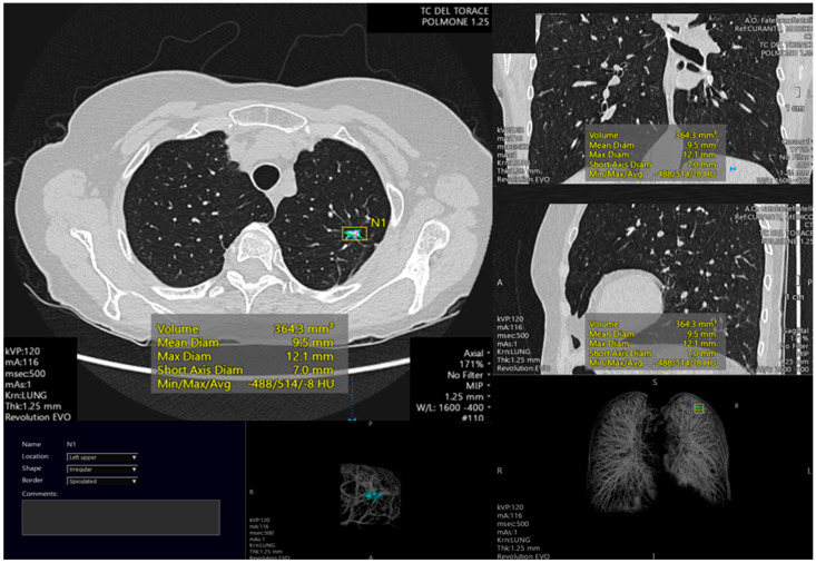
Figure 1.
Example of pulmonary lesion automated segmentation. The lesion is located in the left higher lobe. The automated analysis permits to calculate different lesion parameters such as volume (mm3), mean diameter (mm), maximum diameter (mm), short axis diameter (mm) and density (Hounsfield Units). Also, 3D reconstruction is shown.

