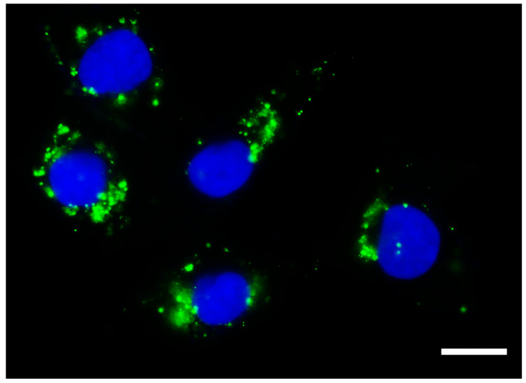
Figure 1.
Exosomes taken up by cancer cells. Photomicrography obtained using high-content screening showing exosomes isolated from the conditioned culture medium of human immature dental pulp stem cells (stained with Vybrant DiO, in green) within the cytoplasm of a cancer cell line derived from metastatic anaplastic thyroid cancer (HTh83). Photomicrograph obtained 24 h after the addition of 50 µg/mL of exosomes. Scale bar 10 µm. Nucleus stained with Hoechst. Total magnification of 100×.

