Figure 4.
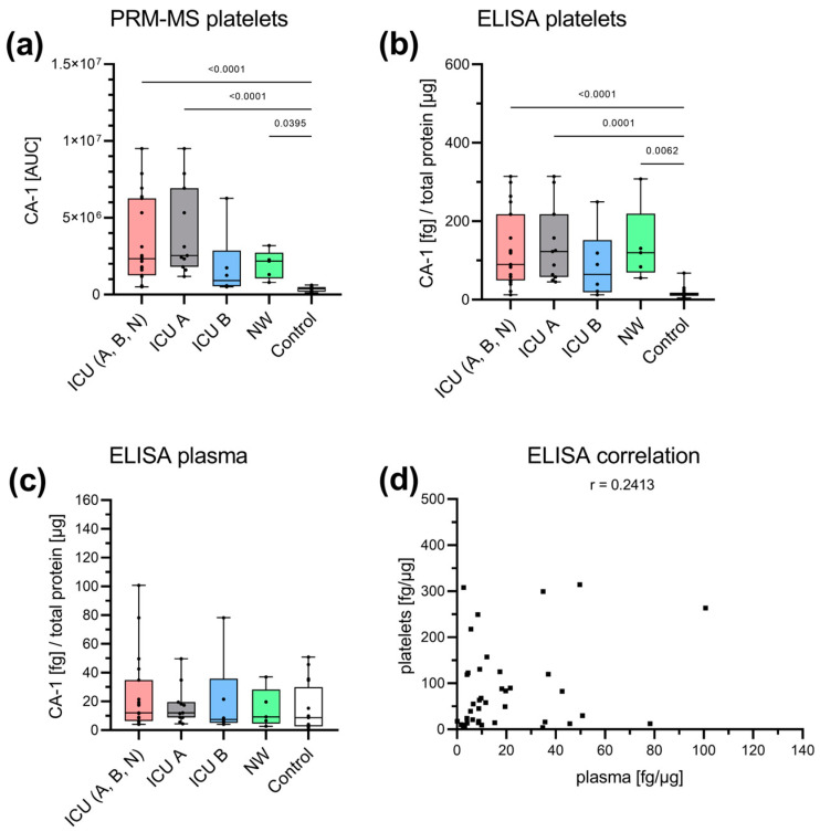
Specific analysis of CA-1. CA-1 was analyzed with PRM-MS and ELISA in both platelets (a,b) and plasma (c). With PRM-MS, CA-1 could only be detected in the platelet lysate. The abundance was significantly higher in the ICU group ((A+B+N) and ICU A), as well as the NW group, than in the control group. There was a tendency toward a decrease in ICU B (survivors) compared with ICU A (non-survivors) and NW. A similar pattern was found with ELISA. No changes were detected in plasma using ELISA. There was no correlation between platelet and plasma concentrations (p = 0.1336) (d). Platelet and plasma readings are presented as boxplots. Data were analyzed using the Kruskal–Wallis test followed by Dunn’s multiple comparison. The correlation was determined according to Spearman. ICU: intensive care unit (ICU A: non-survivors, ICU B: survivors, ICU N: unknown); NW: normal ward.

