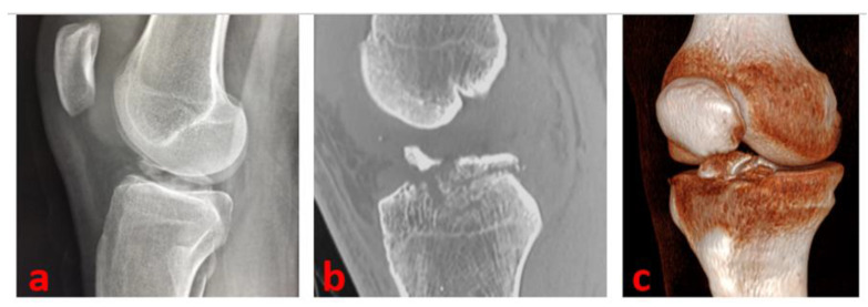
Figure 1.
A true lateral knee radiographic view of the right knee of a 16-year-old male patient of our series shows a Type 3 tibial avulsion fracture (a); CT scans are useful to define the pattern of the fracture and drive the management: CT sagittal view scan (b) and 3D CT reconstruction (c) of the same patient.

