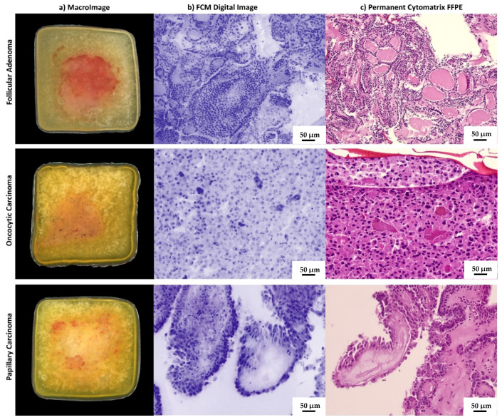
Figure 2.
Imaging of cases from the study. Macro-images of the holder with FNA sample as it is placed within the microscope slot are shown in the column (a). Vivascope digital immediate images converted in pseudocolor are shown in column (b). Conventional H&E-stained sections from FFPE Cytomatrix blocks are shown in column (c). The case from the first line is a follicular adenoma in agreement with histological diagnosis on the surgically removed thyroid gland. The case in line 2 is an oncocytic cell carcinoma, and in line 3 there is a papillary carcinoma, hobnail subtype.

