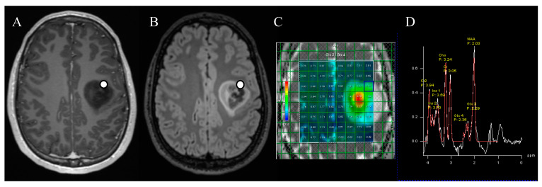
Figure 3.
Marked location of tissue collection for ex vivo 2-HG concentration measurements in axial post-contrast T1-weighted (A) and FLAIR (B) sequences. The patient had a left frontal diffuse glioma, which turned out to be IDH1-mutated. The 2-HG MR spectroscopy (C) was positive, as demonstrated in the metabolite peaks (D) by an elevation of the Glx3 peak compared to the Glx4 peak, attributable to 2-HG.

