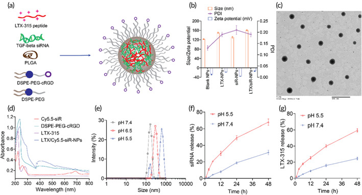
FIGURE 1.

LTX/siR‐NPs preparation and characterization. (a) The representative structure of LTX/siR‐NPs. (b) Particle size distribution and zeta potential of LTX/siR‐NPs were measured by the dynamic light scattering method. (c) The spherical morphology of LTX/siR‐NPs was observed by transmission electron microscopy (scale bar: 500 nm). (d) The UV–VIS spectra of components and LTX/siR‐NPs confirmed the successful load of LTX‐315 and TGF‐β siRNA into the hybrid nanoparticles. (e) The influence of pH conditions on the size of LTX/siR‐NPs. The in vitro accumulative drug‐release profile of (f) siRNA and (g) LTX‐315 at different pH conditions
