FIGURE 2.

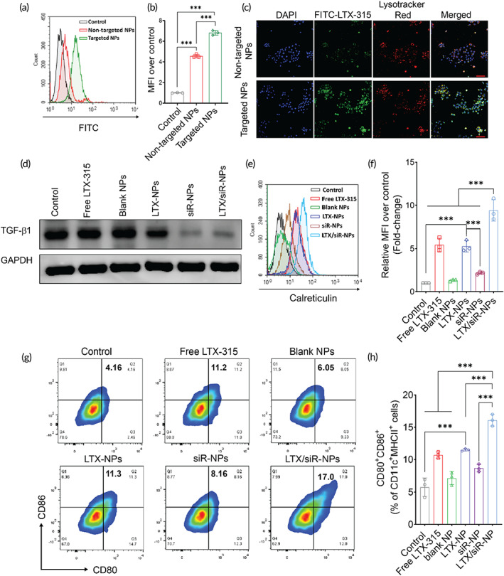
Cellular uptake of LTX/siR‐NPs into 4T1 cells followed by the induction of the immunogenic cell death and reduced expression of TGF‐β1. The internalization of targeted LTX/siR‐NPs (with cRGD conjugation) and nontargeted LTX/siR‐NPs (without cRGD conjugation) into 4T1 cells was confirmed by (a, b) flow cytometry analysis, and (c) confocal laser scanning microscopy (scale bar: 50 nm). (d) Western Blot analysis of TGF‐β1 expression in 4T1 cells after treatment with (1) PBS (Control), (2) free LTX‐315, (3) blank NPs, (4) LTX‐NPs, (5) siR‐NPs, and (6) LTX/siR‐NPs. (e, f) The plasma membrane expression of calreticulin in 4T1 cells after treatment determined by flow cytometry. (g, h) The expression of co‐stimulatory molecules on the surface of BMDCs after the treatments with supernatant of treated cancer cells. Data were presented as mean ± SD (n = 3). **p < 0.01; ***p < 0.001
