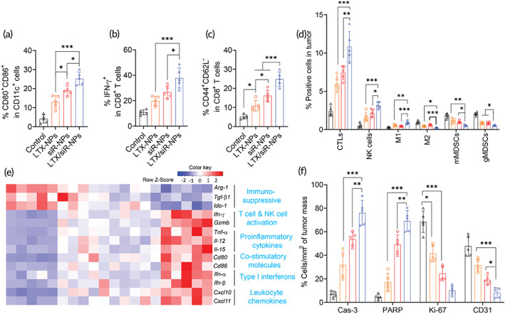
FIGURE 4.

LTX/siR‐NPs re‐shaped the immune cell profile of tumor microenvironment. Flow cytometric analysis of (a) matured DC in tdLNs, (b) IFN‐γ produced by CD8+ T cells and (c) effector memory CD8+ T cells in the spleen, and (d) intratumoral profiles of immune cells of the mice treated with indicated formulations. (e) Heatmap visualization of gene expression in tumors collected from treated mice. (f) Cell percentages (%/mm2 of tumor mass) apoptosis markers (cleaved caspse‐3; cleaved PARP), tumor proliferation (Ki‐67), and angiogenesis marker (CD31) were detected by Immunohistochemical analysis. Data represented as mean ± SD (n = 5). One‐way ANOVA with Tukey's multiple comparison test, *p < 0.05; **p < 0.01; ***p < 0.001
