
FIGURE 3.
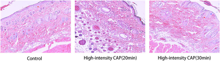
HE staining of erythematous or edematous skin tissues, with enlarged cell gaps indicated by blue arrows and inflammatory cells shown by red arrows. The magnification of these images is 100×. HE, hematoxylin–eosin.
Official websites use .gov
A
.gov website belongs to an official
government organization in the United States.
Secure .gov websites use HTTPS
A lock (
) or https:// means you've safely
connected to the .gov website. Share sensitive
information only on official, secure websites.
HE staining of erythematous or edematous skin tissues, with enlarged cell gaps indicated by blue arrows and inflammatory cells shown by red arrows. The magnification of these images is 100×. HE, hematoxylin–eosin.