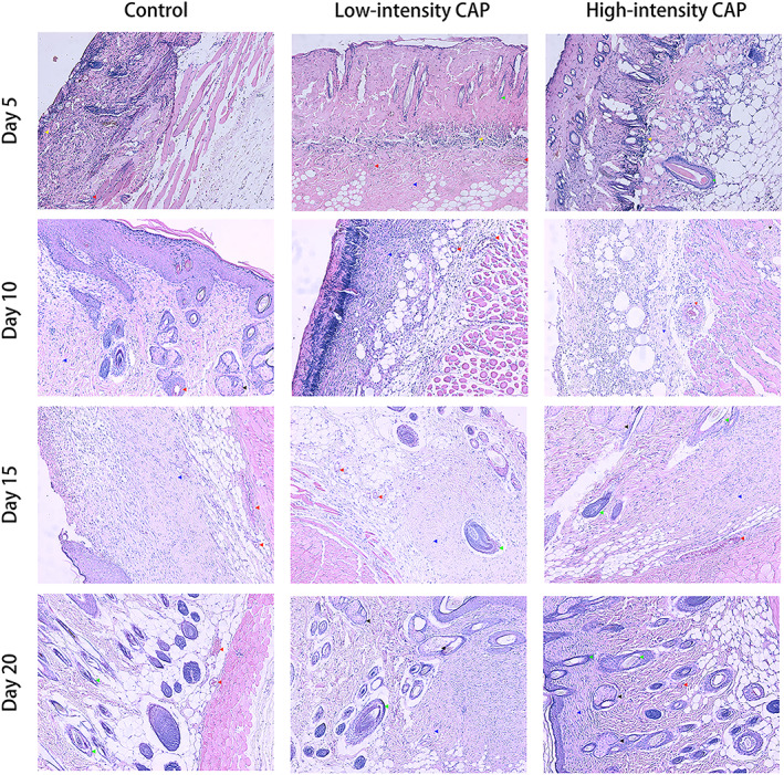
FIGURE 5.

Representative histological features of rat skin: from top to bottom are the images of rat skin on day 5, 10, 15, and 20, respectively, with the control, low‐intensity CAP, and high‐intensity CAP groups, respectively in each row from left to right (yellow, red, green, black, and blue arrows point to inflammatory cells, neovascularization, hair follicles, sebaceous glands, and fibrous components, respectively). The magnification of these images is 100×. CAP, cold atmospheric plasma.
