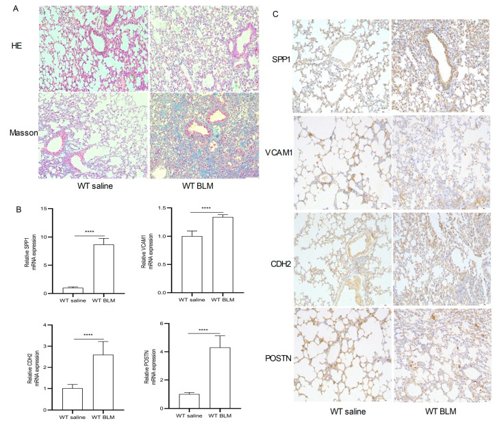
Figure 7.
Pathological staining and RT-qPCR. (A) Hematoxylin–eosin (HE) and Masson Trichrome staining in the WT saline group and WT BLM group (original magnification, ×100). (B) RT-qPCR analysis levels of SPP1, VCAM1, CDH2, and POSTN mRNA expression in the WT saline group and WT BLM group (data represent the mean ± SD, n = 6). **** p < 0.0001. (C) Immunohistochemical analysis of SPP1, VCAM1, CDH2, and POSTN expression in the WT saline group and WT BLM group (original magnification, ×100, n = 4). Abbreviations: BLM, bleomycin; SPP1, Secreted Phosphoprotein 1; VCAM1, Vascular cell adhesion molecule 1; CDH2, Cadherin 2; POSTN, Periostin.

