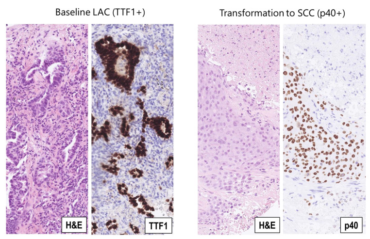
Figure 3.
NSCLC histological phenotypes in case 3. Left panels: Diagnostic biopsy (baseline) from a metastasis to the thoracic lymph nodal station 4R with phenotype of lung adenocarcinoma (LAC); hematoxylin-eosin staining (H&E) shows malignant adeno-papillary structures, which are positive for the immunohistochemical LAC biomarker TTF1 (TTF1). Right panels: Rebiopsy from progressive metastasis in the left lung shows tumor transformation to squamous cell carcinoma (SCC); hematoxylin-eosin staining (H&E) shows a solid, slightly keratinizing tumor tissue, which is positive for the immunohistochemical SCC biomarker p40 (p40). (All magnifications, ×200).

