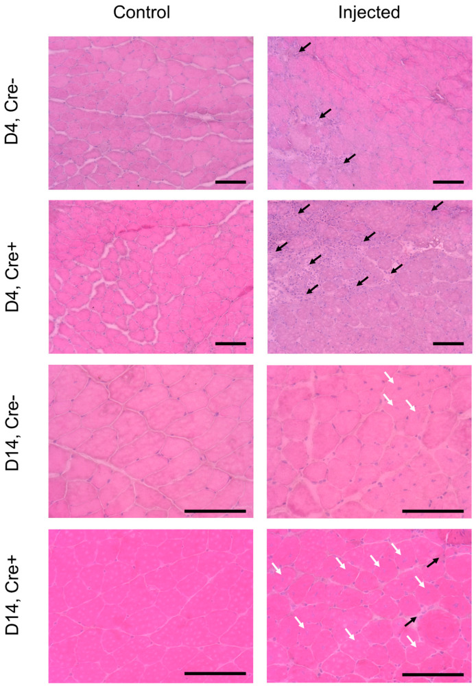
Figure 1.
Increased inflammation detected by morphological and histological investigation in Sept7 conditional knock-down mice. Hematoxylin–eosin staining of a cross section of m. tibialis anterior on 4th and 14th days after BaCl2 injection into Cre− and Cre+ mice. Control panels indicate non-injected legs. Black arrows indicate inflammatory cells. Scale bar is 100 µm. White arrows point to central nuclei.

